. 2024 May 8;23(1):95.
doi: 10.1186/s12943-024-02001-2.

Interleukin-21 receptor signaling promotes metabolic dysfunction-associated steatohepatitis-driven hepatocellular carcinoma by inducing immunosuppressive IgA+ B cells

Affiliations

Interleukin-21 receptor signaling promotes metabolic dysfunction-associated steatohepatitis-driven hepatocellular carcinoma by inducing immunosuppressive IgA+ B cells

Ying Xie et al. Mol Cancer. .

Abstract

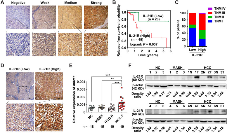
Background: Dysregulation of immune surveillance is tightly linked to the development of metabolic dysfunction-associated steatohepatitis (MASH)-driven hepatocellular carcinoma (HCC); however, its underlying mechanisms remain unclear. Herein, we aimed to determine the role of interleukin-21 receptor (IL-21R) in MASH-driven HCC.

Methods: The clinical significance of IL-21R was assessed in human HCC specimens using immunohistochemistry staining. Furthermore, the expression of IL-21R in mice was assessed in the STAM model. Thereafter, two different MASH-driven HCC mouse models were applied between IL-21R-deficient mice and wild type controls to explore the role of IL-21R in MASH-driven HCC. To further elucidate the potential mechanisms by which IL-21R affected MASH-driven HCC, whole transcriptome sequencing, flow cytometry and adoptive lymphocyte transfer were performed. Finally, flow cytometry, enzyme-linked immunosorbent assay, immunofluorescent staining, chromatin immunoprecipitation assay and western blotting were conducted to explore the mechanism by which IL-21R induced IgA+ B cells.

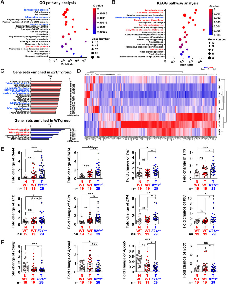
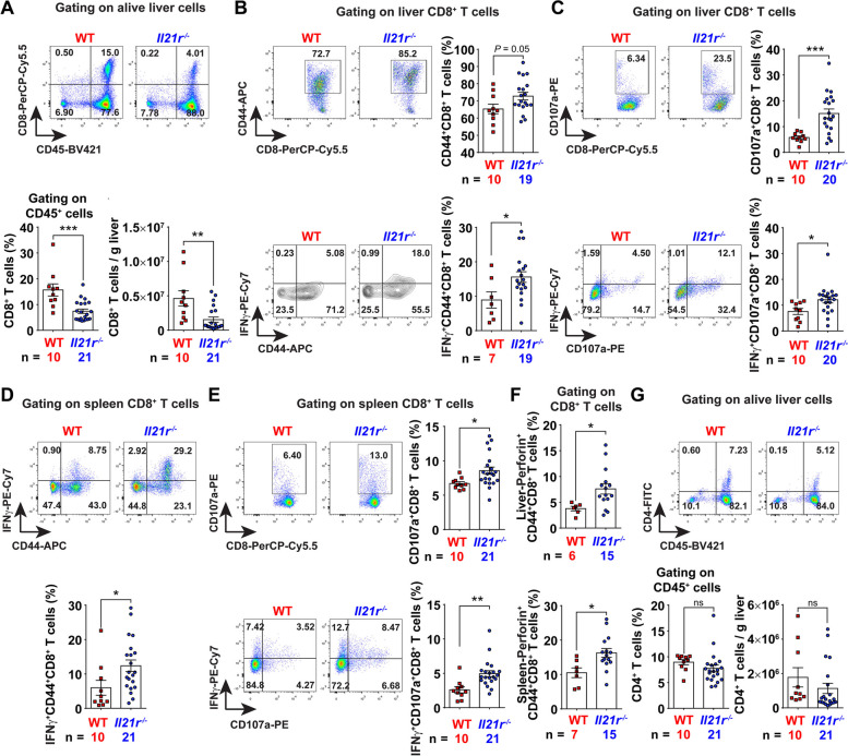
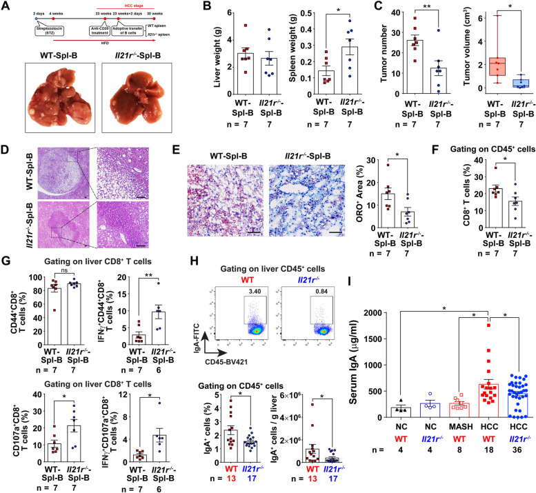
Results: HCC patients with high IL-21R expression exhibited poor relapse-free survival, advanced TNM stage and severe steatosis. Additionally, IL-21R was demonstrated to be upregulated in mouse liver tumors. Particularly, ablation of IL-21R impeded MASH-driven hepatocarcinogenesis with dramatically reduction of lipid accumulation. Moreover, cytotoxic CD8+ T lymphocyte activation was enhanced in the absence of IL-21R due to the reduction of immunosuppressive IgA+ B cells. Mechanistically, the IL-21R-STAT1-c-Jun/c-Fos regulatory axis was activated in MASH-driven HCC and thus promoted the transcription of Igha, resulting in the induction of IgA+ B cells.

Conclusions: IL-21R plays a cancer-promoting role by inducing IgA+ B cells in MASH-driven hepatocarcinogenesis. Targeting IL-21R signaling represents a potential therapeutic strategy for cancer therapy.

Keywords: Cancer-promoting role; IL-21R; IgA+ B cell; MASH-driven HCC.


Conflict of interest statement

The authors declare no competing interests.

Figures

Fig. 1
Fig. 1
The expression of IL-21R is increased in MASH-driven HCC tissues and higher expression of IL-21R is associated with shorter relapse-free survival in HCC patients. A Shown are the representative pictures for immunohistochemistry (IHC) staining with different levels of IL-21R-staining in human HCC tissues. B Higher expression of IL-21R was associated with shorter relapse-free survival in HCC patients. The HCC patients (n = 69) were divided into IL-21R-low and IL-21R-high groups according to the evaluation scores of IHC in human HCC tissues. C The distribution of clinical stages upon diagnosis between HCC patients with low- or high-IL-21R expression. D HCC patients with higher expression of IL-21R exhibited more severe steatosis. E, F The mRNA (E) and protein (F) levels of IL-21R were increased in mouse MASH-driven HCC. The mRNA or protein levels of IL-21R were detected in mouse liver tissue, paracancerous tissue (N), and cancer tissue (T) from mice at normal chow (NC), metabolic dysfunction-associated steatohepatitis (MASH) and hepatocellular carcinoma (HCC) stages by using qRT-PCR or western blotting. β-actin, internal control. Scale bar = 100μm. The number of patients or mice in each group is shown in B and E accordingly. One-way ANOVA was used to determine significance in E. ** P < 0.01, *** P < 0.001
Fig. 2
Fig. 2
Il21r−/− mice exhibit less tumor burden of HCC with decreased lipid droplets. A The body weight gain of IL-21R-deficient (Il21r−/−) mice and wild type (WT) controls in the STAM model. Mice were fed with high-fat diet at four weeks of age, and continuously monitored from four to 27 weeks, and 12 data points were used to analyze weight gain, with 25 mice per time point. B-H Comparison of body weight of endpoint (B), serum ALT levels (C), liver weight, liver/body weight ratio (D), representative pictures of liver (E), number and volume of tumors (F), largest diameter of tumor (G), and number and volume of large tumors (H) between Il21r−/− mice and WT controls at 27 weeks of age in the STAM model. I Shown are the representative pictures for hematoxylin–eosin (HE), Oil Red O (ORO), Sirius Red (SR) and F4/80 immunohistochemistry staining of the liver/tumor tissues between Il21r−/− mice and WT controls at 27 weeks of age in the STAM model. Scale bar = 100μm. J Lipid droplets were significantly decreased in Il21r−/− mice. Lipid droplets and collagen deposition were quantified according to the image analysis of ORO and SR staining, respectively. K, L Triglyceride and cholesterol levels were significantly decreased in the serum or liver tumor extracts of Il21r−/− mice. Serum (K) or liver tumor extracts (L) were analyzed for triglyceride, total cholesterol and free cholesterol. The number of mice in each group is shown in panels accordingly. To determine the significance, two-way ANOVA was used in A while Student's t test was used in other panels accordingly. * P < 0.05, ** P < 0.01, *** P < 0.001, ns, not significant difference
Fig. 3
Fig. 3
Differential genes between the tumor tissues of Il21r−/− mice and wild type controls are enriched in immune, inflammatory and metabolic pathways. A, B GO and KEGG pathway analyses of the differential genes in tumor tissues between Il21r−/− mice and wild type (WT) controls. The horizontal axis shows the enrichment score. The larger the bubble is, the more differential genes are contained in the entry. The smaller the enrichment Q Value is, the greater the degree of significance. In the vertical axis, the pathways in blue font represent immune- and inflammation-related pathways, whereas the pathways in red font represent metabolism-related pathways. C The top hallmark gene sets sorted by normalized enrichment score (NES) are shown to depict gene sets enriched in Il21r−/− mice (upper panel) and WT controls (lower panel) according to gene set enrichment analysis (GSEA). The immune- and inflammation-related gene sets are colored in blue, whereas the metabolism-related gene sets are colored in red. D Heat map depicting the differential expression of 170 immune- and inflammation-related genes in the indicated strains, illustrating the upregulation of most immune- and inflammation-related genes in Il21r−/− mice. E, F The mRNA levels of several immune- and inflammation-related genes (E) and metabolism-related genes (F) were detected in the paracancerous tissue (N), and tumor tissue (T) from Il21r−/− mice and WT controls. For E and F, the number of mice in each group is shown. Student's t test was used to determine significance. * P < 0.05, ** P < 0.01, *** P < 0.001, ns, not significant difference
Fig. 4
Fig. 4
Ablation of IL-21R enhances cytotoxic CD8+ T lymphocyte activation in vivo. A Liver/tumor CD8+ T cells were significantly decreased in Il21r−/− mice. The representative flow cytometry images are shown in the upper panel. In the lower panel, the percentages of CD8+ T cells and the absolute cell number of CD8+ T cells per gram of liver/tumor tissues were analyzed between Il21r−/− mice and wild type (WT) controls. B Effector CD8+ T cells were significantly increased in the liver/tumor tissues of Il21r−/− mice. The percentages of CD8+ T cells positive for CD44 (upper panel), CD44 and IFNγ (lower panel) in the liver/tumor tissues were analyzed between Il21r−/− mice and WT controls. C Degranulating CD8+ T cells were significantly increased in the liver/tumor tissues of Il21r−/− mice. The percentages of CD8+ T cells positive for CD107a (upper panel), CD107a and IFNγ (lower panel) in liver/tumor were analyzed between Il21r−/− mice and WT controls. D, E Both effector (D) and degranulating (E) CD8+ T cells were significantly increased in the spleens of Il21r−/− mice. The percentages of CD8+ T cells positive for CD44 and IFNγ (D), CD107a, CD107a and IFNγ (E) in the spleen were analyzed between Il21r−/− mice and WT controls. F Perforin+CD8+ T cells were significantly increased in Il21r−/− mice. G Ablation of IL-21R did not change the percentage and number of liver/tumor CD4+ T cells. The percentages of CD4+ T cells and the absolute cell number of CD4+ T cells per gram of liver/tumor tissues were analyzed between Il21r−/− mice and WT controls. The gating schemes are indicated above each panel, and the number of mice in each group is shown in the panels accordingly. Student's t test was used to determine significance. * P < 0.05, ** P < 0.01, *** P < 0.001, ns, not significant difference
Fig. 5
Fig. 5
IL-21R-deficient mice have fewer IgA+ B cells, and thus impede MASH-driven HCC development. A Shown are the schematic diagram of adoptive lymphocyte transfer experiment and the representative pictures of the endpoint liver from mice transferred with B cells from Il21r−/− mice (Il21r−/−-Spl-B) or WT controls (WT-Spl-B). B, C Comparison of liver weight, spleen weight (B) and the number and volume of tumors (C) between the mice transferred with B cells from Il21r−/− mice and WT controls. D Shown are the representative pictures for hematoxylin–eosin (HE) staining of liver/tumor tissues between the mice transferred with B cells from Il21r−/− mice and WT controls. E Lipid droplets were significantly decreased in mice transferred with B cells from Il21r−/− mice. Lipid droplets were quantified according to the image analysis of Oil Red O (ORO). Scale bar = 100μm in D and E. F Liver/tumor CD8+ T cells were significantly decreased in the mice transferred with B cells from Il21r−/− mice. G Both effector (upper panel) and degranulating (lower panel) CD8+ T cells were significantly increased in the liver/tumor tissues of mice transferred with B cells from Il21r−/− mice. H Liver/tumor IgA+ B cells were significantly decreased in Il21r−/− mice. The representative flow cytometry images are shown in the upper panel. In the lower panel, the percentages of IgA+ B cells and the absolute cell number of IgA+ B cells per gram of liver/tumor tissues were analyzed between Il21r−/− mice and wild type (WT) controls. I The level of serum IgA was significantly decreased in Il21r−/− mice. The gating schemes are indicated above in FH, and the number of mice in each group is shown in the panels accordingly. Student's t test was used to determine significance. * P < 0.05, ** P < 0.01, ns, not significant difference
Fig. 6
Fig. 6
The IL-21R-STAT1-c-Jun/c-Fos-IgA axis and its implication in MASH-driven HCC. A The B cells isolated from Il21r−/− mice exhibited lower Igha level compared to those isolated from wild type controls. Purified CD19+ B cells by using magnetic beads were obtained from the splenocytes of Il21r−/− mice or wild type (WT) controls, and subsequently subjected to qRT-PCR to detect the mRNA level of Igha. B Ablation of IL-21R decreased the occupancy of c-Jun/c-Fos complex on the promoter of Igha in isolated B cells. The purified CD19+ B cells mentioned in A were subjected to chromatin immunoprecipitation. The chromatin complexes precipitated by the anti-c-Jun or anti-c-Fos antibody and the corresponding isotype-matched IgG were recovered and purified, and subsequently subjected to qRT-PCR using primer sets covering the AP-1 binding sites on the promoter of Igha. The binding sites are indicated above. C Ablation of IL-21R downregulated the protein levels of p-STAT1, c-Jun and c-Fos. The purified CD19+ B cells mentioned in A were subjected to western blotting. β-actin, internal control. D Shown are the schematic diagram of antibody neutralization experiment and the representative pictures of the endpoint liver from mice injected with IL-21R blocking antibody (αIL-21R) or its isotype control (IgG). E The number and volume of liver tumors were significantly decreased in the mice injected with IL-21R antibody compared to those injected with isotype control. F Blocking of IL-21R downregulated the protein levels of IL-21R, p-STAT1, STAT1 and c-Jun in the B cells. Purified CD19+ B cells by using magnetic beads were obtained from the splenocytes of mice injected with IL-21R blocking antibody or its isotype control, and subsequently subjected to western blotting. β-actin, internal control. G Model of IL-21R-STAT1-c-Jun/c-Fos-IgA axis and its implication in MASH-driven HCC. The number of mice in each group is shown in A and E. Student's t test was used to determine significance. * P < 0.05, ** P < 0.01, *** P < 0.001

References

    1. Rinella ME, Lazarus JV, Ratziu V, Francque SM, Sanyal AJ, Kanwal F, et al. A multisociety Delphi consensus statement on new fatty liver disease nomenclature. J Hepatol. 2023;79:1542–1556. doi: 10.1016/j.jhep.2023.06.003. - DOI - PubMed
    1. Huang DQ, El-Serag HB, Loomba R. Global epidemiology of NAFLD-related HCC: trends, predictions, risk factors and prevention. Nat Rev Gastroenterol Hepatol. 2021;18:223–238. doi: 10.1038/s41575-020-00381-6. - DOI - PMC - PubMed
    1. Tan DJH, Ng CH, Lin SY, Pan XH, Tay P, Lim WH, et al. Clinical characteristics, surveillance, treatment allocation, and outcomes of non-alcoholic fatty liver disease-related hepatocellular carcinoma: a systematic review and meta-analysis. Lancet Oncol. 2022;23:521–530. doi: 10.1016/S1470-2045(22)00078-X. - DOI - PMC - PubMed
    1. Febbraio MA, Reibe S, Shalapour S, Ooi GJ, Watt MJ, Karin M. Preclinical Models for Studying NASH-Driven HCC: How Useful Are They? Cell Metab. 2019;29:18–26. doi: 10.1016/j.cmet.2018.10.012. - DOI - PMC - PubMed
    1. Anstee QM, Reeves HL, Kotsiliti E, Govaere O, Heikenwalder M. From NASH to HCC: current concepts and future challenges. Nat Rev Gastroenterol Hepatol. 2019;16:411–428. doi: 10.1038/s41575-019-0145-7. - DOI - PubMed

Publication types

MeSH terms