Review
. 2024 May 8;17(1):31.
doi: 10.1186/s13045-024-01544-7.

Understanding the immunosuppressive microenvironment of glioma: mechanistic insights and clinical perspectives

Affiliations
Review

Understanding the immunosuppressive microenvironment of glioma: mechanistic insights and clinical perspectives

Hao Lin et al. J Hematol Oncol. .

Abstract

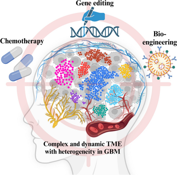
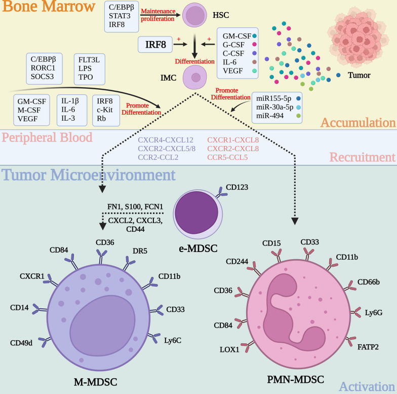
Glioblastoma (GBM), the predominant and primary malignant intracranial tumor, poses a formidable challenge due to its immunosuppressive microenvironment, thereby confounding conventional therapeutic interventions. Despite the established treatment regimen comprising surgical intervention, radiotherapy, temozolomide administration, and the exploration of emerging modalities such as immunotherapy and integration of medicine and engineering technology therapy, the efficacy of these approaches remains constrained, resulting in suboptimal prognostic outcomes. In recent years, intensive scrutiny of the inhibitory and immunosuppressive milieu within GBM has underscored the significance of cellular constituents of the GBM microenvironment and their interactions with malignant cells and neurons. Novel immune and targeted therapy strategies have emerged, offering promising avenues for advancing GBM treatment. One pivotal mechanism orchestrating immunosuppression in GBM involves the aggregation of myeloid-derived suppressor cells (MDSCs), glioma-associated macrophage/microglia (GAM), and regulatory T cells (Tregs). Among these, MDSCs, though constituting a minority (4-8%) of CD45+ cells in GBM, play a central component in fostering immune evasion and propelling tumor progression, angiogenesis, invasion, and metastasis. MDSCs deploy intricate immunosuppressive mechanisms that adapt to the dynamic tumor microenvironment (TME). Understanding the interplay between GBM and MDSCs provides a compelling basis for therapeutic interventions. This review seeks to elucidate the immune regulatory mechanisms inherent in the GBM microenvironment, explore existing therapeutic targets, and consolidate recent insights into MDSC induction and their contribution to GBM immunosuppression. Additionally, the review comprehensively surveys ongoing clinical trials and potential treatment strategies, envisioning a future where targeting MDSCs could reshape the immune landscape of GBM. Through the synergistic integration of immunotherapy with other therapeutic modalities, this approach can establish a multidisciplinary, multi-target paradigm, ultimately improving the prognosis and quality of life in patients with GBM.


Conflict of interest statement

The authors declare that they have no competing interests.

Figures

Fig. 1
Fig. 1
The current challenges of treatment in GBM. Due to its highly dynamic and complex microenvironment components and unique intratumoral heterogeneity, GBM is in urgent need of one or more combination therapies for precise target attacks. These therapies can be drugs, exogenous editing methods, new bioengineering, and so on
Fig. 2
Fig. 2
Molecular mechanism of crosstalk between GBM and systemic immunity. GBM is the most common and lethal brain malignancy in adults. It not only leads to the reprogramming of local immunity in the brain but also affects peripheral immunity to some extent. The microenvironment of GBM is complex, and immune cells are heterogeneous and are mainly composed of MDSCs, microglia, astrocytes, Tregs, blood vessels, and the ECM. The secretion of numerous cytokines, chemokines, and metabolites by GBM can affect the systemic immune system through the blood, lymphatic vessels, and paracrine pathways. Similarly, these channels can also affect the occurrence and development of GBM. OPCs oligodendrocyte progenitor cells; AMPAR α-amino-3-hydroxy-5-methyl-4-isoxazole-propionica
Fig. 3
Fig. 3
Interactions between GBM and cellular components of the TME. The TME, which includes cells and the ECM, is essential for the initiation and progression of GBM. The formation of a GBM immunosuppressive microenvironment mainly depends on the connection between GBM cells and multifarious stromal cells through different metabolites, cytokines, and signaling pathways, forming a huge hybrid immunosuppressive network. The mechanism of immunosuppression is extremely intricate, so eliminating tumors by a single targeted therapy is incredibly difficult. GAM Glioma-associated macrophage/microglia; AHR Aryl hydrocarbon receptor
Fig. 4
Fig. 4
Mechanisms of MDSC generation, recruitment, and activation. HSCs in the BM proliferate and differentiate into IMCs under the stimulation of various signaling pathways, such as the IRF8 signaling pathway. Subsequently, IMCs are recruited and differentiated into MDSCs, including M-MDSCs and PMN-MDSCs, via a variety of chemokines in the PB. Then, MDSCs are activated by a variety of cellular mediators released by tumor cells, thereby exerting immunosuppressive effects and maintaining an immunosuppressive microenvironment. c-Kit Receptor tyrosine kinase; C/EBPβ CCAAT/enhancer binding protein β; CSF-1/M-CSF Macrophage colony-stimulating factor-1; e-MDSCs early-stage myeloid-derived suppressor cells; FATP2 Fatty acid transport protein 2; FCN1 Ficolin 1; FLT3L Fms-related tyrosine kinase 3 ligand; FN1 Fibronectin 1; G-CSF Granulocyte colony-stimulating factor; GM-CSF Granulocyte–macrophage colony-stimulating factor; HSC Hematopoietic stem cell; IL Interleukin; IMC Immature myeloid cells; IRF Interferon regulatory factor; LPS Lipopolysaccharide; M-MDSCs Monocytic myeloid-derived suppressor cells; miRNA Micro RNA; PMN-MDSCs Polymorphonuclear myeloid-derived suppressor cells; Rb Retinoblastoma; RORC1 Receptor-related orphan receptor γ; SOCS3 Suppressor of cytokine signaling 3; STAT Signal transduction and transcription factor; TPO Thrombopoietin; VEGF Vascular endothelial growth factor
Fig. 5
Fig. 5
Immunosuppressive role of MDSC in the TME. Once infiltrated into the tumor, MDSCs can promote tumor progression and exert immunosuppressive effects in a variety of ways. Among them, the most important is the release of multiple cytokines to directly inhibit the activity of CTLs and activate and enhance the function of Tregs, directly inhibiting anti-tumor immunity to create a tumor immunosuppression microenvironment. In addition, it can also inhibit the antigen presentation function of DCs and the tumor-killing function of NK cells and enhance autoimmune suppression through the exosome pathway. Arg1 Arginase 1; COX2 Cyclooxygenase 2; CTL Cytotoxic T cells; DC Dendritic cells; IDO Indoleamine 2,3-dioxygenase 1; IL Interleukin; MDSC Myeloid-derived suppressor cell; miRNA microRNA; MPO Myeloperoxidase; NK cell Natural killer cell; PGE2 Prostaglandin E2; PNT Peroxynitrite; ROS Reactive oxygen species; SLC7A11 Solute carrier family 7 member 11; TGF Transforming growth factor; Treg T regulatory cells; VEGF Vascular endothelial growth factor
Fig. 6
Fig. 6
Existing therapeutic strategies against GBM. Currently, there are various therapeutic strategies for GBM, but single-targeted therapy has poor efficacy, and combining multiple treatments is necessary to achieve therapeutic efficacy. The current view is that the initial treatment consists of surgery, RT, and chemotherapy, followed by a variety of other targeted therapies, including immunotherapy, tumor-related vaccine therapy, virus-killing therapy, engineering-based adjuvant therapy, and TTFields. CAR chimeric antigen receptor; BiTe bispecific T-cell engager; DC dendritic cell; ADC antibody–drug conjugate; TTField tumor treatment field
Fig. 7
Fig. 7
Therapeutic strategies targeting MDSC. Current therapeutic strategies targeting MDSCs can include four steps: suppressing the generation or expansion of MDSCs, depleting the existing MDSCs, restraining the recruitment of MDSCs, and regulating the immunosuppressive function of MDSCs. Akt Protein kinase B; ApoE Apolipoprotein E; ATRA All-trans-retinoicacid; BTK Bruton’s tyrosine kinase; C/EBPβ CCAAT/enhancer binding protein β; CAR-T Chimeric antigen receptor T-Cell immunotherapy; CHK1 Checkpoint kinase 1; CK2 Casein kinase 2; CSF-1 Macrophage colony-stimulating factor-1; Erk Extracellular regulated protein kinases; Fbxw7 F-box and WD-40 domain protein 7; GCN2 General control nonderepressible 2 kinase; IDO Indoleamine2,3-dioxygenase1; IFN-γ Interferon γ; IL Interleukin; iNOS inducible nitric oxide synthase; IRF Interferon regulatory factor; iRGD internalizing RGD; JAK Janus Kinase; LRP8 Low-density lipoprotein receptor-related protein 8; LXRβ Liver X receptor β; MDSC Myeloid-derived suppressor cells; MIF Macrophage migration inhibitory factor; NLRP3 NOD-like receptor thermal protein domain associated protein 3; PGE2 Prostaglandin E2; PI3K Phosphoinositide-3 kinase; STAT Signal transduction and transcription factor; TLR2 Toll-like receptor 2; TMZ Temozolomide; TNF Tumor necrosis factor; TRAIL-R Tumor necrosis factor-related apoptosis-inducing ligand receptor; VEGF Vascular endothelial growth factor
Fig. 8
Fig. 8
Timeline depicting the history of targeted MDSC anti-tumor therapy strategy. ARG1 Arginase 1; ATRA All-trans-retinoic acid; Cox2 Cyclooxygenase 2; GBM Glioblastoma; HDAC Histone deacetylase; IDO Indoleamine 2,3-dioxygenase; M-MDSCs Monocytic myeloid-derived suppressor cells; MDSCs Myeloid-derived suppressor cells; miRNA MicroRNA; NOS Nitric oxide synthase; PMN-MDSCs Polymorphonuclear myeloid-derived suppressor cells; STAT Signal transduction, and transcription factor

Similar articles

Cited by

References

    1. Louis DN, Perry A, Wesseling P, Brat DJ, Cree IA, Figarella-Branger D, et al. The 2021 WHO Classification of Tumors of the Central Nervous System: a summary. Neuro Oncol. 2021;23(8):1231–1251. - PMC - PubMed
    1. De Boeck A, Ahn BY, D'Mello C, Lun X, Menon SV, Alshehri MM, et al. Glioma-derived IL-33 orchestrates an inflammatory brain tumor microenvironment that accelerates glioma progression. Nat Commun. 2020;11(1):4997. - PMC - PubMed
    1. Weller M, van den Bent M, Preusser M, Le Rhun E, Tonn JC, Minniti G, et al. EANO guidelines on the diagnosis and treatment of diffuse gliomas of adulthood. Nat Rev Clin Oncol. 2021;18(3):170–186. - PMC - PubMed
    1. Qazi MA, Vora P, Venugopal C, Sidhu SS, Moffat J, Swanton C, et al. Intratumoral heterogeneity: pathways to treatment resistance and relapse in human glioblastoma. Ann Oncol. 2017;28(7):1448–1456. - PubMed
    1. Sampson JH, Gunn MD, Fecci PE, Ashley DM. Brain immunology and immunotherapy in brain tumours. Nat Rev Cancer. 2020;20(1):12–25. - PMC - PubMed

Publication types