. 2024 Apr 24:15:1378813.
doi: 10.3389/fimmu.2024.1378813. eCollection 2024.

Targeting CEACAM5-positive solid tumors using NILK-2401, a novel CEACAM5xCD47 κλ bispecific antibody

Affiliations

Targeting CEACAM5-positive solid tumors using NILK-2401, a novel CEACAM5xCD47 κλ bispecific antibody

Anja Seckinger et al. Front Immunol. .

Abstract

Background: Blocking the CD47 "don't eat me"-signal on tumor cells with monoclonal antibodies or fusion proteins has shown limited clinical activity in hematologic malignancies and solid tumors thus far. Main side effects are associated with non-tumor targeted binding to CD47 particularly on blood cells.

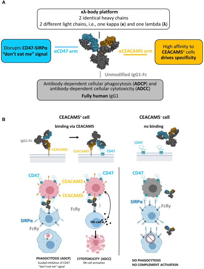
Methods: We present here the generation and preclinical development of NILK-2401, a CEACAM5×CD47 bispecific antibody (BsAb) composed of a common heavy chain and two different light chains, one kappa and one lambda, determining specificity (so-called κλ body format).

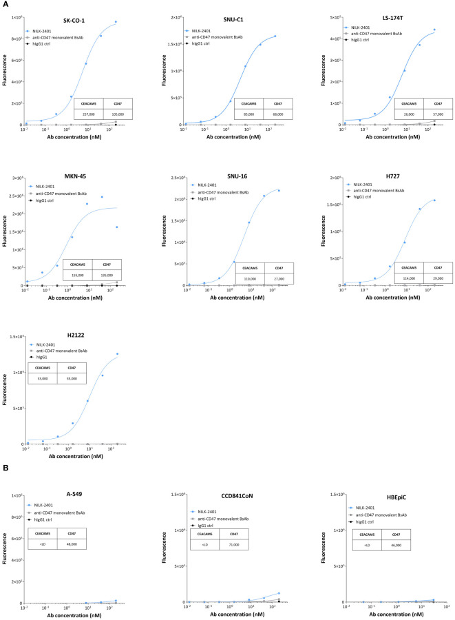
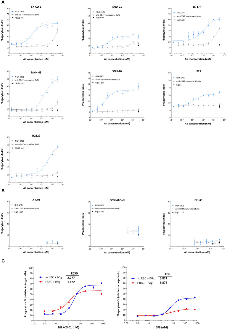
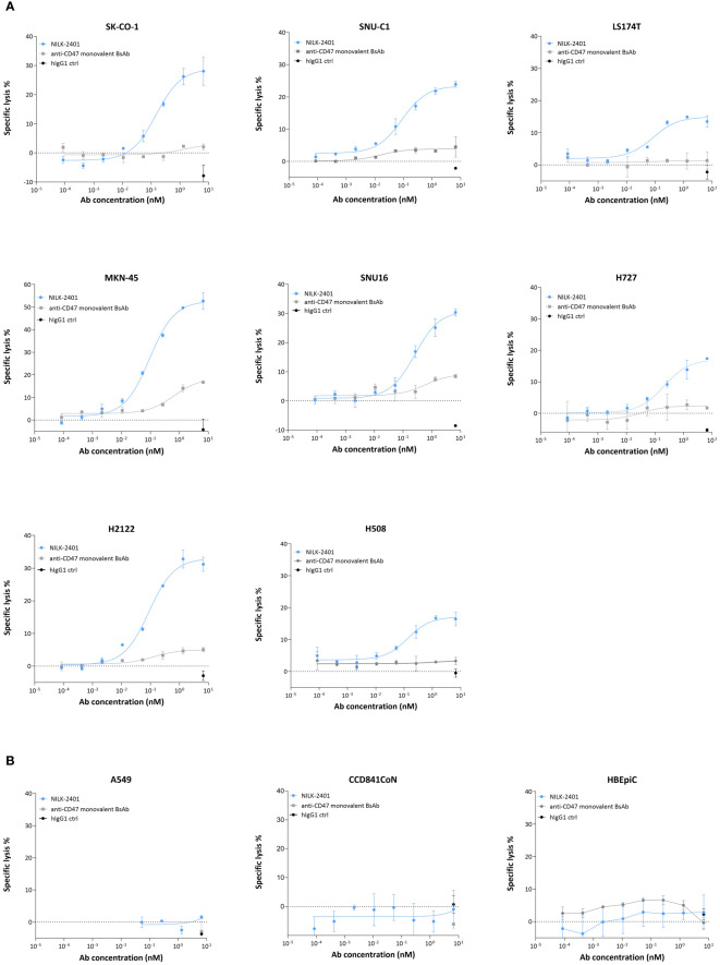
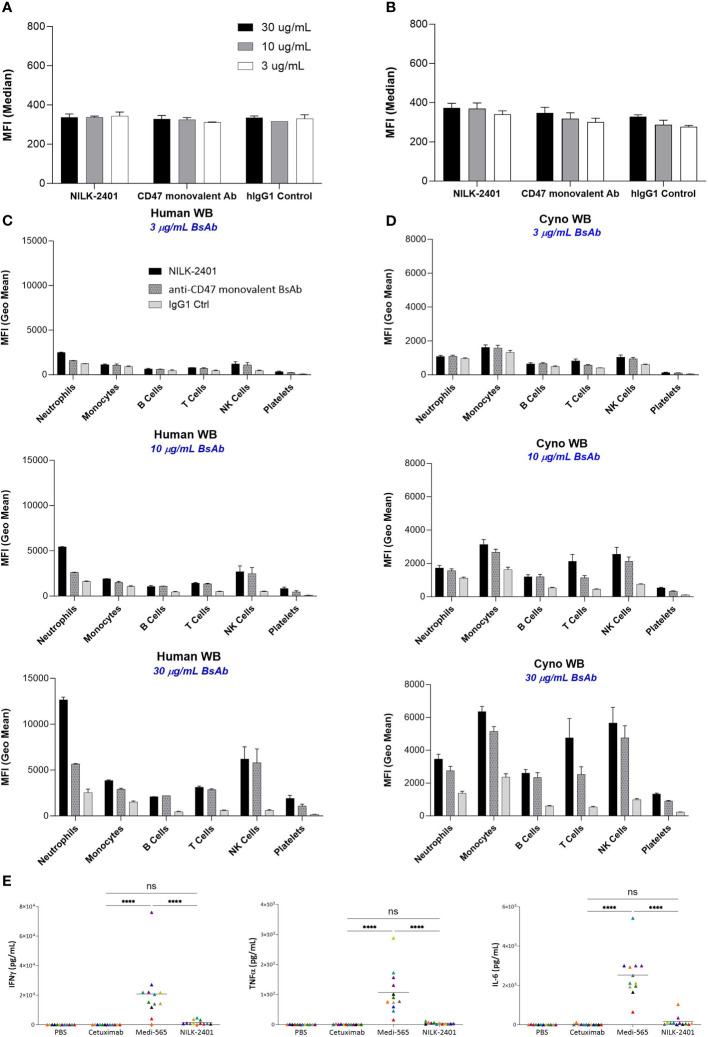
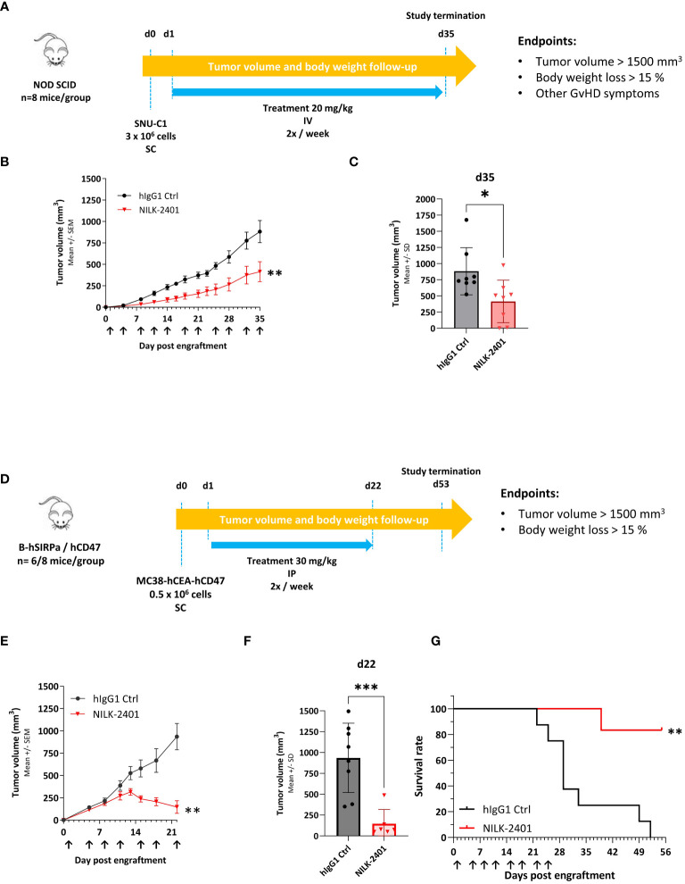
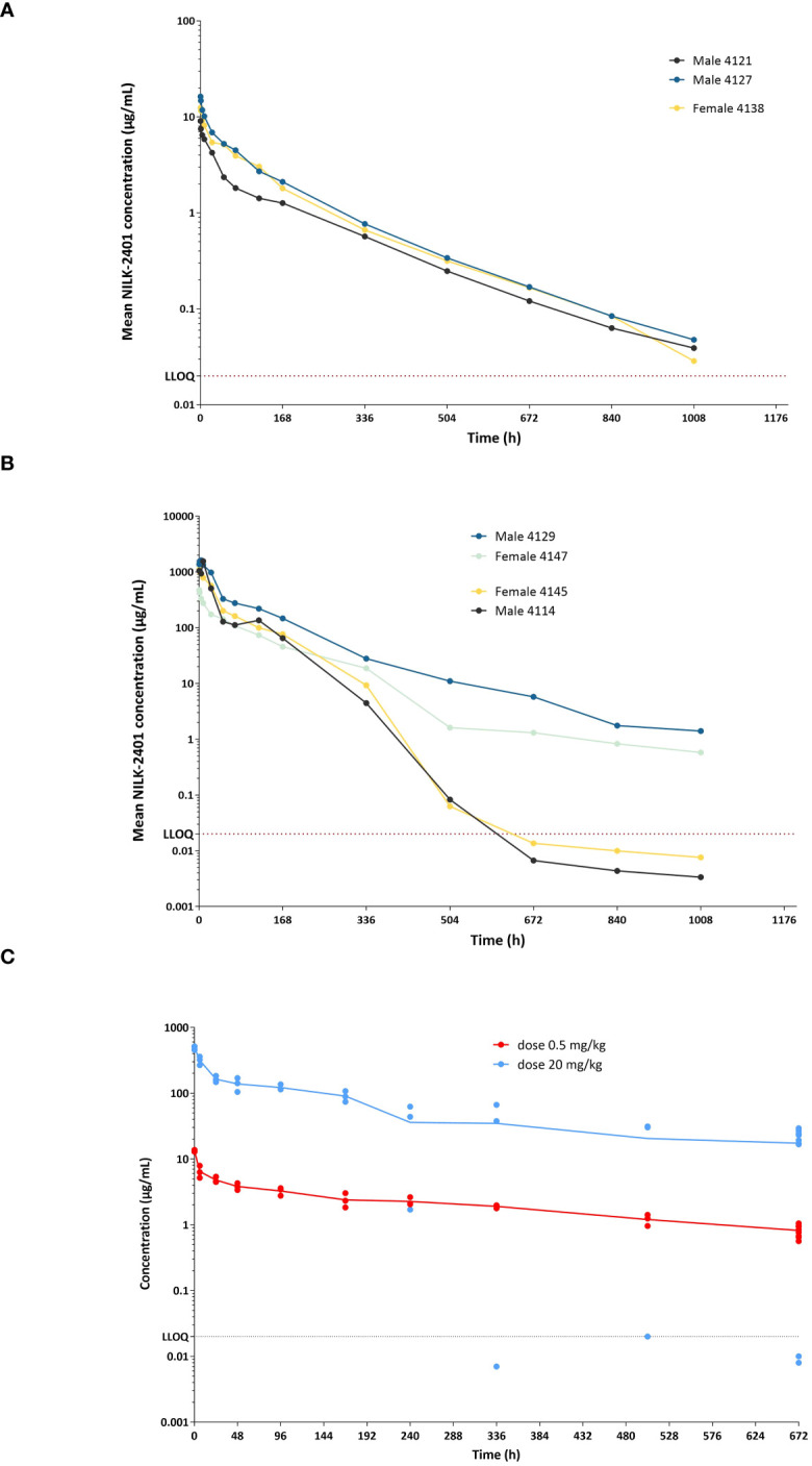
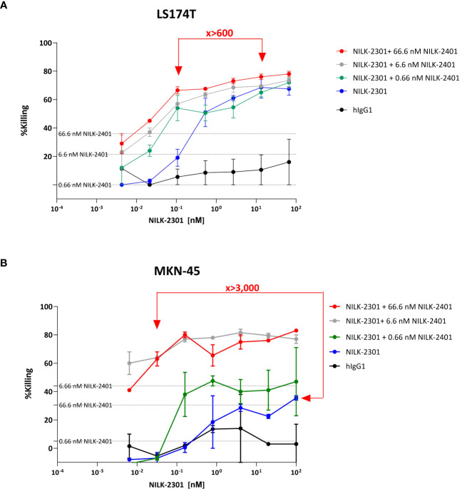
Results: NILK-2401 is a fully human BsAb binding the CEACAM5 N-terminal domain on tumor cells by its lambda light chain arm with an affinity of ≈4 nM and CD47 with its kappa chain arm with an intendedly low affinity of ≈500 nM to enabling tumor-specific blockade of the CD47-SIRPα interaction. For increased activity, NILK-2401 features a functional IgG1 Fc-part. NILK-2401 eliminates CEACAM5-positive tumor cell lines (3/3 colorectal, 2/2 gastric, 2/2 lung) with EC50 for antibody-dependent cellular phagocytosis and antibody-dependent cellular cytotoxicity ranging from 0.38 to 25.84 nM and 0.04 to 0.25 nM, respectively. NILK-2401 binds neither CD47-positive/CEACAM5-negative cell lines nor primary epithelial cells. No erythrophagocytosis or platelet activation is observed. Quantification of the pre-existing NILK-2401-reactive T-cell repertoire in the blood of 14 healthy donors with diverse HLA molecules shows a low immunogenic potential. In vivo, NILK-2401 significantly delayed tumor growth in a NOD-SCID colon cancer model and a syngeneic mouse model using human CD47/human SIRPα transgenic mice and prolonged survival. In cynomolgus monkeys, single doses of 0.5 and 20 mg/kg were well tolerated; PK linked to anti-CD47 and Fc-binding seemed to be more than dose-proportional for Cmax and AUC0-inf. Data were validated in human FcRn TG32 mice. Combination of a CEACAM5-targeting T-cell engager (NILK-2301) with NILK-2401 can either boost NILK-2301 activity (Emax) up to 2.5-fold or allows reaching equal NILK-2301 activity at >600-fold (LS174T) to >3,000-fold (MKN-45) lower doses.

Conclusion: NILK-2401 combines promising preclinical activity with limited potential side effects due to the tumor-targeted blockade of CD47 and low immunogenicity and is planned to enter clinical testing.

Keywords: CD47; CEACAM5; bispecific antibody; immunotherapy; solid cancer.


Conflict of interest statement

AS and DH declare employment at LamKap Bio Group. VB, VM, BD, FR, LC, AV, NB, ER, KM, CB, CM, FJ-B, and YP declare employment at Light Chain Bioscience - Novimmune SA. SS-P declares consultancy work for Light Chain Bioscience - Novimmune SA and LamKap Bio. The authors declare that this study received funding from LamKap Bio beta. The funder or its employees had the following involvement in the study: study design, data analysis, decision to publish, and preparation of the manuscript. The author(s) declared that they were an editorial board member of Frontiers, at the time of submission. This had no impact on the peer review process and the final decision.

Figures

Figure 1
Figure 1
NILK-2401. (A) Schematic depiction of the bispecific antibody, aiming at blocking the CD47 “don’t eat me” signal which allows tumor cells to escape immune surveillance through its interaction with signal regulatory protein alpha (SIRPα) on phagocytes. (B) Envisioned mechanism of action: 1) Initial binding of NILK-2401 to CEACAM5+ tumor cells driven by the high affinity anti-CEACAM5 arm. 2) Co-engagement and blockade of CD47 on tumor cells driven by the low affinity anti-CD47 arm. 3) Fc-mediated effector cell recruitment, fully functional IgG1 Fc. 4) Tumor cell killing via antibody-dependent cellular phagocytosis (ADCP) and antibody-dependent cellular cytotoxicity (ADCC).
Figure 2
Figure 2
Binding of NILK-2401 to CEACAM5-positive and -negative cell lines as well as primary epithelial cells. Dose-dependent binding profile of NILK-2401 on (A) CEACAM5-expressing cancer cell lines SK-CO-1, SNU-C1, LS174T, MKN-45, SNU-16, H727, and H2122. (B) Absence of binding was observed for CEACAM5-negative cell line, A549 and to CCD841CoN, isolated from normal colon, as well as HBEpiC, isolated from normal bronchial tissue. BsAb, bispecific antibody; ctrl, control. Graph is representative of two independent experiments. Acquisition was performed on at least 10,000 cells, one simplicate per condition.
Figure 3
Figure 3
Phagocytosis activity of NILK-2401. Dose-dependent phagocytic activity of NILK-2401 towards (A) CEACAM5-expressing cancer cell lines SK-CO-1, SNU-C1, LS174T, MKN-45, SNU-16, H727, and H2122. (B) Absence of phagocytosis was observed for the CEACAM5-negative cell line A549 and to CCD841CoN, isolated from normal colon, as well as HBEpiC, isolated from normal bronchial tissue. (C) Concentration-response curves of phagocytosis induced by NILK-2401 (left panel) and 5F9 control antibody (right panel) in the presence of red blood cells (RBCs) and IgG excess (IVIg). Exemplary data for 1/4 donors with corresponding EC50 values are shown. Data show mean ± standard deviation. BsAb, bispecific antibody; ctrl, control.
Figure 4
Figure 4
ADCC activity of NILK-2401. Dose-dependent ADCC activity of NILK-4201 towards (A) CEACAM5-expressing cancer cell lines SK-CO-1, SNU-C1, LS174T, MKN-45, SNU-16, H727, and H2122. (B) Absence of killing was observed for the CEACAM5-negative cell line A549 and CCD841CoN, isolated from normal colon, as well as HBEpiC, isolated from normal bronchial tissue. Effector (PBMCs): target ratio is 50:1. Exemplary data for one out of at least five donors. Data show mean ± standard deviation.
Figure 5
Figure 5
NILK-2401 binding to various cell populations from human and cynomolgus whole blood as well as cytokine release. Average MFI values for NILK-2401 binding to (A) human and (B) cynomolgus red blood cells (RBCs) as well as NILK-2401 binding (black bars) to neutrophils, monocytes, B-cells, T-cells, NK cells, and platelets from (C) human (n=2) and (D) cynomolgus (n=4) whole blood (WB) samples, compared to anti-CD47 monovalent control (ctrl) Ab (dashed grey bars) and IgG1 isotype control (light grey bars). Antibodies were tested at 3 µg/mL, 10 µg/mL, or 30 µg/mL. See also Supplementary Table S3 . (E) Cytokine release profile of NILK-2401 using human whole blood from 12 healthy donors. Following 24 h of incubation with 330 nM of NILK-2401, Cetuximab or Medi-565 (330 nM), IFNγ (top panel), IL-6 (middle panel), and TNFα (bottom panel) were measured in supernatants using MSD technology. Each individual color point represents one donor. Phosphate-buffered saline (PBS), anti-EGFR mAb Cetuximab and CEACAM5xCD3 BiTe Medi-565 were used as controls. A two-way ANOVA test was performed for statistical significance. ****P<0.0001. BsAb, bispecific antibody; ns, not significant. Data show mean ± standard deviation.
Figure 6
Figure 6
NILK-2401 in vivo anti-tumor activity. (A) SNU-C1 xenograft model. NOD SCID mice were subcutaneously (SC) injected with 3 x 10⁶ SNU-C1 tumor cells (d0) and received twice a week intravenous (IV) injection of 20 mg/kg of the specified treatment (n=8 mice per group). (B) Tumor volume average over time. Data show mean ± SEM. (C) Tumor volume on d35 post engraftment; arrows indicate days of treatment injections. Data show mean ± SD. Statistical analysis was performed on the area under curve (AUC, between d0 and d35) and the tumor volume average using unpaired t-test. (D) Transgenic mouse model. hCD47/hSIRPα transgenic mice were SC injected with 0.5x10⁶ MC38-hCEA-hCD47 tumor cells (d0) and received twice a week intraperitoneal injection of 30 mg/kg of NILK-2401 (n=6) or vehicle control (ctrl; n=8) starting from d1. (E) Tumor volume average over time. Data show mean ± SEM. (F) Tumor volume 22 days post engraftment. Data show mean ± SD. Arrows indicate days of treatment injections. (G) Survival curve till the end of the experiment (day 53 post engraftment). For statistical analysis, unpaired t-test was performed on the area under curve (AUC at d22; (E) and the tumor volume average (F). A Log-rank (Mantel-Cox) test was performed for survival analysis (G). *P<0.05, **P<0.01, ***P<0.001. SD, standard deviation; SEM, standard error of the mean.
Figure 7
Figure 7
NILK-2401 concentrations versus time in cynomolgus monkeys and Tg32 human FcRn mice. Male and female cynomolgus monkeys received a single IV administration at (A) 0.5 mg/kg or (B) 20 mg/kg, respectively. NILK-2401 serum concentrations were quantified at different time-points following the IV injection using a fit-for-purpose PK assay. Each line corresponds to a different animal, with full lines and dashed lines corresponding to female and male animals, respectively. The horizontal black dashed line corresponds to the LLOQ (lower limit of quantification). (C) PK profile was also analyzed in Tg32 human FcRn mice following a single injection of NILK-2401 at 0.5 mg/kg (light blue dots) and 20 mg/kg (red dots). PK parameters are summarized in Supplementary Tables S4 and S5 .
Figure 8
Figure 8
NILK-2401 plus NILK-2301 combination activity. “Mixed killing assay” using (A) LS174T and (B) MKN-45 as target cells, respectively. Cells were exposed to NILK-2301 alone (blue curve) or NILK-2301 plus NILK-2401 at 0.66 nM (green curve), 6.6 nM (grey curve), or 66.6 nM (red curve), respectively. Cells without treatment (pink curve) or those treated with human IgG1 control antibody (hIgG1; black curve) were used as comparators. NILK-2401 can boost NILK-2301 activity, e.g., equivalent activity of NILK-2401 plus NILK-2301 combination therapy at >600-fold (LS174T) to >3,000-fold MKN-45) lower doses compared to NILK-2301 monotherapy, i.e., ≈0.02 vs. 13.3 and, ≈0.03 vs 100 nM, were found. Data show mean ± SEM.

Similar articles

Cited by

References

    1. Weiner GJ. Building better monoclonal antibody-based therapeutics. Nat Rev Cancer. (2015) 15:361–70. doi: 10.1038/nrc3930 - DOI - PMC - PubMed
    1. Zhukovsky EA, Morse RJ, Maus MV. Bispecific antibodies and CARs: generalized immunotherapeutics harnessing T cell redirection. Curr Opin Immunol. (2016) 40:24–35. doi: 10.1016/j.coi.2016.02.006 - DOI - PMC - PubMed
    1. June CH, O'Connor RS, Kawalekar OU, Ghassemi S, Milone MC. CAR T cell immunotherapy for human cancer. Science. (2018) 359:1361–5. doi: 10.1126/science.aar6711 - DOI - PubMed
    1. Sharma P, Allison JP. The future of immune checkpoint therapy. Science. (2015) 348:56–61. doi: 10.1126/science.aaa8172 - DOI - PubMed
    1. Matlung HL, Szilagyi K, Barclay NA, van den Berg TK. The CD47-SIRPalpha signaling axis as an innate immune checkpoint in cancer. Immunol Rev. (2017) 276:145–64. doi: 10.1111/imr.12527 - DOI - PubMed

Publication types

MeSH terms