

Tissue and cellular localization of condensed tannins in poplar roots and potential association with nitrogen uptake
- PMID: 38721337
- PMCID: PMC11076728
- DOI: 10.3389/fpls.2024.1388549
Tissue and cellular localization of condensed tannins in poplar roots and potential association with nitrogen uptake
Abstract
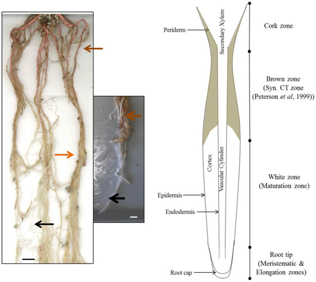
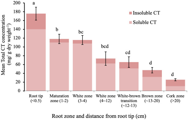
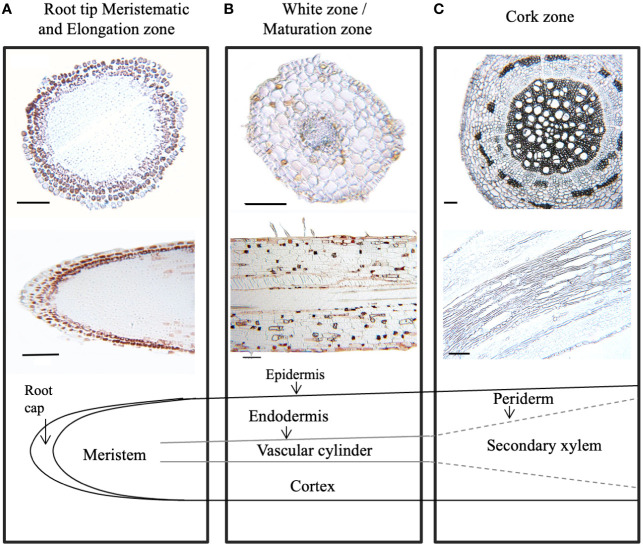
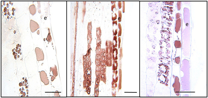
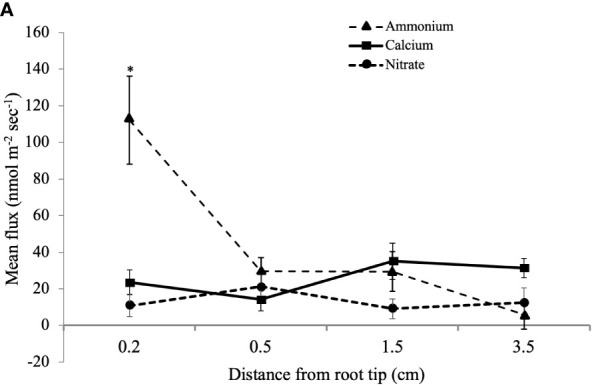
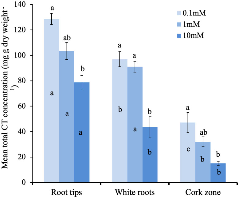
Condensed tannins are common in vegetative tissues of woody plants, including in roots. In hybrid poplar (Populus tremula x alba; also known as P. x canescens) CT assays indicated they were most concentrated in younger white roots and at the root tip. Furthermore, CT-specific staining of embedded tissue sections demonstrated accumulation in root cap cells and adjacent epidermal cells, as well as a more sporadic presence in cortex cells. In older, brown roots as well as roots with secondary growth (cork zone), CT concentration was significantly lower. The insoluble fraction of CTs was greatest in the cork zone. To determine if CT accumulation correlates with nutrient uptake in poplar roots, a microelectrode ion flux measurement (MIFE™) system was used to measure flux along the root axis. Greatest NH4 + uptake was measured near the root tip, but NO3- and Ca2+ did not vary along the root length. In agreement with earlier work, providing poplars with ample nitrogen led to higher accumulation of CTs across root zones. To test the functional importance of CTs in roots directly, CT-modified transgenic plants could be important tools.
Keywords: 4-dimethylaminocinnamaldehyde (DMACA); Populus; flavonoid; microelectrode ion flux measurement (MIFE); proanthocyanidin; root cap.
Copyright © 2024 Westley, Ma, Hawkins and Constabel.
Conflict of interest statement
The authors declare that the research was conducted in the absence of any commercial or financial relationships that could be construed as a potential conflict of interest.
Figures
Similar articles
-
Anatomical patterns of condensed tannin in fine roots of tree species from a cool-temperate forest.Ann Bot. 2021 Jul 28;128(1):59-71. doi: 10.1093/aob/mcab022. Ann Bot. 2021. PMID: 33608716 Free PMC article.
-
Net fluxes of ammonium and nitrate in association with H+ fluxes in fine roots of Populus popularis.Planta. 2013 Apr;237(4):919-31. doi: 10.1007/s00425-012-1807-7. Epub 2012 Nov 20. Planta. 2013. PMID: 23179443
-
Physiological characteristics and RNA sequencing in two root zones with contrasting nitrate assimilation of Populus × canescens.Tree Physiol. 2020 Oct 7;40(10):1392-1404. doi: 10.1093/treephys/tpaa071. Tree Physiol. 2020. PMID: 32542375
-
Phenylalanine as a nitrogen source induces root growth and nitrogen-use efficiency in Populus × canescens.Tree Physiol. 2018 Jan 1;38(1):66-82. doi: 10.1093/treephys/tpx109. Tree Physiol. 2018. PMID: 29036367
-
Nitrogen metabolism of two contrasting poplar species during acclimation to limiting nitrogen availability.J Exp Bot. 2013 Nov;64(14):4207-24. doi: 10.1093/jxb/ert234. Epub 2013 Aug 20. J Exp Bot. 2013. PMID: 23963674 Free PMC article.
References
-
- Bryant J. P., Chapin F. S., Klein D. R. (1983). Carbon nutrient balance of boreal plants in relation to vertebrate herbivory. Oikos 40, 357–368. doi: 10.2307/3544308 - DOI
-
- Chowdhury J., Ferdous J., Lihavainen J., Albrectsen B. R., Lundberg-Felten J. (2023). Fluorogenic properties of 4-dimethylaminocinnamaldehyde (DMACA) enable high resolution imaging of cell-wall-bound proanthocyanidins in plant root tissues. Front. Plant Sci. 13. doi: 10.3389/fpls.2022.1060804 - DOI - PMC - PubMed
LinkOut - more resources
Full Text Sources
Research Materials
Miscellaneous