Review
. 2024 Apr 22;12(2):36.
doi: 10.21037/atm-23-1774. Epub 2024 Apr 18.

Wide-bore polyester sutures may create sufficient collagen for cure of prolapse/incontinence: a work in progress

Affiliations
Review

Wide-bore polyester sutures may create sufficient collagen for cure of prolapse/incontinence: a work in progress

Ahmet Akin Sivaslioglu et al. Ann Transl Med. .

Abstract

The main thrust of the Integral Theory Paradigm (ITP) is that inadequate ligament collagen causes pelvic organ prolapses (POP) and pelvic symptoms, a concept validated by multiple publications which cured POP and bladder/bowel/pain dysfunctions by collagen-creating slings. Sling surgery for surgical cure of these conditions was eliminated in the United States, Europe and other regulatory jurisdictions by banning all mesh products (including tapes) in about 2017. The aim of this work was to inform of the progress of a highly promising alternative method for collage creation for ligament repair: wide-bore polyester sutures accurately applied to weak ligaments. The scientific rationale for the wide-bore polyester plication method was a revisit and analysis of prior Instron testing data from a rejected polyester aortic graft from a doctoral thesis. The analysis indicated that the collagen produced by No. 2 polyester sutures would be sufficient to repair weakened pelvic ligaments. The surgical methodology consisted of application of wide-bore No. 2 or No. 3 polyester sutures to existing vaginal surgical techniques such as cardinal/uterosacral ligament (CL/USL) repair in the Fothergill operation, deep transversus perinei (DTP) ligamentous supports of the perineal body (PB) and uniquely, pubourethral ligament (PUL) repair for stress urinary incontinence (SUI). No vaginal tissue was excised. These operations are now being performed in several centres around the world. Because of this, the results detailed below are indicative only, and necessarily incomplete, as they are only from these units. Twelve month data (n=35) for SUI cure (83%) following PUL repair by the urethral ligament plication (ULP) operation has been submitted for publication; POP quantification (POPQ) points Ba, C, Bp, D were significantly improved at 6 weeks postoperative review following repair of CLs (cystocele) and USLs (uterine/apical prolapse) (n=56): deep transverse perinei ligament repair (descending perineal syndrome "DPS") (n=4) were cured at 6-12 months review. Though numbers are few, in the context of DPS being considered incurable, these numbers are significant. Except for the ULP operation, the techniques for cystocele, uterine prolapse, perineocele were essentially evolved versions of the Fothergill and standard PB repairs without any vaginal or ligament excisions. Though promising, more extensive and longer-term results are clearly required before this wide-bore polyester ligament repair method can become mainstream.

Keywords: Polyester sutures; collagen; descending perineal syndrome (DPS); pelvic organ prolapses (POP); stress urinary incontinence (SUI).


Conflict of interest statement

Conflicts of Interest: All authors have completed the ICMJE uniform disclosure form (available at https://atm.amegroups.com/article/view/10.21037/atm-23-1774/coif). The series “Integral Theory Paradigm” was commissioned by the International Society for Pelviperineology without any funding or sponsorship. The authors have no other conflicts of interest to declare.

Figures

Figure 1
Figure 1
Experimental animal study (canine) 2 weeks after removal of collagen-creating tapes. White arrows indicate the significant artificial collagenous neoligament formed in reaction to the tapes. Reused from Petros P. The female pelvic floor function, dysfunction and management according to the Integral Theory. 3rd ed. Heidelberg: Springer Berlin; 2010. With permission from Peter Petros; retains ownership of the copyright. V, vagina; B, bladder; U, uterus.
Figure 2
Figure 2
The TFS shortens and reinforces loose or damaged ligaments. The TFS sling is placed transversely, so it has very minimal contact with the vagina. It directly re-attaches organs to the skeleton, mimicking exactly the 3-level pelvic organ support system: PUL, ATFP, CL, USL, deep transversus perinei part of PB. Insert: TFS anchor and tape polypropylene anchor 11 mm × 4 mm sits on a stainless-steel applicator. A lightweight macropore 7 mm wide monofilament tape passes through a one-way system at the anchor base which shortens and tensions the damaged ligament. Reused with permission from Central European Journal of Urology (7). PUL, pubourethral ligament; ATFP, arcus tendineus fascia pelvis; CL, cardinal ligament; USL, uterosacral ligament; PB, perineal body; TFS, Tissue Fixation System.
Figure 3
Figure 3
Pathogenesis of SUI and cure by PUL suturing (ULP operation). Weak “PUL” cannot support the distal vagina and posterior urethral wall. These are stretched down to open the urethra from “C” to “O”, by the posterior muscle forces, “LP” and conjoint “LMA”. The polyester suture holds “PUL” together, reinforces it with new collagen and prevents urethral opening, much like the hemostat test support at midurethra. Reused from Petros P. The female pelvic floor function, dysfunction and management according to the Integral Theory. 3rd ed. Heidelberg: Springer Berlin; 2010. With permission from Peter Petros; retains ownership of the copyright. PS, pubic symphysis; PCM, pubococcygeus muscle; PUL, pubourethral ligaments; LP, levator plate; LMA, longitudinal muscles of the anus; C, closed; O, open; SUI, stress urinary incontinence; ULP, urethral ligament plication; H, suburethral vaginal hammock.
Figure 4
Figure 4
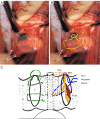
ULP operation. Surgical binding of loose PUL via two parallel incisions in the distal vaginal sulci. (A) Original live anatomical dissection of “PUL” (left incision) during a two incision IVS midurethral sling operation. The tape measure overlies the urethra. The left paraurethral sulcus has been incised along its length and opened out laterally with forceps. “EUL” attaches the external meatus to the anterior part of the PS. The “PUL” originates behind the “PS” from its lower posterior part. Coming down from the “PS”, the “PUL” splits into two parts, medial “M” to insert into the side of the midurethra and its lateral branch “L”, attaches to the pubococcygeus muscle (not seen) then comes down to attach to the vagina “V”. (B) No. 2 or No. 3 polyester sutures bind both branches of the “PUL” to fascias attached to the pubic bone, “EUL”, urethra, vagina and “PCM”, essentially as performed in the original operation. (C) shows the opened out parallel incisions in both sulci, extending from “EUL” to the bladder neck. On the right, the pathway of the No. 2 polyester sutures is shown, from the medial branch of the “PUL”, to the descending branch of “PUL”, then the “EUL”, then, “PCM”. On the left, Si and S2 show suturing of the vaginal incision. Reused from Petros P. The female pelvic floor function, dysfunction and management according to the Integral Theory. 3rd ed. Heidelberg: Springer Berlin; 2010. With permission from Peter Petros; retains ownership of the copyright. EUL, external urethral ligament; PUL, pubourethral ligaments; PCM, pubococcygeus muscle; ULP, urethral ligament plication; IVS, intravaginal slingplasty; PS, pubic symphysis; T, tension-free skin sutures; U, urethra.
Figure 5
Figure 5
Cervix at 10 cm, full dilatation. The head is at the upper end of the birth canal. The “USL” and “CL” are very significantly stretched as is the vaginal attachment “VAG” to “CL” which attaches to the anterior wall of the cervix. Reused from Petros P. The female pelvic floor function, dysfunction and management according to the Integral Theory. 3rd ed. Heidelberg: Springer Berlin; 2010. With permission from Peter Petros; retains ownership of the copyright. USL, uterosacral ligament; CL, cardinal ligament; VAG, vagina; ATFP, arcus tendinous fascia pelvis; CX, cervix; S, sacrum.
Figure 6
Figure 6
Pathogenesis and repair of transverse defect cystocele. The “CL” and the “PCF” attachment of vagina to “CL” have torn under pressure of the head and prolapse downwards. Unsupported by “CL”, “PCF” and the overlying bladder base prolapse downwards as a transverse defect cystocele. A No. 2 polyester suture brings together the ruptured edges “r” without tension. Reused from Petros P. The female pelvic floor function, dysfunction and management according to the Integral Theory. 3rd ed. Heidelberg: Springer Berlin; 2010. With permission from Peter Petros; retains ownership of the copyright. CL, cardinal ligament; PCF, pubocervical fascial.
Figure 7
Figure 7
Pathogenesis and surgical techniques for major or minor uterine prolapse. (A) Major uterine prolapse. Weak “USLs” elongate with effort, resulting in uterine or apical prolapse. Note how “CL” also elongate with major uterine prolapse. Reused from Petros P. The female pelvic floor function, dysfunction and management according to the Integral Theory. 3rd ed. Heidelberg: Springer Berlin; 2010. With permission from Peter Petros; retains ownership of the copyright. (B) USL plication methods. Where required, the uterus or apex are grasped and pushed back into their correct anatomical position. Left image: a 5 cm transverse incision (red broken lines) is made at the enterocele bulge. It is opened up by a speculum (broken lines). Two No 2 polyester sutures S1 and S2 are inserted at “A” and “B” and brought together. Middle image: for a longitudinal incision (and also transverse), “USLs” are located and sutured together right to the cervix over a 5 cm length. Right image: when the prolapse is very large, and “USLs” thin, the “USLs” may need to be individually plicated so as to optimize the amount of collagen from the No. 2 polyester sutures, which are then joined “USLs” by interrupted polyester sutures (arrows). Published with permission from Peter Petros: Personal Collection; retains ownership of copyright. USLs, uterosacral ligaments; CL, cardinal ligament; CX, cervix.
Figure 8
Figure 8
Re-assignment of excess vaginal epithelium using continuous sutures. This suture can be interrupted or continuous. A No. 00 vicryl suture is placed 1–2 mm into the cut epithelium “e”, then taken into the deep fascia “f” on one side then to “f” on the other side. At this point, the forefinger and middle finger are placed around the suture, and the epithelium is pushed down onto the fascia. The suture continues into the epithelium “e” on the other side and then tied. Published with permission from Peter Petros: Personal Collection; retains ownership of copyright.
Figure 9
Figure 9
Pathogenesis of perineal structures. So as to exit, the head must stretch the structures at the outlet which may be damaged (brackets): the attachment of levators to the symphysis (dislocation) “PUL” (stress urinary incontinence); not shown are perineal body and deep transversus perinei (rectocele, DPS). Reused from Petros P. The female pelvic floor function, dysfunction and management according to the Integral Theory. 3rd ed. Heidelberg: Springer Berlin; 2010. With permission from Peter Petros; retains ownership of the copyright. ATFP, arcus tendineus fascia pelvis; PUL, pubourethral ligament; DPS, descending perineal syndrome.
Figure 10
Figure 10
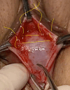
A 5 cm full thickness transverse incision is made just inside the hymenal ring. With descending perineal syndrome, the serosa and smooth muscle wall of the rectum is ruptured, and the rectal mucosa is spread to become adherent to the vagina and “DTP”. Careful dissection is required to separate the rectal mucosa from vagina, perineal body, descending rami. Two to three interrupted sutures are applied to close the smooth muscle layer of the rectum. The “DTP” ligaments are identified and sutured with No. 2 polyester sutures as shown. As the “DTP” shortens, the perineal bodies fold inwards and are gently approximated with vicryl sutures. The vagina is sutured without excision of tissue. Reused from Petros P. The female pelvic floor function, dysfunction and management according to the Integral Theory. 3rd ed. Heidelberg: Springer Berlin; 2010. With permission from Peter Petros; retains ownership of the copyright. DTP, deep transversus perinei; A, anus; OF, obturator fossa.
Video S1
Video S1
Video abstract.
Video S2
Video S2
Anatomy of PUL repair (PPP) for cure of SUI. PUL, pubourethral ligament; PPP, pubourethral ligament plication procedure; SUI, stress urinary incontinence.
Video S3
Video S3
Surgery PUL repair (PPP) for cure of SUI (Surgeon Akin Sivaslioglu). PUL, pubourethral ligament; PPP, pubourethral ligament plication procedure; SUI, stress urinary incontinence.
Video S4
Video S4
CL repair for cystocele transverse incision (Surgeons Ray Hodgson Peter Petros). CL, cardinal ligament; USL, uterosacral ligament.
Video S5
Video S5
CL and USL repair for cystocele and uterine prolapse longitudinal incision (Surgeon Xiuli Sun). CL, cardinal ligament; USL, uterosacral ligament.
Video S6
Video S6
USL transverse incision (Surgeons Ray Hodgson and Peter Petros). USL, uterosacral ligament.
Video S7
Video S7
Repair of perineocele (dissection).
Video S8
Video S8
Surgery for repair of perineocele.

References

    1. Petros PE, Ulmsten U. An Integral Theory of female urinary incontinence. Acta Ob-stet Gynecol Scan 1990;69 Suppl 153:7-31. 10.1111/j.1600-0412.1990.tb08027.x - DOI - PubMed
    1. Petros PE, Ulmsten U, Papadimitriou J. The Autogenic Neoligament procedure: A technique for planned formation of an artificial neo-ligament. Acta Obstet Gynecol Scand 1990;69 Suppl 153:43-51. 10.1111/j.1600-0412.1990.tb08031.x - DOI - PubMed
    1. Petros PE, Ulmsten U. The combined intravaginal sling and tuck operation: An ambulatory procedure for stress and urge incontinence. Acta Obstet Gynecol Scand 1990;69 Suppl 153:53-9. 10.1111/j.1600-0412.1990.tb08032.x - DOI - PubMed
    1. Ulmsten U, Petros P. Intravaginal slingplasty (IVS): An ambulatory surgical procedure for treatment of female urinary incontinence. Scand J Urol Nephrol 1995;29:75-82. 10.3109/00365599509180543 - DOI - PubMed
    1. Toozs-Hobson P, Cardozo L, Hillard T. Managing pain after synthetic mesh implants in pelvic surgery. Eur J Obstet Gynecol Reprod Biol 2019;234:49-52. 10.1016/j.ejogrb.2018.12.037 - DOI - PubMed