Review
. 2024 Apr 22;12(2):23.
doi: 10.21037/atm-23-1877. Epub 2024 Apr 18.

Structural, functional, and dysfunctional pelvic anatomy

Affiliations
Review

Structural, functional, and dysfunctional pelvic anatomy

Bernard Liedl et al. Ann Transl Med. .

Abstract

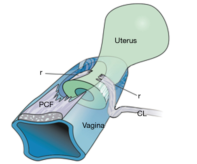
The structural basis of the Integral Theory is holistic. Four main pelvic muscles interact holistically with five main pelvic ligaments to maintain pelvic organ structure and function. The vagina is structurally weak. The support it provides to the bladder base is contingent on being stretched by opposite pelvic muscle forces, much like a trampoline. Its main role is to transmit muscle forces to facilitate continence, evacuation and control of urgency. Therefore, as an organ that cannot regenerate, the vagina should be conserved, and not excised. The ligaments provide the main structural support for the organs and are the most vulnerable part of the anatomical system to injury because their structural collagen is depolymerized prior to labour, and stretched during labour. Further ligament weakening occurs after menopause due to collagen breakdown. Hence, collagen loss is the main cause of organ prolapse and lower urinary tract symptoms (LUTS). The strengthening of damaged ligaments, whether surgically or non-surgically, can improve or cure symptoms and prolapse. Because collagen loss in ligaments is a principal cause of dysfunction in older women, collagen-creating techniques are advised: precisely inserted tapes to create neoligaments, or wide-bore No. 2 or No. 3 polyester ligament sutures instead of dissolvable sutures.

Keywords: Pelvic organ prolapse; collagen; ligaments; lower urinary tract symptoms surgery (LUTS surgery); pelvic floor muscles.


Conflict of interest statement

Conflicts of Interest: All authors have completed the ICMJE uniform disclosure form (available at https://atm.amegroups.com/article/view/10.21037/atm-23-1877/coif). The series “Integral Theory Paradigm” was commissioned by the International Society for Pelviperineology without any funding or sponsorship. M.N. reports stocks and stock options at Momentis and Femselect. The authors have no other conflicts of interest to declare.

Figures

Figure 1
Figure 1
Structural and functional anatomy of the pelvic floor. Broken lines represent the “open” position of the bladder base/urethra and anorectum during evacuation. Reused from (1). Permission from Peter Petros, who retains ownership of the copyright. CX, cervix; N, urothelial stretch receptors; R, rectum; U, urethra. Bone: PS, pubic symphysis; S, sacrum. Suspensory ligaments: PUL, pubourethral ligament; ATFP, arcus tendineus fascia pelvis; USL, uterosacral ligament; CL, cardinal ligament. Muscles: PCM, pubococcygeus muscle; LP, levator plate; LMA, conjoint longitudinal muscle of the anus; PRM, puborectalis muscle. Supporting fascia: PCF, pubocervical fascia; RVF, rectovaginal fascia. Perineal anchoring structures: PB, perineal body; EAS, external anal sphincter.
Figure 2
Figure 2
The muscles. Anatomical specimen from a female cadaver, cut away from its bony insertions. The bladder and vagina have been excised at the level of bladder neck. Reused from (1). With permission from Peter Petros; retains ownership of the copyright. U, urethra; V, vagina; R, rectum; PCM, pubococcygeus muscle sweeping behind R to merge with the contralateral side and ileococcygeus to form the LP; LP, levator plate; PRM, puborectalis muscle; PUL, insertion of pubourethral ligament into PCM; EAS, external anal sphincter; LMA, conjoint longitudinal muscle of the anus.
Figure 3
Figure 3
PB. The lower part of the vagina has been cut away. The PB is suspended from the pubic rami by the DTP ligaments. These are 4 cm long. They insert behind the descending rami, exactly between the upper 2/3 and lower 1/3. Reused from (1). With permission from Peter Petros; retains ownership of the copyright. OF, obturator fossa; PB, perineal body; DTP, deep transverse perineal; A, anus.
Figure 4
Figure 4
Upper end of the birth canal. Reused from (1). With permission from Peter Petros; retains ownership of the copyright. ATFP, arcus tendineus fascia pelvis; CL, cardinal ligament; CX, cervix; USL, uterosacral ligament; VAG, pubocervical fascia of vagina attached to CL and anterior cervix; S, sacrum.
Figure 5
Figure 5
Transverse defect cystocele causation. Excessive pressure by the head may “r” the attachments of the CL and fascial layer of the vagina (PCF) to the CL and cervix. Note how the torn CL prolapses to the side of the cervix. The bladder overlying the vagina descends much like a trapdoor as a cystocele (transverse defect). Reused from (1). With permission from Peter Petros; retains ownership of the copyright. r, rupture; PCF, pubocervical fascia; CL, cardinal ligament.
Figure 6
Figure 6
Causation of uterine prolapse. A 3rd or 4th degree uterine prolapse is caused by elongated USLs and usually, CLs also. Reused from (1). With permission from Peter Petros; retains ownership of the copyright. CL, cardinal ligament; USL, uterosacral ligament.
Figure 7
Figure 7
Structural damage at birth canal outlet. “levators” = collagenous insertions of the pubococcygeus muscle and puborectalis muscle to the symphysis. Reused from (1). With permission from Peter Petros; retains ownership of the copyright. ATFP, arcus tendineus fascia pelvis; PUL, pubourethral ligament.
Figure 8
Figure 8
4D ultrasound of levator hiatus avulsion. View from above of the levator hiatus (arrows). Asterisk denotes a right-sided avulsion of pubococcygeus and/or puborectalis from its bony insertion point as a consequence of overdistension (see Figure 7). The pelvic muscles are outlined by broken lines. Reused from (1). With permission from Peter Petros; retains ownership of the copyright. U, urethra; V, vagina; R, rectum.
Figure 9
Figure 9
Pathogenesis of DPS. The PB has been stretched, narrowed, and flattened; the rectal serosa has been broken and rectal mucosa (labelled “rectum”) has emerged through the broken serosa and spread laterally to adhere to the vagina and PB. The DTP ligaments are elongated and displaced to cause DPS. Reused from (1).With permission from Peter Petros; retains ownership of the copyright. DPS, descending perineal syndrome; OF, obturator fossa; A, anus; DTP, deep transversus perinei; PB, perineal body.
Figure 10
Figure 10
Anatomical pathway from damaged ligaments lead to a symptom-based diagnostic algorithm. (A) Head descending down the vaginal birth canal (blue). As the head descends down the vaginal birth canal, it damages the vagina and ligaments at 4 levels, shaded in red, (circles numbered 1–4). The circles and numbers 1–4, causatively correlate with specific prolapses and symptoms in the right figure, also numbered 1–4 in the three columns. The numbers 1–4 also correlate anatomically with the figure above the algorithm. (B) The diagnostic algorithm summarizes the causative relationship between anatomical damage of ligaments and vagina (circles 1–4, left figure), to prolapse and symptoms in three anatomical zones (numbers 1–4, right figure). The height of the bar indicates incidence of the symptom in that zone. Reused from (1). With permission from Peter Petros; retains ownership of the copyright. ATFP, arcus tendineus fascia pelvis; B, bladder; CL, cardinal ligament; PB, perineal body; PS, pubic symphysis; PUL, pubourethral ligament; S, sacrum; USL, uterosacral ligament; UT, urinary tract.
Figure 11
Figure 11
USL causation of rectal intussusception. Anterior rectal wall intussusception caused by elongation of the USLs attached to the lateral walls of the rectum. Reused from (1). With permission from Peter Petros; retains ownership of the copyright. SSL, sacrospinous ligament; USL, uterosacral ligament.
Video S1
Video S1
Video abstract.
Video S2
Video S2
Bladder closure US Three directional forces close the urethra distally and at the bladder neck.
Video S3
Video S3
Anorectal closure US Three directional forces and PRM close the anorectal angle.
Video S4
Video S4
Micturition PCM relaxes, LP/LMA open out the posterior urethral wall.
Video S5
Video S5
Defecation PRM relaxes. LP/LMA open out the anorectal angle.

References

    1. Petros P. The female pelvic floor function, dysfunction and management according to the Integral Theory. 3rd ed. Heidelberg: Springer Berlin; 2010.
    1. Petros PE, Ulmsten U. An Integral Theory of female urinary incontinence. Acta Obstet Gynecol Scand 1990;69 Suppl 153:1-79. 10.1111/j.1600-0412.1990.tb08027.x - DOI - PubMed
    1. Petros PE, Swash M. The Musculo-Elastic Theory of anorectal function and dysfunction. Pelviperineology 2008;27:89-121.
    1. Petros PE. The pubourethral ligaments--an anatomical and histological study in the live patient. Int Urogynecol J Pelvic Floor Dysfunct 1998;9:154-7. 10.1007/BF02001085 - DOI - PubMed
    1. Wagenlehner FM, Del Amo E, Santoro GA, et al. Live anatomy of the perineal body in patients with third-degree rectocele. Colorectal Dis 2013;15:1416-22. 10.1111/codi.12333 - DOI - PubMed