Review
. 2024 Apr 22;12(2):25.
doi: 10.21037/atm-23-1883. Epub 2024 Apr 18.

A brief physiology and pathophysiology of the anorectum based on the Integral Theory paradigm

Affiliations
Review

A brief physiology and pathophysiology of the anorectum based on the Integral Theory paradigm

Darren M Gold et al. Ann Transl Med. .

Abstract

The remit of this review is confined to the experimental scientific works and surgeries based on the Integral Theory paradigm. The video abstract summarizes the anorectal function, how ligaments cause dysfunction and cure of fecal incontinence and obstructed defecation by ligament repair. Anorectal function is reflex and binary, with cortical and peripheral components. The same three oppositely acting reflex muscle forces which open and close the bladder, contract against the pubourethral (PUL) and uterosacral (USL) ligaments: (I) to close the anorectum for continence when the puborectalis muscle (PRM) contracts forwards; (II) to open the anorectum prior to evacuation when the PRM relaxes; (III) to stretch the rectum in opposite directions to support the anorectal stretch receptors "N" to prevent premature activation of the defecation reflex, (fecal urgency). Weak or loose PULs or USLs may cause dysfunction of closure, of evacuation, and inability to control the defecation reflex (fecal urgency). Repair of the PUL and USL can improve or cure these dysfunctions. The perineal body (PB) acts as an anatomical support for the distal vagina, anorectum and external anal sphincter (EAS). It serves as an anchoring point for the forward action of the pubococcygeus muscle (PCM), which tensions the anterior rectal wall during closure and defecation. Bladder and bowel dysfunction have a similar pathogenesis, ligament laxity, mainly pubourethral and uterosacral, with added PB damage for anorectal dysfunction. PB damage can cause obstructive defecation and descending perineal syndrome (DPS). Repair of damaged PUL and USL can restore the closure and evacuation functions of both bladder an anorectum. DPS can be cured by repair of the PB's suspensory ligaments, deep transversus perinei.

Keywords: Rectum; anus; binary control; fecal incontinence (FI); obstructed defecation.


Conflict of interest statement

Conflicts of Interest: All authors have completed the ICMJE uniform disclosure form (available at https://atm.amegroups.com/article/view/10.21037/atm-23-1883/coif). The series “Integral Theory Paradigm” was commissioned by the International Society for Pelviperineology without any funding or sponsorship. The authors have no other conflicts of interest to declare.

Figures

Figure 1
Figure 1
Integrated structural anatomy of the pelvic organs, ligaments and muscles. Bone: PS, S. Suspensory ligaments: PUL, ATFP, USL, CL. Anorectal stretch receptors N, which respond to pressure of rectal contents to activate the defecation reflex (felt as urge to defecate); “N” at bladder base, urothelial stretch receptors which activate the micturition reflex (urinary urge) and rectum. Muscles: PCM, LP, LMA, PRM. Supporting fascia: PCF, RVF. Perineal anchoring structures: PB, EAS. Reused from Petros P. The female pelvic floor function, dysfunction and management according to the Integral Theory. 3rd edition. Heidelberg: Springer Berlin; 2010. With permission from Peter Petros; retains ownership of the copyright. U, uterus; R, rectum; CX ring, cervical ring; PS, pubic symphysis; S, sacrum; PUL, pubourethral ligament; ATFP, arcus tendineus fascia pelvis; USL, uterosacral ligament; CL, cardinal ligament; PCM, pubococcygeus muscle; LP, levator plate; LMA, conjoint longitudinal muscle of the anus; PRM, puborectalis muscle; PCF, pubocervical fascia; RVF, rectovaginal fascia; PB, perineal body; EAS, external anal sphincter.
Figure 2
Figure 2
PB. The lower part of the vagina has been cut away. The “PB” is suspended from the pubic rami by the transverse perineal ligaments “DTP”. These are 4 cm long. They are inserted behind the descending rami, exactly between the upper 2/3 and lower 1/3. Reused from Petros P. The female pelvic floor function, dysfunction and management according to the Integral Theory. 3rd edition. Heidelberg: Springer Berlin; 2010. With permission from Peter Petros; retains ownership of the copyright. OF, obturator fossa; PB, perineal body; DTP, deep transversus perinei; A, anus.
Figure 3
Figure 3
Binary cortico/peripheral control of bladder and bowel. Binary control of bladder and bowel is virtually identical. “N” are the stretch receptors in the bladder and anorectum which send afferent impusles of fullness to the brain to compute activation by the micturition or defecation reflexes. Broken lines below the urethra and behind the rectum indicate “open” (evacuation position). Anatomical damage to any part of the system may interfere with the binary control of all bladder and anorectal closure and opening functions. Reused from Petros P. The female pelvic floor function, dysfunction and management according to the Integral Theory. 3rd edition. Heidelberg: Springer Berlin; 2010. With permission from Peter Petros; retains ownership of the copyright. LP, levator plate; LMA, conjoint longitudinal muscle of the anus; USL, uterosacral ligament; CX, cervix; CL, cardinal ligament; ATFP, arcus tendineus fascia pelvis; PUL, pubourethral ligament; PCM, pubococcygeus muscle.
Figure 4
Figure 4
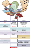
Symptom-based diagnostic algorithm. The ligaments fall naturally into the 3 zones of causation: PUL, CL, ATFP, USL, PB. Anterior zone: urethral meatus to bladder neck. Middle zone: bladder neck to cervix. Posterior zone: cervix to perineal body. Boxes are ticked with questionnaire answers, even if a symptom occurs only “sometimes”. The prolapses in the algorithm correlate with ligament damage in the columns. Symptom groupings in the columns help deduce which ligaments cause which symptoms. The broken lines indicate the open positions of the bladder and bowel. Bladder and bowel symptoms which co-occur, as in the posterior zone are all potentially curable by USL repair, with or without perineal body repair for rectocele. Reused from Petros P. The female pelvic floor function, dysfunction and management according to the Integral Theory. 3rd edition. Heidelberg: Springer Berlin; 2010. With permission from Peter Petros; retains ownership of the copyright. U, uterus; R, rectum; N, bladder base stretch receptors; CX ring, cervical ring; PS, pubic symphysis; S, sacrum; PUL, pubourethral ligament; ATFP, arcus tendineus fascia pelvis; USL, uterosacral ligament; CL, cardinal ligament; PCM, pubococcygeus muscle; LP, levator plate; LMA, conjoint longitudinal muscle of the anus; PRM, puborectalis muscle; PCF, pubocervical fascia; RVF, rectovaginal fascia; PB, perineal body; EAS, external anal sphincter.
Figure 5
Figure 5
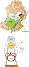
Anterior rectal wall intussusception caused by uterine prolapse. Top figure: sagittal view. Elongation of USL. Bottom figure: view from above. USLs splay laterally as they lengthen with the prolapse, as does the anterior rectal wall which invaginates to cause intussusception. Adapted and reused from Petros P. The female pelvic floor function, dysfunction and management according to the Integral Theory. 3rd edition. Heidelberg: Springer Berlin; 2010. With permission from Peter Petros; retains ownership of the copyright. USL, uterosacral ligament; SSL, sacrospinous ligament.
Figure 6
Figure 6
Mechanism of double incontinence (stress urinary and stress fecal incontinence). A hemostat (red arrow) is placed in the midurethral position in the vagina. It acts as an anchor for the PUL ligaments against which the “PCM” and “LP” contract to control urine and fecal loss on effort. “F” (small arrows), represents the “LP/LMA” rotational vector forces which stretch the rectum around a contracted “PRM” to close the anorectum. Reused with permission from Pelviperineology (6). PS, pubic symphysis; PUL, pubourethral ligaments; PCM, pubococcygeus muscle; R, rectum; PRM, puborectalis muscle; LMA, conjoint longitudinal muscle of the anus; LP, levator plate.
Figure 7
Figure 7
DPS. The perineal body “PB” has been stretched, narrowed and flattened; the rectal serosa has been broken and rectal mucosa (rectum) has spread laterally to adhere to the vagina, PB, and the DTPs (10). Reused from Petros P. The female pelvic floor function, dysfunction and management according to the Integral Theory. 3rd edition. Heidelberg: Springer Berlin; 2010. With permission from Peter Petros; retains ownership of the copyright. OF, obturator fossa; DTP, deep transversus perinei ligament; PB, perineal body; A, anus; DPS, descending perineal syndrome.
Figure 8
Figure 8
Anorectal angle formation. Anorectal angle. If “PUL” and “USL” are competent, the opposite muscle forces, “PCM”, “LP/LMA” are in balance, and the anorectal angle “A” is normal. The small yellow arrow represents unbalanced forward action of the “PRM” when the “LP/LMA” weaken because of a weak “USL”. Reused from Petros P. The female pelvic floor function, dysfunction and management according to the Integral Theory. 3rd edition. Heidelberg: Springer Berlin; 2010. With permission from Peter Petros; retains ownership of the copyright. PUL, pubourethral ligament; PCM, pubococcygeus muscle; ATFP, arcus tendineus fascia pelvis; PB, perineal body; RVF, rectovaginal fascia; PRM, puborectalis muscle; EAS, external anal sphincter; USL, uterosacral ligament; LP, levator plate; LMA, conjoint longitudinal muscle of the anus.
Figure 9
Figure 9
Transperineal ultrasound prior to PUL and USL reconstruction. Acute anorectal angle at rest and straining. Note excessive indentation of the posterior rectal wall at rest by LP. On straining (non-evacuatory), there is descent of the posterior rectal wall, but no significant change in the anorectal angle. Dotted lines demonstrate rectal wall. Reused with permission from Pelviperineology (11). LP, levator plate; PUL, pubourethral ligament; USL, uterosacral ligament.
Figure 10
Figure 10
Transperineal ultrasound after PUL and USL reconstruction. Postoperative perineal ultrasound, at rest and straining. Note restoration of normal anatomy “at rest”, and also, a more acute A/R angle on straining following repair with PUL and USL slings. During straining (non-evacuatory), an upward/forward force seems to have been exerted on to “p” (posterior rectal wall). This force at “p”, can only be from the PRM. It lifts the rectum forwards and upwards towards the upper part of the “U” and bladder neck; meanwhile, the anorectal angle has become more acute by the rectum being pulled downward and backwards around “p” (PRM). This sequence of events supports those detailed for anorectal closure in Figure 1. The vertical white line “U” indicates urethra; a&p indicate the anterior and posterior walls of the rectum. Dotted lines demonstrate rectal wall. Reused with permission from Pelviperineology (11). PS, pubic symphysis; B, bladder; LP, levator plate; PUL, pubourethral ligament; USL, uterosacral ligament; A/R, anorectum; PRM, puborectalis muscle.
Figure 11
Figure 11
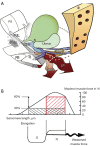
Striated muscles require firm insertion points for optimum contractility. (A) Three-dimensional view from above. The uterus has prolapsed to first degree. The “USLs” have elongated by “E”. The “LP” and “LMA” have also effectively lengthened because they contract against the “USLs”. The rectum also has descended, by virtue of its attachments laterally to the elongated “USL”. The wavy shape of the “LP” and “LMA” indicate diminution of contractile strength. (B) Gordon’s law (22): a striated muscle contracts optimally over a short normal length only (“N”, red square). Elongation of the muscle results in rapid loss of contractile force (“E”, black rectangle), from 80% to 30%. Adapted and reused from Petros P. The female pelvic floor function, dysfunction and management according to the Integral Theory. 3rd edition. Heidelberg: Springer Berlin; 2010. With permission from Peter Petros; retains ownership of the copyright. PS, pubic symphysis; PUL, pubourethral ligament; PCM, pubococcygeus muscle; ATFP, arcus tendineus fascia pelvis; N, bladder base stretch receptors; R, rectum; PB perineal body; EAS, external anal sphincter; USL, uterosacral ligament; LP, levator plate; LMA, conjoint longitudinal muscle of the anus; S, sacrum.
Video S1
Video S1
Video abstract.
Video S2
Video S2
Anorectal closure. Ultrasound.
Video S3
Video S3
Defecation sagittal view. X-ray defecography.
Video S4
Video S4
Lateral distension of rectal walls during defecation. CT scan. By permission Dr. Ilario Froehner Jr.

References

    1. Petros P, Swash M. The Musculo-Elastic Theory of anorectal function and dysfunction. Pelviperineology 2008;27:89-93.
    1. Petros P, Quaghebeur J, Wyndaele JJ. Defining urge as an uncontrolled micturition explains pathogenesis, informs cure and helps solve the burgeoning OAB crisis. Neurourol Urodyn 2022;41:1281-92. 10.1002/nau.24990 - DOI - PMC - PubMed
    1. Petros P, Swash M, Bush M, et al. Defecation 1: Testing a hypothesis for pelvic striated muscle action to open the anorectum. Tech Coloproctol 2012;16:437-43. 10.1007/s10151-012-0861-2 - DOI - PubMed
    1. Bush M, Petros P, Swash M, et al. Defecation 2: Internal anorectal resistance is a critical factor in defecatory disorders. Tech Coloproctol 2012;16:445-50. 10.1007/s10151-012-0860-3 - DOI - PubMed
    1. Farag A. Use of the Hagen-Poiseuille law: a new mathematical approach for the integration and evaluation of anorectal physiological testing in patients with faecal incontinence and pelvic dyschezia and in normal controls. Eur Surg Res 1998;30:279-89. 10.1159/000008588 - DOI - PubMed