

Diagnostic accuracy of ultrasound screening for fetal structural abnormalities during the first and second trimester of pregnancy in low-risk and unselected populations
- PMID: 38721874
- PMCID: PMC11079979
- DOI: 10.1002/14651858.CD014715.pub2
Diagnostic accuracy of ultrasound screening for fetal structural abnormalities during the first and second trimester of pregnancy in low-risk and unselected populations
Abstract
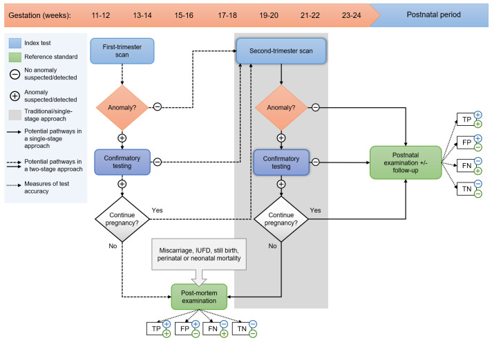
Background: Prenatal ultrasound is widely used to screen for structural anomalies before birth. While this is traditionally done in the second trimester, there is an increasing use of first-trimester ultrasound for early detection of lethal and certain severe structural anomalies.
Objectives: To evaluate the diagnostic accuracy of ultrasound in detecting fetal structural anomalies before 14 and 24 weeks' gestation in low-risk and unselected pregnant women and to compare the current two main prenatal screening approaches: a single second-trimester scan (single-stage screening) and a first- and second-trimester scan combined (two-stage screening) in terms of anomaly detection before 24 weeks' gestation.
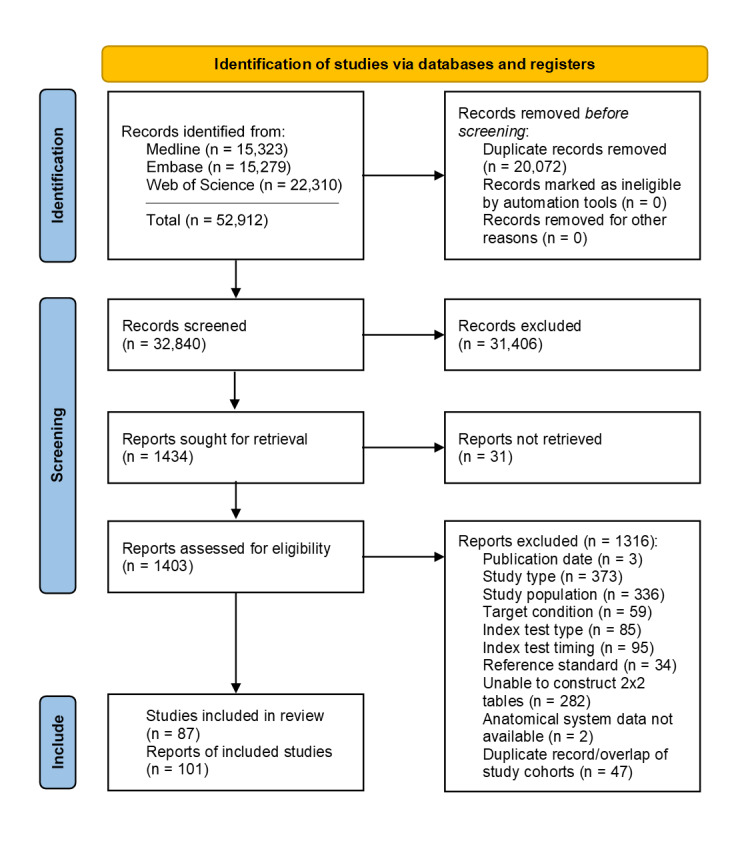
Search methods: We searched MEDLINE, EMBASE, Science Citation Index Expanded (Web of Science), Social Sciences Citation Index (Web of Science), Arts & Humanities Citation Index and Emerging Sources Citation Index (Web of Science) from 1 January 1997 to 22 July 2022. We limited our search to studies published after 1997 and excluded animal studies, reviews and case reports. No further restrictions were applied. We also screened reference lists and citing articles of each of the included studies.
Selection criteria: Studies were eligible if they included low-risk or unselected pregnant women undergoing a first- and/or second-trimester fetal anomaly scan, conducted at 11 to 14 or 18 to 24 weeks' gestation, respectively. The reference standard was detection of anomalies at birth or postmortem.
Data collection and analysis: Two review authors independently undertook study selection, quality assessment (QUADAS-2), data extraction and evaluation of the certainty of evidence (GRADE approach). We used univariate random-effects logistic regression models for the meta-analysis of sensitivity and specificity.
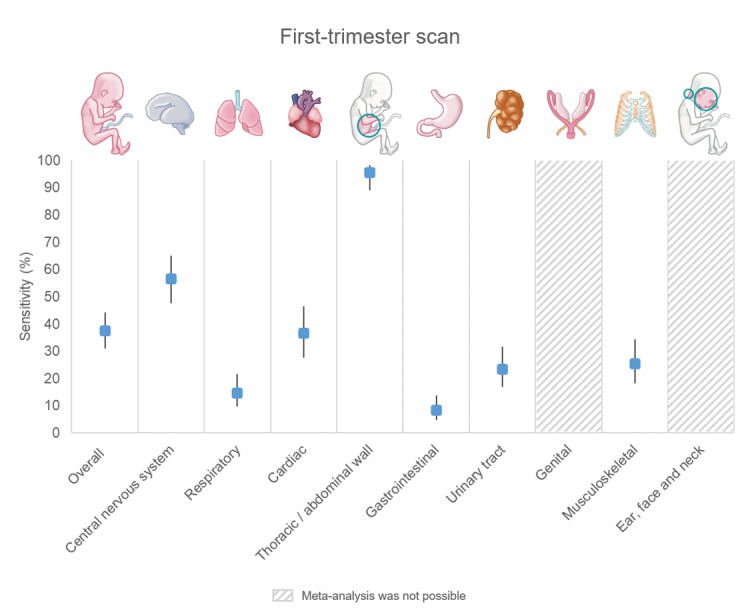
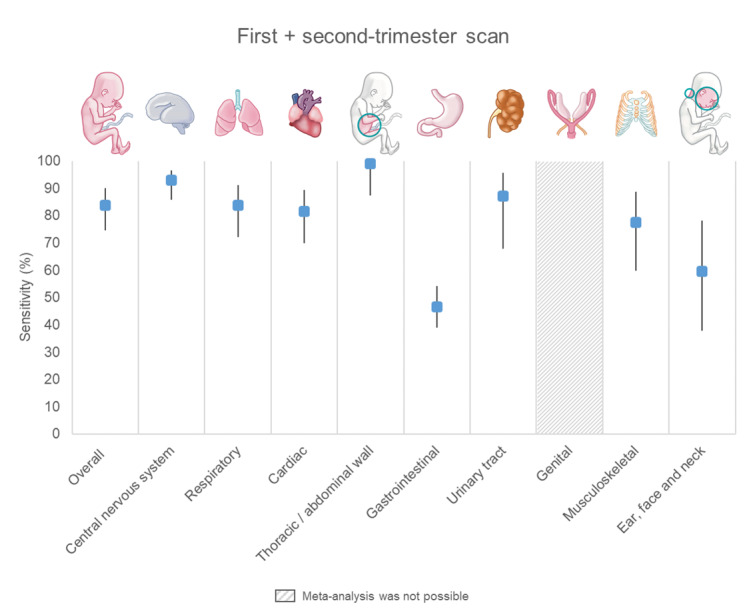
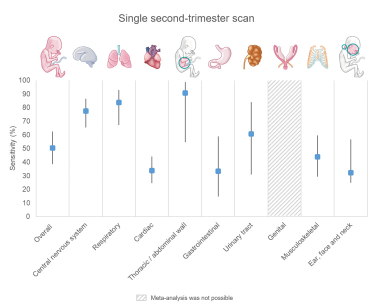
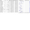
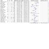
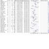
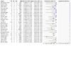
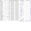
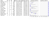
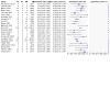
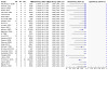
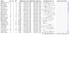
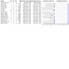
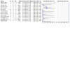
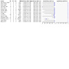
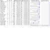
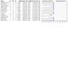
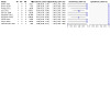
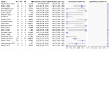
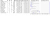
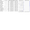
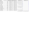
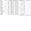
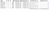
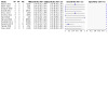
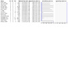
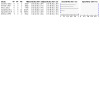
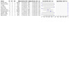
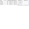
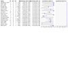
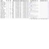
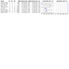
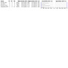
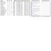
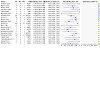
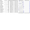
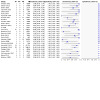
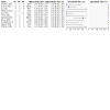
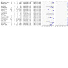
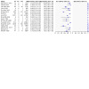
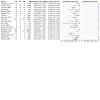
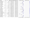
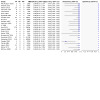
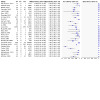
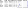
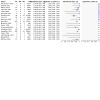
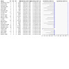
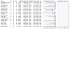
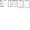
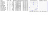
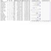
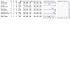
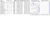
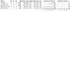
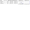
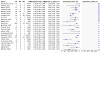
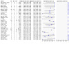
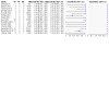
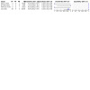
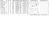
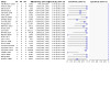
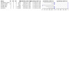
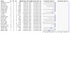
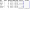
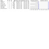
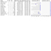
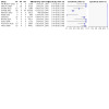
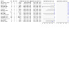
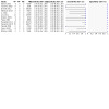
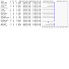
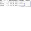
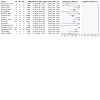
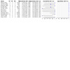
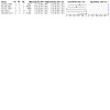
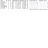
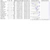
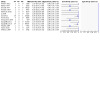
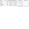
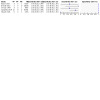
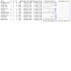
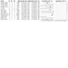
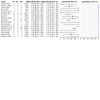
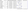
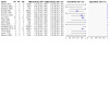
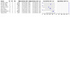
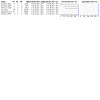
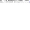
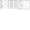
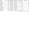
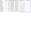
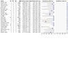
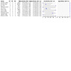
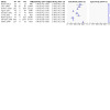
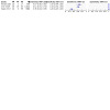
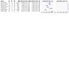
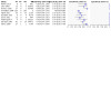
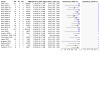
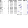
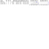
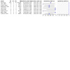
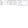
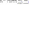
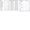
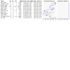
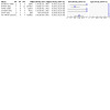
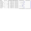
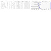
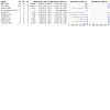
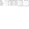
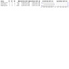
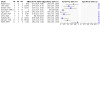
Main results: Eighty-seven studies covering 7,057,859 fetuses (including 25,202 with structural anomalies) were included. No study was deemed low risk across all QUADAS-2 domains. Main methodological concerns included risk of bias in the reference standard domain and risk of partial verification. Applicability concerns were common in studies evaluating first-trimester scans and two-stage screening in terms of patient selection due to frequent recruitment from single tertiary centres without exclusion of referrals. We reported ultrasound accuracy for fetal structural anomalies overall, by severity, affected organ system and for 46 specific anomalies. Detection rates varied widely across categories, with the highest estimates of sensitivity for thoracic and abdominal wall anomalies and the lowest for gastrointestinal anomalies across all tests. The summary sensitivity of a first-trimester scan was 37.5% for detection of structural anomalies overall (95% confidence interval (CI) 31.1 to 44.3; low-certainty evidence) and 91.3% for lethal anomalies (95% CI 83.9 to 95.5; moderate-certainty evidence), with an overall specificity of 99.9% (95% CI 99.9 to 100; low-certainty evidence). Two-stage screening had a combined sensitivity of 83.8% (95% CI 74.7 to 90.1; low-certainty evidence), while single-stage screening had a sensitivity of 50.5% (95% CI 38.5 to 62.4; very low-certainty evidence). The specificity of two-stage screening was 99.9% (95% CI 99.7 to 100; low-certainty evidence) and for single-stage screening, it was 99.8% (95% CI 99.2 to 100; moderate-certainty evidence). Indirect comparisons suggested superiority of two-stage screening across all analyses regarding sensitivity, with no significant difference in specificity. However, the certainty of the evidence is very low due to the absence of direct comparisons.
Authors' conclusions: A first-trimester scan has the potential to detect lethal and certain severe anomalies with high accuracy before 14 weeks' gestation, despite its limited overall sensitivity. Conversely, two-stage screening shows high accuracy in detecting most fetal structural anomalies before 24 weeks' gestation with high sensitivity and specificity. In a hypothetical cohort of 100,000 fetuses, the first-trimester scan is expected to correctly identify 113 out of 124 fetuses with lethal anomalies (91.3%) and 665 out of 1776 fetuses with any anomaly (37.5%). However, 79 false-positive diagnoses are anticipated among 98,224 fetuses (0.08%). Two-stage screening is expected to correctly identify 1448 out of 1776 cases of structural anomalies overall (83.8%), with 118 false positives (0.1%). In contrast, single-stage screening is expected to correctly identify 896 out of 1776 cases before 24 weeks' gestation (50.5%), with 205 false-positive diagnoses (0.2%). This represents a difference of 592 fewer correct identifications and 88 more false positives compared to two-stage screening. However, it is crucial to acknowledge the uncertainty surrounding the additional benefits of two-stage versus single-stage screening, as there are no studies directly comparing them. Moreover, the evidence supporting the accuracy of first-trimester ultrasound and two-stage screening approaches primarily originates from studies conducted in single tertiary care facilities, which restricts the generalisability of the results of this meta-analysis to the broader population.
Copyright © 2024 The Cochrane Collaboration. Published by John Wiley & Sons, Ltd.
Conflict of interest statement
Marieke Buijtendijk was supported by a PhD fellowship funded by a TKI‐LSH Dutch Top Sector Life Sciences & Health consortium grant, where Samsung Medison South Korea was a contributor to the pool of funds supporting the fellowship. Samsung loans ultrasound equipment to the department for conducting pre‐clinical research projects. Additionally, she was invited to present during a webinar on embryology and first‐trimester 3D ultrasound imaging organised by the American Institute for Ultrasound in Medicine (AIUM) in collaboration with NeuroLogica, a subsidiary of Samsung Electronics, for which the department received payment.
Bo Bet and Mariska Leeflang received no funding and report no conflicts of interest.
Harsha Shah has received funding from Samsung Medison South Korea to support travel costs for research in three‐dimensional ultrasound. She received payments for lectures/presentations from NeuroLogica Corporation first‐trimester 3D ultrasound imaging.
Tom Reuvekamp, Timothy Goring, Daniël Docter and Melanie Timmerman received no funding and report no conflicts of interest.
Yousif Dawood received funding from the AMC PhD scholarship (University of Amsterdam) for four years for the conception of a microfocus computed tomography (micro‐CT) based three‐dimensional atlas of human fetal development. There is no conflict of interest.
Malou Lugthart, Bente Berends and Jacqueline Limpens do not receive funding. There is no conflict of interest.
Eva Pajkrt received funding from ZonMW for the Pessary or Cerclage to prevent preterm delivery in women with short cervical length and a history of preterm birth (PC) study (study number: 837002406; Koullali 2017) and receives money from ZonMW for the Expectant Management versus Induction of Labor (PROMEXIL) Follow‐up trial (study number: 843002826; de Ruigh 2021). She was appointed the Minister of Health, Welfare and Sport and the Minister of Security and Justice to the role of chairperson of the review commission for late pregnancy termination and termination of life of newborns (appointed on 27 January 2016) and has been reimbursed expenses for attending meetings.
Maurice van den Hoff obtained funding for a PhD fellowship through a TKI‐LSH Dutch Top Sector Life Sciences & Health consortium grant, where Samsung was a contributor to the pool of funds supporting the fellowship. Samsung loans ultrasound equipment to the department for conducting pre‐clinical research projects. Dr. van den Hoff is Past‐Chair of the European Society of Cardiology (ESC) Working Group Development, Anatomy and Pathology. From September 2012 onward, he held various roles, including board member, treasurer, vice chair and chair. During this time, he received support for travel expenses to attend ESC meetings.
Bernadette de Bakker declares no conflicts of interests.
Figures


Update of
References
References to studies included in this review
Abu‐Rustum 2010 {published data only}
-
- Abu-Rustum RS, Daou L, Abu-Rustum SE. Role of first-trimester sonography in the diagnosis of aneuploidy and structural fetal anomalies. Journal of Ultrasound in Medicine 2010;29(10):1445-52. - PubMed
Andrew 2015 {published data only}
-
- Andrew C, Gopal S, Ramachandran H, Suvika M. First trimester ultrasound: addition of anatomical screening adds value to the examination: a retrospective case series. Journal of Evolution of Medical and Dental Sciences 2015;4(16):2690-9.
Bardi 2022 {published data only}
Batra 2014 {published data only}
-
- Batra M, Gopinathan KK, Balakrishnan B, Gopinath P, Kuriakose R, Gupta J. The detection rate of cardiac anomalies at 11-13+6 week scan using four chamber view and three vessel view. Journal of Fetal Medicine 2014;1(2):95-8.
Becker 2012 {published data only}
-
- Becker R, Schmitz L, Kilavuz S, Stumm M, Wegner R D, Bittner U. 'Normal' nuchal translucency: a justification to refrain from detailed scan? Analysis of 6858 cases with special reference to ethical aspects. Prenatal Diagnosis 2012;32(6):550-6. - PubMed
Bister 2011 {published data only}
-
- Bister D, Set P, Cash C, Coleman N, Fanshawe T. Incidence of facial clefts in Cambridge, United Kingdom. European Journal of Orthodontics 2011;33(4):372-6. - PubMed
Biswas 2013 {published data only}
-
- Biswas J, De K, Dasgupta S, Ghosh B, Sanyal P, Mukherjee A. Prenatal ultrasound screening to diagnose congenital anomalies among one thousand unselected women and their pregnancy outcome. Journal of Evolution of Medical and Dental Sciences 2013;2(13):1966-71.
Bodin 2018 {published data only}
Boyd 2000‐1 {published data only}
-
- Boyd PA, Wellesley DG, De Walle HE, Tenconi R, Garcia-Minaur S, Zandwijken GR, et al. Evaluation of the prenatal diagnosis of neural tube defects by fetal ultrasonographic examination in different centres across Europe. Journal of Medical Screening 2000;7(4):169-74. - PubMed
Boyd 2000‐10 {published data only}
-
- Boyd PA, Wellesley DG, De Walle HE, Tenconi R, Garcia-Minaur S, Zandwijken GR, et al. Evaluation of the prenatal diagnosis of neural tube defects by fetal ultrasonographic examination in different centres across Europe. Journal of Medical Screening 2000;7(4):169-74. - PubMed
Boyd 2000‐11 {published data only}
-
- Boyd PA, Wellesley DG, De Walle HE, Tenconi R, Garcia-Minaur S, Zandwijken GR, et al. Evaluation of the prenatal diagnosis of neural tube defects by fetal ultrasonographic examination in different centres across Europe. Journal of Medical Screening 2000;7(4):169-74. - PubMed
Boyd 2000‐12 {published data only}
-
- Boyd PA, Wellesley DG, De Walle HE, Tenconi R, Garcia-Minaur S, Zandwijken GR, et al. Evaluation of the prenatal diagnosis of neural tube defects by fetal ultrasonographic examination in different centres across Europe. Journal of Medical Screening 2000;7(4):169-74. - PubMed
Boyd 2000‐2 {published data only}
-
- Boyd PA, Wellesley DG, De Walle HE, Tenconi R, Garcia-Minaur S, Zandwijken GR, et al. Evaluation of the prenatal diagnosis of neural tube defects by fetal ultrasonographic examination in different centres across Europe. Journal of Medical Screening 2000;7(4):169-74. - PubMed
Boyd 2000‐3 {published data only}
-
- Boyd PA, Wellesley DG, De Walle HE, Tenconi R, Garcia-Minaur S, Zandwijken GR, et al. Evaluation of the prenatal diagnosis of neural tube defects by fetal ultrasonographic examination in different centres across Europe. Journal of Medical Screening 2000;7(4):169-74. - PubMed
Boyd 2000‐4 {published data only}
-
- Boyd PA, Wellesley DG, De Walle HE, Tenconi R, Garcia-Minaur S, Zandwijken GR, et al. Evaluation of the prenatal diagnosis of neural tube defects by fetal ultrasonographic examination in different centres across Europe. Journal of Medical Screening 2000;7(4):169-74. - PubMed
Boyd 2000‐5 {published data only}
-
- Boyd PA, Wellesley DG, De Walle HE, Tenconi R, Garcia-Minaur S, Zandwijken GR, et al. Evaluation of the prenatal diagnosis of neural tube defects by fetal ultrasonographic examination in different centres across Europe. Journal of Medical Screening 2000;7(4):169-74. - PubMed
Boyd 2000‐6 {published data only}
-
- Boyd PA, Wellesley DG, De Walle HE, Tenconi R, Garcia-Minaur S, Zandwijken GR, et al. Evaluation of the prenatal diagnosis of neural tube defects by fetal ultrasonographic examination in different centres across Europe. Journal of Medical Screening 2000;7(4):169-74. - PubMed
Boyd 2000‐7 {published data only}
-
- Boyd PA, Wellesley DG, De Walle HE, Tenconi R, Garcia-Minaur S, Zandwijken GR, et al. Evaluation of the prenatal diagnosis of neural tube defects by fetal ultrasonographic examination in different centres across Europe. Journal of Medical Screening 2000;7(4):169-74. - PubMed
Boyd 2000‐8 {published data only}
-
- Boyd PA, Wellesley DG, De Walle HE, Tenconi R, Garcia-Minaur S, Zandwijken GR, et al. Evaluation of the prenatal diagnosis of neural tube defects by fetal ultrasonographic examination in different centres across Europe. Journal of Medical Screening 2000;7(4):169-74. - PubMed
Boyd 2000‐9 {published data only}
-
- Boyd PA, Wellesley DG, De Walle HE, Tenconi R, Garcia-Minaur S, Zandwijken GR, et al. Evaluation of the prenatal diagnosis of neural tube defects by fetal ultrasonographic examination in different centres across Europe. Journal of Medical Screening 2000;7(4):169-74. - PubMed
Boyd 2011 {published data only}
-
- Boyd PA, Tonks AM, Rankin J, Rounding C, Wellesley D, Draper ES. Monitoring the prenatal detection of structural fetal congenital anomalies in England and Wales: register-based study. Journal of Medical Screening 2011;18(1):2-7. - PubMed
Brown 2021 {published data only}
-
- Brown I, Rolnik DL, Fernando S, Menezes M, Ramkrishna J, Costa FD, et al. Ultrasound findings and detection of fetal abnormalities before 11 weeks of gestation. Prenatal Diagnosis 2021;41(13):1675-84. - PubMed
Carvalho 2002 {published data only}
-
- Carvalho MH, Brizot M L, Lopes LM, Chiba CH, Miyadahira S, Zugaib M. Detection of fetal structural abnormalities at the 11-14 week ultrasound scan. Prenatal Diagnosis 2002;22(1):1-4. - PubMed
Chelli 2009 {published data only}
-
- Chelli D, Gaddour I, Najar I, Boudaya F, Zouaoui B, Sfar E, et al. First trimester ultrasound: an early screening tool for fetal structural and chromosomic abnormalities. [French]. Tunisie Medicale 2009;87(12):857-62. - PubMed
Chen 2009 {published data only}
-
- Chen M, Leung TY, Sahota DS, Fung TY, Chan LW, Law LW, et al. Ultrasound screening for fetal structural abnormalities performed by trained midwives in the second trimester in a low-risk population--an appraisal. Acta Obstetricia et Gynecologica Scandinavica 2009;88(6):713-9. - PubMed
Choudry 2009 {published data only}
-
- Choudhry MS, Rahman N, Boyd P, Lakhoo K. Duodenal atresia: associated anomalies, prenatal diagnosis and outcome. Pediatric Surgery International 2009;25(8):727-30. - PubMed
Dane 2007 {published data only}
-
- Dane B, Dane C, Sivri D, Kiray M, Cetin A, Yayla M. Ultrasound screening for fetal major abnormalities at 11-14 weeks. Acta Obstetricia et Gynecologica Scandinavica 2007;86(6):666-70. - PubMed
Del Bianco 2006 {published data only}
-
- Del Bianco A, Russo S, Lacerenza N, Rinaldi M, Rinaldi G, Nappi L, et al. Four chamber view plus three-vessel and trachea view for a complete evaluation of the fetal heart during the second trimester. Journal of Perinatal Medicine 2006;34(4):309-12. - PubMed
de Robertis 2017 {published data only}
-
- De Robertis V, Rembouskos G, Fanelli T, Volpe G, Muto B, Volpe P. The three-vessel and trachea view (3VTV) in the first trimester of pregnancy: an additional tool in screening for congenital heart defects (CHD) in an unselected population. Prenatal Diagnosis 2017;37(7):693-8. - PubMed
Drukker 2020 {published data only}
-
- Drukker L, Cavallaro A, Salim I, Ioannou C, Impey L, Papageorghiou A T. How often do we incidentally find a fetal abnormality at the routine third-trimester growth scan? A population-based study. American Journal of Obstetrics & Gynecology 2020;223(6):919.e1-13. - PubMed
Drysdale 2002 {published data only}
-
- Drysdale K, Ridley D, Walker K, Higgins B, Dean T. First-trimester pregnancy scanning as a screening tool for high-risk and abnormal pregnancies in a district general hospital setting. Journal of Obstetrics & Gynaecology 2002;22(2):159-65. - PubMed
Ebrashy 2019 {published data only}
-
- Ebrashy A, Aboulghar M, Elhodiby M, El-Dessouky S H, Elsirgany S, Gaafar H M, et al. Fetal heart examination at the time of 13 weeks scan: a 5 years' prospective study. Journal of Perinatal Medicine 2019;47(8):871-8. - PubMed
Erenel 2019 {published data only}
-
- Erenel H, Karslı MF, Özel A, Korkmaz SO, Arısoy R, Saltık L, et al. The importance of four-chamber and three-vessel (3-V) views in the screening of fetal cardiac anomalies in the first trimester. Perinatal Journal 2019;27(3):169-75.
Fleurke‐Rozema 2014 {published data only}
-
- Fleurke-Rozema JH, Vogel TA, Voskamp BJ, Pajkrt E, den Berg PP, Beekhuis JR, et al. Impact of introduction of mid-trimester scan on pregnancy outcome of open spina bifida in The Netherlands. Ultrasound in Obstetrics & Gynecology 2014;43(5):553-6. - PubMed
Fleurke‐Rozema 2016 {published data only}
-
- Fleurke-Rozema JH, de Kamp K, Bakker MK, Pajkrt E, Bilardo CM, Snijders RJ. Prevalence, diagnosis and outcome of cleft lip with or without cleft palate in The Netherlands. Ultrasound in Obstetrics & Gynecology 2016;48(4):458-63. - PubMed
Fleurke‐Rozema 2017 {published data only}
-
- Fleurke-Rozema H, de Kamp K, Bakker M, Pajkrt E, Bilardo C, Snijders R. Prevalence, timing of diagnosis and pregnancy outcome of abdominal wall defects after the introduction of a national prenatal screening program. Prenatal Diagnosis 2017;37(4):383-8. - PubMed
Galindo 2011 {published data only}
-
- Galindo A, Herraiz I, Escribano D, Lora D, Melchor JC, la Cruz J. Prenatal detection of congenital heart defects: a survey on clinical practice in Spain. Fetal Diagnosis & Therapy 2011;29(4):287-95. - PubMed
Gallot 2007 {published data only}
-
- Gallot D, Boda C, Ughetto S, Perthus I, Robert-Gnansia E, Francannet C, et al. Prenatal detection and outcome of congenital diaphragmatic hernia: a French registry-based study. Ultrasound in Obstetrics & Gynecology 2007;29(3):276-83. - PubMed
Gireada 2022 {published data only}
Gornall 2003 {published data only}
-
- Gornall AS, Budd JL, Draper ES, Konje JC, Kurinczuk JJ. Congenital cystic adenomatoid malformation: accuracy of prenatal diagnosis, prevalence and outcome in a general population. Prenatal Diagnosis 2003;23(12):997-1002. - PubMed
Grande 2012 {published data only}
-
- Grande M, Arigita M, Borobio V, Jimenez J M, Fernandez S, Borrell A. First-trimester detection of structural abnormalities and the role of aneuploidy markers. Ultrasound in Obstetrics & Gynecology 2012;39(2):157-63. - PubMed
Grandjean 1999 {published data only}
-
- Grandjean H, Larroque D, Levi S. The performance of routine ultrasonographic screening of pregnancies in the Eurofetus Study. American Journal of Obstetrics & Gynecology 1999;181(2):446-54. - PubMed
Gu 2011 {published data only}
-
- Gu Y, Hu YL, Ru T, Yang Y, Dai CY, Yang LJ. Combination prenatal ultrasound screening in early and middle stage of pregnancy for detecting fetal abnormalities. [Chinese]. Chinese Journal of Medical Imaging Technology 2011;27(10):2087-90.
Hafner 2003 {published data only}
-
- Hafner E, Schuller T, Metzenbauer M, Schuchter K, Philipp K. Increased nuchal translucency and congenital heart defects in a low-risk population. Prenatal Diagnosis 2003;23(12):985-9. - PubMed
Hartge 2011 {published data only}
-
- Hartge DR, Weichert J, Krapp M, Germer U, Gembruch U, Axt-Fliedner R. Results of early foetal echocardiography and cumulative detection rate of congenital heart disease. Cardiology in the Young 2011;21(5):505-17. - PubMed
Hautala 2019 {published data only}
-
- Hautala J, Gissler M, Ritvanen A, Tekay A, Pitkanen-Argillander O, Stefanovic V, et al. The implementation of a nationwide anomaly screening programme improves prenatal detection of major cardiac defects: an 11-year national population-based cohort study. British Journal of Obstetrics & Gynaecology 2019;126(7):864-73. - PubMed
He 2016 {published data only}
-
- He H, Wan J, Yu L, Sui S, Guo W. Three-dimensional ultrasound in diagnosis of fetal limb deformities. [Chinese]. Chinese Journal of Medical Imaging Technology 2016;32(11):1714-8.
Hernadi 1997 {published data only}
-
- Hernadi L, Torocsik M. Screening for fetal anomalies in the 12th week of pregnancy by transvaginal sonography in an unselected population. Prenatal Diagnosis 1997;17(8):753-9. - PubMed
Hildebrand 2010 {published data only}
-
- Hildebrand E, Selbing A, Blomberg M. Comparison of first and second trimester ultrasound screening for fetal anomalies in the southeast region of Sweden. Acta Obstetricia et Gynecologica Scandinavica 2010;89(11):1412-9. - PubMed
Iliescu 2013 {published data only}
-
- Iliescu D, Tudorache S, Comanescu A, Antsaklis P, Cotarcea S, Novac L, et al. Improved detection rate of structural abnormalities in the first trimester using an extended examination protocol. Ultrasound in Obstetrics & Gynecology 2013;42(3):300-9. - PubMed
Ingale 2017 {published data only}
-
- Ingale SY, Menon SS. To correlate the incidence of congenital anomalies on antenatal scan and those detected in postnatal period in Krishna Institute of Medical Sciences, Karad. Journal of Evolution of Medical and Dental Sciences 2017;6(5):395-7.
Jacobsen 2011 {published data only}
-
- Jakobsen TR, Sogaard K, Tabor A. Implications of a first trimester Down syndrome screening program on timing of malformation detection. Acta Obstetricia et Gynecologica Scandinavica 2011;90(7):728-36. - PubMed
Johnson 1997 {published data only}
-
- Johnson SP, Sebire NJ, Snijders RJ, Tunkel S, Nicolaides KH. Ultrasound screening for anencephaly at 10-14 weeks of gestation. Ultrasound in Obstetrics & Gynecology 1997;9(1):14-6. - PubMed
Kenkhuis 2018 {published data only}
-
- Kenkhuis MJA, Bakker M, Bardi F, Fontanella F, Bakker MK, Fleurke-Rozema JH, et al. Effectiveness of 12-13-week scan for early diagnosis of fetal congenital anomalies in the cell-free DNA era. Ultrasound in Obstetrics & Gynecology 2018;51(4):463-9. - PubMed
Krapp 2011 {published data only}
-
- Krapp M, Ludwig A, Axt-Fliedner R, Kreiselmaier P. First trimester fetal echocardiography: which planes and defects can be displayed during the daily routine in a prenatal medicine unit? Ultraschall in der Medizin 2011;32(4):362-6. - PubMed
Lee 1998 {published data only}
-
- Lee K, Kim SY, Choi SM, Kim JS, Lee BS, Seo K, et al. Effectiveness of prenatal ultrasonography in detecting fetal anomalies and perinatal outcome of anomalous fetuses. Yonsei Medical Journal 1998;39(4):372-82. - PubMed
Leiroz 2021 {published data only}
-
- Leiroz R, Aquino MA, Santos KP, Monteiro MDC, Aires TSF, Araujo Junior E, et al. Accuracy of the mid-trimester ultrasound scan in the detection of fetal congenital anomalies in a reference center in Northeastern Brazil. Journal of Gynecology Obstetrics and Human Reproduction 2021;50(10):102225. - PubMed
Li 2008 {published data only}
-
- Li Q, Guan B, Li D. Detection of fetal structural abnormalities by early pregnancy ultrasound in China. International Journal of Gynaecology & Obstetrics 2008;100(3):277-8. - PubMed
Liao 2021 {published data only}
-
- Liao Y, Wen H, Ouyang S, Yuan Y, Bi J, Guan Y, et al. Routine first-trimester ultrasound screening using a standardized anatomical protocol. American Journal of Obstetrics & Gynecology 2021;224(4):396.e1-396.e15. - PubMed
Maarse 2011 {published data only}
-
- Maarse W, Pistorius LR, Van Eeten WK, Breugem CC, Kon M, Van den Boogaard MJ, et al. Prenatal ultrasound screening for orofacial clefts. Ultrasound in Obstetrics & Gynecology 2011;38(4):434-9. - PubMed
McAuliffe 2005 {published data only}
-
- McAuliffe FM, Fong KW, Toi A, Chitayat D, Keating S, Johnson JA. Ultrasound detection of fetal anomalies in conjunction with first-trimester nuchal translucency screening: a feasibility study. American Journal of Obstetrics & Gynecology 2005;193(3 Pt 2):1260-5. - PubMed
Natu 2014 {published data only}
-
- Natu N, Wadnere N, Yadav S, Kumar R. Utility of first trimester anomaly scan in screening of congenital abnormalities in low and high risk pregnancies. JK Science 2014;16(4):151-5.
Novotna 2012 {published data only}
-
- Novotna M, Haslik L, Svabik K, Zizka Z, Belosovicova H, Brestak M, et al. Detection of fetal major structural anomalies at the 11-14 ultrasound scan in an unselected population. Ceska Gynekologie 2012;77(4):330-5. - PubMed
Ogge 2006 {published data only}
-
- Ogge G, Gaglioti P, Maccanti S, Faggiano F, Todros T. Prenatal screening for congenital heart disease with four-chamber and outflow-tract views: a multicenter study. Ultrasound in Obstetrics & Gynecology 2006;28(6):779-84. - PubMed
Orlandi 2014 {published data only}
-
- Orlandi E, Rossi C, Perino A, Musico G, Orlandi F. Simplified first-trimester fetal cardiac screening (four chamber view and ventricular outflow tracts) in a low-risk population. Prenatal Diagnosis 2014;34(6):558-63. - PubMed
Oztekin 2009 {published data only}
-
- Oztekin O, Oztekin D, Tinar S, Adibelli Z. Ultrasonographic diagnosis of fetal structural abnormalities in prenatal screening at 11-14 weeks. Diagnostic & Interventional Radiology 2009;15(3):221-5. - PubMed
Petousis 2020 {published data only}
-
- Petousis S, Sotiriadis A, Margioula-Siarkou C, Tsakiridis I, Christidis P, Kyriakakis M, et al. Detection of structural abnormalities in fetuses with normal karyotype at 11-13 weeks using the anatomic examination protocol of the International Society of Ultrasound in Obstetrics and Gynecology (ISUOG). Journal of Maternal-Fetal & Neonatal Medicine 2020;33(15):2581-7. - PubMed
Pilalis 2012 {published data only}
-
- Pilalis A, Basagiannis C, Eleftheriades M, Faros E, Troukis E, Armelidou E, et al. Evaluation of a two-step ultrasound examination protocol for the detection of major fetal structural defects. Journal of Maternal-Fetal & Neonatal Medicine 2012;25(9):1814-7. - PubMed
Rydberg 2017 {published data only}
-
- Rydberg C, Tunon K. Detection of fetal abnormalities by second-trimester ultrasound screening in a non-selected population. Acta Obstetricia et Gynecologica Scandinavica 2017;96(2):176-82. - PubMed
Sainz 2020 {published data only}
-
- Sainz JA, Gutierrez L, Garcia-Mejido J, Ramos Z, Bonomi MJ, Fernandez-Palacin A, et al. Early fetal morphological evaluation (11-13 + 6 weeks) accomplished exclusively by transabdominal imaging and following routine midtrimester fetal ultrasound scan recommendations. Since when can it be performed? Journal of Maternal-Fetal & Neonatal Medicine 2020;33(7):1140-50. - PubMed
Saltvedt 2006 {published data only}
-
- Saltvedt S, Almstrom H, Kublickas M, Valentin L, Grunewald C. Detection of malformations in chromosomally normal fetuses by routine ultrasound at 12 or 18 weeks of gestation-a randomised controlled trial in 39,572 pregnancies. British Journal of Obstetrics & Gynaecology 2006;113(6):664-74. - PubMed
Salvador 2005 {published data only}
-
- Salvador J, Borrell A, Lladonosa A. Increasing detection rates of birth defects by prenatal ultrasound leading to apparent increasing prevalence. Lessons learned from the population-based registry of birth defects of Barcelona. Prenatal Diagnosis 2005;25(11):991-6. - PubMed
Sepulveda 2013 {published data only}
-
- Sepulveda W, Wong AE. First trimester screening for holoprosencephaly with choroid plexus morphology ('butterfly' sign) and biparietal diameter. Prenatal Diagnosis 2013;33(13):1233-7. - PubMed
Smrcek 2006 {published data only}
-
- Smrcek JM, Berg C, Geipel A, Fimmers R, Axt-Fliedner R, Diedrich K, et al. Detection rate of early fetal echocardiography and in utero development of congenital heart defects. Journal of Ultrasound in Medicine 2006;25(2):187-96. - PubMed
Souka 2006 {published data only}
-
- Souka AP, Pilalis A, Kavalakis I, Antsaklis P, Papantoniou N, Mesogitis S, et al. Screening for major structural abnormalities at the 11- to 14-week ultrasound scan. American Journal of Obstetrics & Gynecology 2006;194(2):393-6. - PubMed
Srisupundit 2006 {published data only}
-
- Srisupundit K, Tongsong T, Sirichotiyakul S, Chanprapaph P. Fetal structural anomaly screening at 11-14 weeks of gestation at Maharaj Nakorn Chiang Mai Hospital. Journal of the Medical Association of Thailand 2006;89(5):588-93. - PubMed
Stefos 1999 {published data only}
-
- Stefos T, Plachouras N, Sotiriadis A, Papadimitriou D, Almoussa N, Navrozoglou I, et al. Routine obstetrical ultrasound at 18-22 weeks: our experience on 7,236 fetuses. Journal of Maternal-Fetal Medicine 1999;8(2):64-9. - PubMed
Syngelaki 2011 {published data only}
-
- Syngelaki A, Chelemen T, Dagklis T, Allan L, Nicolaides K H. Challenges in the diagnosis of fetal non-chromosomal abnormalities at 11-13 weeks. Prenatal Diagnosis 2011;31(1):90-102. - PubMed
Syngelaki 2019 {published data only}
-
- Syngelaki A, Hammami A, Bower S, Zidere V, Akolekar R, Nicolaides K H. Diagnosis of fetal non-chromosomal abnormalities on routine ultrasound examination at 11-13 weeks' gestation. Ultrasound in Obstetrics & Gynecology 2019;54(4):468-76. - PubMed
Taipale 2004 {published data only}
-
- Taipale P, Ammala M, Salonen R, Hiilesmaa V. Two-stage ultrasonography in screening for fetal anomalies at 13-14 and 18-22 weeks of gestation. Acta Obstetricia et Gynecologica Scandinavica 2004;83(12):1141-6. - PubMed
Takita 2016 {published data only}
-
- Takita H, Hasegawa J, Arakaki T, Nakamura M, Hamada S, Tokunaka M, et al. Usefulness of antenatal ultrasound fetal morphological assessments in the first and second trimester: a study at a single Japanese university hospital. Journal of Medical Ultrasonics 2016;43(1):57-62. - PubMed
Tiechl 2021 {published data only}
-
- Tiechl J, Abdel Azim S, Leitner K, Berger A, Mutz-Dehbalaie I, Goebel G, et al. Screening for open spina bifida in a routine clinical setting at the first-trimester scan: a prospective multicentre cohort study. Fetal Diagnosis & Therapy 2021;48(2):96-102. - PubMed
Timbolschi 2015 {published data only}
-
- Timbolschi D, Schaefer E, Monga B, Fattori D, Dott B, Favre R, et al. Neural tube defects: the experience of the registry of congenital malformations of Alsace, France, 1995-2009. Fetal Diagnosis & Therapy 2015;37(1):6-17. - PubMed
Tudorache 2016 {published data only}
-
- Tudorache S, Ungureanu A, Dragusin R C, Sorop FM, Cara ML, Iliescu DG. First trimester diagnostic accuracy of a two-dimensional simplified ultrasound technique in congenital heart diseases and great arteries anomalies. Obstetrica si Ginecologie 2016;64(3):165-76.
Tydeman 2013 {published data only}
-
- Tydeman G, Clegg I, Brown S. Detection rates of the Fetal Anomaly Screening Programme (FASP) 11 key conditions in one unit: is the recommended annual audit of any value? Prenatal Diagnosis 2013;33(9):910-2. - PubMed
van Velzen 2016 {published data only}
-
- Velzen CL, Clur SA, Rijlaarsdam ME, Bax CJ, Pajkrt E, Heymans MW, et al. Prenatal detection of congenital heart disease--results of a national screening programme. British Journal of Obstetrics & Gynaecology 2016;123(3):400-7. - PubMed
Vayna 2018 {published data only}
-
- Vayna AM, Veduta A, Duta S, Panaitescu A M, Stoica S, Buinoiu N, et al. Diagnosis of fetal structural anomalies at 11 to 14 weeks. Journal of Ultrasound in Medicine 2018;37(8):2063-73. - PubMed
Vellamkondu 2017 {published data only}
Vimpelli 2006 {published data only}
-
- Vimpelli T, Huhtala H, Acharya G. Fetal echocardiography during routine first-trimester screening: a feasibility study in an unselected population. Prenatal Diagnosis 2006;26(5):475-82. - PubMed
Waern 2021 {published data only}
Wang 2013 {published data only}
-
- Wang L, Wu QQ, Chen Y, Ma YQ, Yao L, Li M. Ultrasound screening of fetal structural abnormalities by standard ultrasound views during the first trimester. Chinese Medical Journal 2013;126(5):986-7. - PubMed
Wayne 2002 {published data only}
-
- Wayne C, Cook K, Sairam S, Hollis B, Thilaganathan B. Sensitivity and accuracy of routine antenatal ultrasound screening for isolated facial clefts. British Journal of Radiology 2002;75(895):584-9. - PubMed
Whitlow 1999 {published data only}
-
- Whitlow BJ, Chatzipapas IK, Lazanakis ML, Kadir RA, Economides DL. The value of sonography in early pregnancy for the detection of fetal abnormalities in an unselected population. British Journal of Obstetrics & Gynaecology 1999;106(9):929-36. - PubMed
Wu 2009 {published data only}
-
- Wu Q, Li M, Ju L, Zhang W, Yang X, Yan Y, et al. Application of the 3-vessel view in routine prenatal sonographic screening for congenital heart disease. Journal of Ultrasound in Medicine 2009;28(10):1319-24. - PubMed
Yu 2011‐1 {published data only}
-
- Yu Z, Xi Y, Ding W, Han S, Cao L, Zhu C, et al. Congenital heart disease in a Chinese hospital: pre- and postnatal detection, incidence, clinical characteristics and outcomes. Pediatrics International 2011;53(6):1059-65. - PubMed
Yu 2011‐2 {published data only}
-
- Yu Z, Xi Y, Ding W, Han S, Cao L, Zhu C, et al. Congenital heart disease in a Chinese hospital: pre- and postnatal detection, incidence, clinical characteristics and outcomes. Pediatrics International 2011;53(6):1059-65. - PubMed
Yu 2011‐3 {published data only}
-
- Yu Z, Xi Y, Ding W, Han S, Cao L, Zhu C, et al. Congenital heart disease in a Chinese hospital: pre- and postnatal detection, incidence, clinical characteristics and outcomes. Pediatrics International 2011;53(6):1059-65. - PubMed
Yu 2011‐4 {published data only}
-
- Yu Z, Xi Y, Ding W, Han S, Cao L, Zhu C, et al. Congenital heart disease in a Chinese hospital: pre- and postnatal detection, incidence, clinical characteristics and outcomes. Pediatrics International 2011;53(6):1059-65. - PubMed
Zhang 2020 {published data only}
-
- Zhang N, Dong H, Wang P, Wang Z, Wang Y, Guo Z. The value of obstetric ultrasound in screening fetal nervous system malformation. World Neurosurgery 2020;138:645-53. - PubMed
Zhang 2021 {published data only}
Zheng 2014 {published data only}
-
- Zheng J, Jiao Y, Wang HF, Xiong Y, Lin Q, Xu FH, et al. Ultrasonographic diagnosis of fetal limb abnormalities at 11-13 weeks of gestation. [Chinese]. Chinese Journal of Medical Imaging Technology 2014;30(8):1230-3.
References to studies excluded from this review
Abu‐Rustum 2010a {published data only}
-
- Abu-Rustum RS, Daou L, Abu-Rustum SE. Role of ultrasonography in early gestation in the diagnosis of congenital heart defects. Journal of Ultrasound in Medicine 2010;29(5):817-21. - PubMed
Anumba 2005 {published data only}
-
- Anumba DO, Scott JE, Plant ND, Robson SC. Diagnosis and outcome of fetal lower urinary tract obstruction in the northern region of England. Prenatal Diagnosis 2005;25(1):7-13. - PubMed
Becker 2005 {published data only}
-
- Becker R, Albig M, Gasiorek-Wiens A, Entezami M, Knoll T, Wegner R D. The potential of first trimester anomaly scan and first trimester fetal echocardiography as screening procedures in a medium risk population. Journal of Obstetrics and Gynecology of India 2005;55(3):228-30.
Becker 2006 {published data only}
-
- Becker R, Wegner RD. Detailed screening for fetal anomalies and cardiac defects at the 11-13-week scan. Ultrasound in Obstetrics & Gynecology 2006;27(6):613-8. - PubMed
Boyd 1998 {published data only}
-
- Boyd PA, Chamberlain P, Hicks NR. 6-year experience of prenatal diagnosis in an unselected population in Oxford, UK. Lancet 1998;352(9140):1577-81. - PubMed
Boyd 2011a {published data only}
-
- Boyd PA, Tonks AM, Rankin J, Rounding C, Wellesley D, Draper ES, et al. Monitoring the prenatal detection of structural fetal congenital anomalies in England and Wales: register-based study. Journal of Medical Screening 2011;18(1):2-7. - PubMed
Boyd 2012 {published data only}
-
- Boyd PA, Rounding C, Chamberlain P, Wellesley D, Kurinczuk JJ. The evolution of prenatal screening and diagnosis and its impact on an unselected population over an 18-year period. British Journal of Obstetrics & Gynaecology 2012;119(9):1131-40. - PubMed
Brantberg 2017 {published data only}
-
- Brantberg A, Blaas HG, Haugen SE, Eik-Nes SH. Esophageal obstruction-prenatal detection rate and outcome. Ultrasound in Obstetrics & Gynecology 2007;30(2):180-7. - PubMed
Braz 2018 {published data only}
-
- Braz P, Machado A, Matias Dias, C. The impact of prenatal diagnosis on congenital anomaly outcomes: Data from 1997 to 2016. European Journal of Medical Genetics 2018;61(9):508-12. - PubMed
Brenes 2003 {published data only}
-
- Brenes J, Asenjo JE, Gomez M, Costales C, Santos J Ga, De Marco E, et al. Evaluation of prenatal diagnosis of fetal cardiopathies. [Spanish]. Toko-Ginecologia Practica 2003;62(668):188-95.
Bullen 2001 {published data only}
-
- Bullen PJ, Rankin JM, Robson SC. Investigation of the epidemiology and prenatal diagnosis of holoprosencephaly in the North of England. American Journal of Obstetrics & Gynecology 2001;184(6):1256-62. - PubMed
Byrne 2018 {published data only}
-
- Byrne JJ, Morgan JL, Twickler DM, McIntire DD, Dashe JS. Utility of follow-up standard sonography for fetal anomaly detection. American Journal of Obstetrics & Gynecology 2020;222(6):615.e1-9. - PubMed
Campana 2010 {published data only}
-
- Campana H, Ermini M, Aiello HA, Krupitzki H, Castilla EE, Lopez-Camelo JS. Prenatal sonographic detection of birth defects in 18 hospitals from South America. Journal of Ultrasound in Medicine 2010;29(2):203-12. - PubMed
Cash 2001 {published data only}
-
- Cash C, Set P, Coleman N. The accuracy of antenatal ultrasound in the detection of facial clefts in a low-risk screening population. Ultrasound in Obstetrics & Gynecology 2001;18(5):432-6. - PubMed
Cedergren 2006 {published data only}
-
- Cedergren M, Selbing A. Detection of fetal structural abnormalities by an 11-14-week ultrasound dating scan in an unselected Swedish population. Acta Obstetricia et Gynecologica Scandinavica 2006;85(8):912-5. - PubMed
Chatzipapas 1999 {published data only}
-
- Chatzipapas IK, Whitlow BJ, Economides DL. The 'Mickey Mouse' sign and the diagnosis of anencephaly in early pregnancy. Ultrasound in Obstetrics & Gynecology 1999;13(3):196-9. - PubMed
Chelemen 2011 {published data only}
-
- Chelemen T, Syngelaki A, Maiz N, Allan L, Nicolaides K H. Contribution of ductus venosus Doppler in first-trimester screening for major cardiac defects. Fetal Diagnosis & Therapy 2011;29(2):127-34. - PubMed
Chen 2019 {published data only}
-
- Chen FC, Bacovsky A, Entezami M, Henrich W. Nearly half of all severe fetal anomalies can be detected by first-trimester screening in experts' hands. Journal of Perinatal Medicine 2019;47(6):619-24. - PubMed
Chu 2017 {published data only}
-
- Chu C, Yan Y, Ren Y, Li X, Gui Y. Prenatal diagnosis of congenital heart diseases by fetal echocardiography in second trimester: a Chinese multicenter study. Acta Obstetricia et Gynecologica Scandinavica 2017;96(4):454-63. - PubMed
Chua 2000 {published data only}
-
- Chua LK, Rose DH. The detection rate of major and common foetal heart abnormalities in routine prenatal ultrasonography. Clinical Radiology 2000;55(11):897.
Clausen 2022 {published data only}
-
- Clausen AB, Garne E, Damkjaer M. Live birth prevalence of atrioventricular septal defect after the implementation of new prenatal screening guidelines. Danish Medical Journal 2022;69(2):13. - PubMed
Clementi 2000 {published data only}
-
- Clementi M, Tenconi R, Bianchi F, Stoll C. Evaluation of prenatal diagnosis of cleft lip with or without cleft palate and cleft palate by ultrasound: experience from 20 European registries. EUROSCAN study group. Prenatal Diagnosis 2000;20(11):870-5. - PubMed
Colosi 2015 {published data only}
Delmas 2017 {published data only}
-
- Delmas HL, Kohler M, Doray B, Lemery D, Francannet C, Quistrebert J, et al. Congenital unilateral renal agenesis: prevalence, prenatal diagnosis, associated anomalies. Data from two birth-defect registries. Birth Defects Research 2017;109(15):1204-11. - PubMed
Duta 2021 {published data only}
-
- Duta S, Veduta A, Vayna AM, Panaitescu A, Nedelea F, Peltecu G. The outcome of structural heart defects diagnosed in the first trimester of pregnancy. Journal of Maternal-Fetal & Neonatal Medicine 2021;34(9):1389-94. - PubMed
Economides 1998 {published data only}
-
- Economides DL, Braithwaite JM. First trimester ultrasonographic diagnosis of fetal structural abnormalities in a low risk population. British Journal of Obstetrics & Gynaecology 1998;105(1):53-7. - PubMed
Eleftheriades 2012 {published data only}
-
- Eleftheriades M, Tsapakis E, Sotiriadis A, Manolakos E, Hassiakos D, Botsis D. Detection of congenital heart defects throughout pregnancy; impact of first trimester ultrasound screening for cardiac abnormalities. Journal of Maternal-Fetal & Neonatal Medicine 2012;25(12):2546-50. - PubMed
Ensing 2014 {published data only}
-
- Ensing S, Kleinrouweler CE, Maas SM, Bilardo CM, Van der Horst CM, Pajkrt E. Influence of the 20-week anomaly scan on prenatal diagnosis and management of fetal facial clefts. Ultrasound in Obstetrics & Gynecology 2014;44(2):154-9. - PubMed
Eurenius 1999 {published data only}
-
- Eurenius K, Axelsson O, Cnattingius S, Eriksson L, Norsted T. Second trimester ultrasound screening performed by midwives; sensitivity for detection of fetal anomalies. Acta Obstetricia et Gynecologica Scandinavica 1999;78(2):98-104. - PubMed
Everwijn 2018 {published data only}
-
- Everwijn SMP, Nisselrooij AEL, Rozendaal L, Clur SAB, Pajkrt E, Hruda J, et al. The effect of the introduction of the three-vessel view on the detection rate of transposition of the great arteries and tetralogy of Fallot. Prenatal Diagnosis 2018;38(12):951-7. - PubMed
Ficara 2020 {published data only}
-
- Ficara A, Syngelaki A, Hammami A, Akolekar R, Nicolaides K H. Value of routine ultrasound examination at 35-37 weeks' gestation in diagnosis of fetal abnormalities. Ultrasound in Obstetrics & Gynecology 2020;55(1):75-80. - PubMed
Forci 2020 {published data only}
Fricke 2021 {published data only}
-
- Fricke K, Bhat M, Avdikos V, Asp A, Brodszki J, Thurn L. High prenatal detection rates of complex congenital heart defects (CHD). Lakartidningen 2021;118(11):17. - PubMed
Garne 2001 {published data only}
-
- Garne E, Eurocat Working Group. Prenatal diagnosis of six major cardiac malformations in Europe--a population based study. Acta Obstetricia et Gynecologica Scandinavica 2001;80(3):224-8. - PubMed
Garne 2002 {published data only}
-
- Garne E, Haeusler M, Barisic I, Gjergja R, Stoll C, Clementi M, et al. Congenital diaphragmatic hernia: evaluation of prenatal diagnosis in 20 European regions. Ultrasound in Obstetrics & Gynecology 2002;19(4):329-33. - PubMed
Grandjean 1998 {published data only}
-
- Grandjean H, Larroque D, Levi S, Eurofetus Team. Sensitivity of routine ultrasound screening of pregnancies in the Eurofetus database. In: Levi S, Chervenak F A, editors(s). Ultrasound Screening for Fetal Anomalies: Is It Worth It?: Screening Revisited after the Eurofetus Data. 847 edition. New York: New York Acad Sciences, 1998:118-24. - PubMed
Haeusler 2002 {published data only}
-
- Haeusler MC, Berghold A, Stoll C, Barisic I, Clementi M, Group Euroscan Study. Prenatal ultrasonographic detection of gastrointestinal obstruction: results from 18 European congenital anomaly registries. Prenatal Diagnosis 2002;22(7):616-23. - PubMed
Hafner 1998 {published data only}
-
- Hafner E, Scholler J, Schuchter K, Sterniste W, Philipp K. Detection of fetal congenital heart disease in a low-risk population. Prenatal Diagnosis 1998;18(8):808-15. - PubMed
Hunter 2000 {published data only}
Iliescu 2014 {published data only}
-
- Iliescu DG, Cara ML, Tudorache S, Antsaklis P, Novac LV, Antsaklis A, et al. Agenesis of ductus venosus in sequential first and second trimester screening. Prenatal Diagnosis 2014;34(11):1099-105. - PubMed
Itsukaichi 2018 {published data only}
-
- Itsukaichi M, Serikawa T, Yoshihara K, Suzuki H, Haino K, Yamaguchi M, et al. Effectiveness of fetal cardiac screening for congenital heart disease using a combination of the four-chamber view and three-vessel view during the second trimester scan. Journal of Obstetrics & Gynaecology Research 2018;44(1):49-53. - PubMed
Johnson 2012 {published data only}
-
- Johnson N, Bishop K, Trotman H, Reid M. Congenital abnormalities at a tertiary center in Jamaica: an 18-month maternal-fetal medicine experience. Journal of Maternal-Fetal & Neonatal Medicine 2012;25(6):687-91. - PubMed
Jorgensen 2015 {published data only}
-
- Jorgensen DE, Vejlstrup N, Jorgensen C, Maroun LL, Steensberg J, Hessellund A, et al. Prenatal detection of congenital heart disease in a low risk population undergoing first and second trimester screening. Prenatal Diagnosis 2015;35(4):325-30. - PubMed
Kansal 2022 {published data only}
-
- Kansal K, Mittal M, Gupta A. Detection of congenital structural anomalies by mid trimester ultrasonography. Journal of Cardiovascular Disease Research 2022;13(1):1671-7.
Khoo 2008 {published data only}
-
- Khoo NS, Van Essen P, Richardson M, Robertson T. Effectiveness of prenatal diagnosis of congenital heart defects in South Australia: a population analysis 1999-2003. Australian & New Zealand Journal of Obstetrics & Gynaecology 2008;48(6):559-63. - PubMed
Laurichesse 2017 {published data only}
-
- Laurichesse Delmas H, Kohler M, Doray B, Lemery D, Francannet C, et al. Congenital unilateral renal agenesis: prevalence, prenatal diagnosis, associated anomalies. Data from two birth-defect registries. Birth Defects Research 2017;109(15):1204-11. - PubMed
Levi 2003 {published data only}
-
- Levi S, Zhang WH, Alexander S, Viart P, Grandjean H. Short-term outcome of isolated and associated congenital heart defects in relation to antenatal ultrasound screening. Ultrasound in Obstetrics and Gynecology 2003;21(6):532-8. - PubMed
Li 2019 {published data only}
Liao 2016 {published data only}
-
- Liao YM, Li SL, Luo GY, Wen HX, Ouyang SY, Chen CY, et al. Routine screening for fetal limb abnormalities in the first trimester. Prenatal Diagnosis 2016;36(2):117-26. - PubMed
Liao 2021a {published data only}
-
- Liao Y, Wen H, Luo G, Ouyang S, Bi J, Yuan Y, et al. Fetal open and closed spina bifida on a routine scan at 11 weeks to 13 weeks 6 days. Journal of Ultrasound in Medicine 2021;40(2):237-47. - PubMed
Liou 2011 {published data only}
-
- Liou JD, Huang YH, Hung TH, Hsieh CL, Hsieh TT, Lo LM. Prenatal diagnostic rates and postnatal outcomes of fetal orofacial clefts in a Taiwanese population. International Journal of Gynaecology & Obstetrics 2011;113(3):211-4. - PubMed
Long 1998 {published data only}
-
- Long G, Sprigg A. A comparative study of routine versus selective fetal anomaly ultrasound scanning. Journal of Medical Screening 1998;5(1):6-10. - PubMed
Ma 2018 {published data only}
-
- Ma Y, Zhang J, Yang X. Clinical application of standardized prenatal ultrasound screening during 11-13 weeks gestation. [Chinese]. Chinese Journal of Medical Imaging Technology 2018;34(1):86-9.
Magripes 1998 {published data only}
-
- Magriples U, Copel JA. Accurate detection of anomalies by routine ultrasonography in an indigent clinic population. American Journal of Obstetrics & Gynecology 1998;179(4):978-81. - PubMed
Marek 2011 {published data only}
-
- Marek J, Tomek V, Skovranek J, Povysilova V, Samanek M. Prenatal ultrasound screening of congenital heart disease in an unselected national population: a 21-year experience. Heart 2011;97(2):124-30. - PubMed
Michailidis 2001 {published data only}
-
- Michailidis GD, Economides DL. Nuchal translucency measurement and pregnancy outcome in karyotypically normal fetuses. Ultrasound in Obstetrics & Gynecology 2001;17(2):102-5. - PubMed
Minnella 2020 {published data only}
-
- Minnella GP, Crupano FM, Syngelaki A, Zidere V, Akolekar R, Nicolaides KH. Diagnosis of major heart defects by routine first-trimester ultrasound examination: association with increased nuchal translucency, tricuspid regurgitation and abnormal flow in ductus venosus. Ultrasound in Obstetrics & Gynecology 2020;55(5):637-44. - PubMed
Nakling 2005 {published data only}
-
- Nakling J, Backe B. Routine ultrasound screening and detection of congenital anomalies outside a university setting. Acta Obstetricia et Gynecologica Scandinavica 2005;84(11):1042-8. - PubMed
Nikkila 2006 {published data only}
-
- Nikkila A, Rydhstroem H, Kallen B, Jorgensen C. Ultrasound screening for fetal anomalies in southern Sweden: a population-based study. Acta Obstetricia et Gynecologica Scandinavica 2006;85(6):688-93. - PubMed
Ou 2022 {published data only}
Pavlicek 2020 {published data only}
-
- Pavlicek J, Piegzova A, Klaskova E, Kapralova S, Palatova A, Spacek R, et al. Development, effectiveness, and current possibilities in prenatal detection of congenital heart defects. Cor Et Vasa 2020;62(1):21-8.
Pitukkijronnakorn 2009 {published data only}
-
- Pitukkijronnakorn S, Chittacharoen A, Jetsawangsri T, Panburana P, Jaovisidha A, Roungsipragarn R, et al. The value of mid-trimester routine ultrasonographic screening in antenatal detection of congenital malformations. Journal of the Medical Association of Thailand 2009;92(6):748-53. - PubMed
Poggenpoel 2012 {published data only}
-
- Poggenpoel EJ, Geerts LT, Theron GB. The value of adding a universal booking scan to an existing protocol of routine mid-gestation ultrasound scan. International Journal of Gynecology & Obstetrics 2012;116(3):201-5. - PubMed
Queisser‐Luft 1998 {published data only}
-
- Queisser-Luft A, Stopfkuchen H, Stolz G, Schlaefer K, Merz E. Prenatal diagnosis of major malformations: quality control of routine ultrasound examinations based on a five-year study of 20,248 newborn fetuses and infants. Prenatal Diagnosis 1998;18(6):567-76. - PubMed
Sainz 2015 {published data only}
-
- Sainz JA, Zurita MJ, Guillen I, Borrero C, Garcia-Mejido J, Almeida C, et al. Prenatal screening of congenital heart defects in population at low risk of congenital defects. A reality today. Anales de Pediatria 2015;82(1):27-34. - PubMed
Stoll 1998 {published data only}
-
- Stoll C, Alembik Y, Dott B, Meyer MJ, Pennerath A, Peter MO, et al. Evaluation of prenatal diagnosis of congenital heart disease. Prenatal Diagnosis 1998;18(8):801-7. - PubMed
Stoll 2000a {published data only}
-
- Stoll C, Dott B, Alembik Y, Roth M. Evaluation of prenatal diagnosis of cleft lip/palate by foetal ultrasonographic examination. Annales de Genetique 2000;43(1):11-4. - PubMed
Stoll 2000b {published data only}
-
- Stoll C, Wiesel A, Queisser-Luft A, Froster U, Bianca S, Clementi M. Evaluation of the prenatal diagnosis of limb reduction deficiencies. EUROSCAN Study Group. Prenatal Diagnosis 2000;20(10):811-8. - PubMed
Stoll 2001 {published data only}
-
- Stoll C, Garne E, Clementi M, Group Euroscan Study. Evaluation of prenatal diagnosis of associated congenital heart diseases by fetal ultrasonographic examination in Europe. Prenatal Diagnosis 2001;21(4):243-52. - PubMed
Stoll 2002 {published data only}
-
- Stoll C, Dott B, Alembik Y, De Geeter B. Evaluation and evolution during time of prenatal diagnosis of congenital heart diseases by routine fetal ultrasonographic examination. Annales de Genetique 2002;45(1):21-7. - PubMed
Tabor 2003 {published data only}
-
- Tabor A, Zdravkovic M, Perslev A, Moller LK, Pedersen BL. Screening for congenital malformations by ultrasonography in the general population of pregnant women: factors affecting the efficacy. Acta Obstetricia et Gynecologica Scandinavica 2003;82(12):1092-8. - PubMed
Taipale 2003 {published data only}
-
- Taipale P, Ammala M, Salonen R, Hiilesmaa V. Learning curve in ultrasonographic screening for selected fetal structural anomalies in early pregnancy. Obstetrics & Gynecology 2003;101(2):273-8. - PubMed
Tegnander 2006 {published data only}
-
- Tegnander E, Eik-Nes SH. The examiner's ultrasound experience has a significant impact on the detection rate of congenital heart defects at the second-trimester fetal examination. Ultrasound in Obstetrics & Gynecology 2006;28(1):8-14. - PubMed
Tegnander 2006a {published data only}
-
- Tegnander E, Williams W, Johansen OJ, Blaas HG, Eik-Nes SH. Prenatal detection of heart defects in a non-selected population of 30,149 fetuses--detection rates and outcome. Ultrasound in Obstetrics & Gynecology 2006;27(3):252-65. - PubMed
Todros 1997 {published data only}
-
- Todros T, Faggiano F, Chiappa E, Gaglioti P, Mitola B, Sciarrone A. Accuracy of routine ultrasonography in screening heart disease prenatally. Gruppo Piemontese for Prenatal Screening of Congenital Heart Disease. Prenatal Diagnosis 1997;17(10):901-6. - PubMed
Trines 2013 {published data only}
-
- Trines J, Fruitman D, Zuo KJ, Smallhorn JF, Hornberger LK, Mackie AS. Effectiveness of prenatal screening for congenital heart disease: assessment in a jurisdiction with universal access to health care. Canadian Journal of Cardiology 2013;29(7):879-85. - PubMed
Tudorache 2016a {published data only}
Tudorache 2016b {published data only}
-
- Tudorache S, Cara ML, Burada F, Simionescu C, Dragoescu AN, Iliescu DG. First trimester diagnostic accuracy of two-dimensional and four-dimensional ultrasound in major congenital heart diseases. Obstetrica si Ginecologie 2016;64(4):221-32.
van Allen 2006 {published data only}
-
- Van Allen MI, Boyle E, Thiessen P, McFadden D, Cochrane D, Chambers GK, et al. The impact of prenatal diagnosis on neural tube defect (NTD) pregnancy versus birth incidence in British Columbia. Journal of Applied Genetics 2006;47(2):151-8. - PubMed
van Dorsten 1998 {published data only}
-
- VanDorsten JP, Hulsey TC, Newman RB, Menard MK. Fetal anomaly detection by second-trimester ultrasonography in a tertiary center. American Journal of Obstetrics & Gynecology 1998;178(4):742-9. - PubMed
van Velzen 2015 {published data only}
-
- Velzen CL, Haak MC, Reijnders G, Rijlaarsdam ME, Bax CJ, Pajkrt E, et al. Prenatal detection of transposition of the great arteries reduces mortality and morbidity. Ultrasound in Obstetrics & Gynecology 2015;45(3):320-5. - PubMed
Vavilala 2011 {published data only}
-
- Vavilala S, Geeta K. The 11-13 weeks scan: where do we stand? A 5-year review at Fernandez Hospital. International Journal of Infertility and Fetal Medicine 2011;2(2):65-9.
Westin 2006 {published data only}
-
- Westin M, Saltvedt S, Bergman G, Kublickas M, Almstrom H, Grunewald C, et al. Routine ultrasound examination at 12 or 18 gestational weeks for prenatal detection of major congenital heart malformations? A randomised controlled trial comprising 36,299 fetuses. British Journal of Obstetrics & Gynaecology 2006;113(6):675-82. - PubMed
Wiesel 2005 {published data only}
-
- Wiesel A, Queisser-Luft A, Clementi M, Bianca S, Stoll C, Group Euroscan Study. Prenatal detection of congenital renal malformations by fetal ultrasonographic examination: an analysis of 709,030 births in 12 European countries. European Journal of Medical Genetics 2005;48(2):131-44. - PubMed
Wong 2004 {published data only}
-
- Wong SF, Welsh A, Chan FY. Outcome of a routine ultrasound screening program in a tertiary center in Australia. International Journal of Gynaecology & Obstetrics 2004;87(2):153-4. - PubMed
Additional references
ACOG 2016
-
- ACOG. Practice bulletin no. 163: screening for fetal aneuploidy. Obstetrics & Gynecology 2016;127:e123-37. - PubMed
AIUM 2013
-
- American Institute of Ultrasound in Medicine. AIUM practice guideline for the performance of fetal echocardiography. Journal of Ultrasound in Medicine 2013;32:1067-82. - PubMed
Alldred 2012
Alldred 2017a
-
- Alldred SK, Takwoingi Y, Guo B, Pennant M, Deeks JJ, Neilson JP, et al. First and second trimester serum tests with and without first trimester ultrasound tests for Down's syndrome screening. Cochrane Database of Systematic Reviews 2017, Issue 3. Art. No: CD012599. [DOI: 10.1002/14651858.CD012599] - DOI - PMC - PubMed
Alldred 2017b
-
- Alldred SK, Takwoingi Y, Guo B, Pennant M, Deeks JJ, Neilson JP, et al. First trimester ultrasound tests alone or in combination with first trimester serum tests for Down's syndrome screening. Cochrane Database of Systematic Reviews 2017, Issue 3. Art. No: CD012600. [DOI: 10.1002/14651858.CD012600] - DOI - PMC - PubMed
Audibert 2017
-
- Audibert F, De Bie I, Johnson J-A, Okun N, Wilson DR, Armour C, et al. No. 348-joint SOGC-CCMG guideline: update on prenatal screening for fetal aneuploidy, fetal anomalies, and adverse pregnancy outcomes. Journal of Obstetrics and Gynaecology Canada 2017;39(9):805-17. - PubMed
Badeau 2017
-
- Badeau M, Lindsay C, Blais J, Nshimyumukiza L, Takwoingi Y, Langlois S, et al. Genomics‐based non‐invasive prenatal testing for detection of fetal chromosomal aneuploidy in pregnant women. Cochrane Database of Systematic Reviews 2017, Issue 11. Art. No: CD011767. [DOI: 10.1002/14651858.CD011767.pub2] - DOI - PMC - PubMed
Bardi 2019
Boyd 2010
-
- Boyd P, Garne E, Cariati E, Strigini F, Bianchi F, Pierini A. EUROCAT (European Surveillance of Congenital Anomalies) Special Report: Prenatal Screening Policies in Europe. EUROCAT Central Registry, Newtownabbey: University of Ulster 2010.
Boyle 2018
Brent 2004
-
- Brent RL. Environmental causes of human congenital malformations: the pediatrician’s role in dealing with these complex clinical problems caused by a multiplicity of environmental and genetic factors. Pediatrics 2004;113:957-68. - PubMed
Bricker 2000
-
- Bricker L, Garcia J, Henderson J, Mugford M, Neilson J, Roberts T, et al. Ultrasound screening in pregnancy: a systematic review of the clinical effectiveness, cost-effectiveness and women’s views. Health Technology Assessment 2000;4:7-28. - PubMed
Buijtendijk 2021
-
- Buijtendijk M, Shah H, Lugthart MA, Dawood Y, Limpens J, Bakker BS, et al. Diagnostic accuracy of ultrasound screening for fetal structural abnormalities during the first and second trimester of pregnancy in low‐risk and unselected populations. Cochrane Database of Systematic Reviews 2021, Issue 7. Art. No: CD014715. [DOI: 10.1002/14651858.CD014715] - DOI - PMC - PubMed
Campbell 2013
Crane 1994
-
- Crane JP, LeFevre ML, Winborn RC, Evans JK, Ewigman BG, Bain RP, et al. A randomized trial of prenatal ultrasonographic screening: impact on the detection, management, and outcome of anomalous fetuses. The RADIUS Study Group. American Journal of Obstetrics & Gynecology 1994;171(2):392-9. - PubMed
de Ruigh 2021
-
- Ruigh AA, Simons NE, Van 't Hooft J, Wassenaer-Leemhuis AG, Aarnoudse-Moens CSH, Wely M, et al. Child outcomes after induction of labour or expectant management in women with preterm prelabour rupture of membranes between 34 and 37 weeks of gestation: study protocol of the PPROMEXIL Follow-up trial. A long-term follow-up study of the randomised controlled trials PPROMEXIL and PPROMEXIL-2. BMJ Open 2021;11(6):e046046. - PMC - PubMed
Deeks 2005
-
- Deeks JJ, Macaskill P, Irwig L. The performance of tests of publication bias and other sample size effects in systematic reviews of diagnostic test accuracy was assessed. Journal of Clinical Epidemiology 2005;58:882‐93. - PubMed
Donofrio 2014
-
- Donofrio MT, Moon-Grady AJ, Hornberger LK, Copel JA, SklanskyMS, Abuhamad A, et al. Diagnosis and treatment of fetal cardiac disease: a scientific statement from the American Heart Association. Circulation 2014;129:2183–242. - PubMed
Everwijn 2018
-
- Everwijn SMP, Nisselrooij AEL, Rozendaal L, Clur SAB, Hrunda J, Linskens IH, et al. The effect of the introduction of the three‐vessel view on the detection rate of transposition of the great arteries and tetralogy of Fallot. Prenatal Diagnosis 2018;38:951-7. - PubMed
Gao 2010
-
- Gao Y, Raj JU. Regulation of the pulmonary circulation in the fetus and newborn. Physiological Reviews 2010;90(4):1291-335. - PubMed
Karim 2017
-
- Karim JN, Roberts NW, Salomon LJ, Papageorghiou AT. Systematic review of first-trimester ultrasound screening for detection of fetal structural anomalies and factors that affect screening performance. Ultrasound in Obstetrics & Gynecology 2017;50:429–41. - PubMed
King 2007
-
- King TC. 7 - Cardiovascular Pathology. In: King TC, editors(s). Elsevier's Integrated Pathology. Philadelphia: Mosby, 2007:169-95.
Korenromp 2005
-
- Korenromp MJ, Christiaens GC, den Bout J, Mulder EJ, Hunfeld JA, Bilardo CM, et al. Long‐term psychological consequences of pregnancy termination for fetal abnormality: a cross‐sectional study. Prenatal Diagnosis 2005;25:253-60. - PubMed
Koullali 2017
-
- Koullali B, Kempen LEM, Zijl MD, Naaktgeboren CA, Schuit E, Bekedam DJ, et al. A multi-centre, non-inferiority, randomised controlled trial to compare a cervical pessary with a cervical cerclage in the prevention of preterm delivery in women with short cervical length and a history of preterm birth – PC study. BMC Pregnancy and Childbirth 2017;17(1):215. - PMC - PubMed
Lavelanet 2018
Lewis 2018
Liu 2016
Macaskill 2010
-
- Macaskill P, Gatsonis C, Deeks JJ, Harbord RM, Takwoingi Y. Chapter 10: Analysing and Presenting Results. In: Deeks JJ, Bossuyt PM, Gatsonis C (editors), Cochrane Handbook for Systematic Reviews of Diagnostic Test Accuracy Version 1.0. The Cochrane Collaboration, 2010. Available from methods.cochrane.org/sdt/handbook-dta-reviews.
March Of Dimes 2006
-
- Christianson A, Howson CP, Modell B. March of Dimes: global report on birth defects. www.marchofdimes.org (accessed 12 March 2020).
Mol 1999
-
- Mol BW, Lijmer JG, Meulen J, Pajkrt E, Bilardo CM, Bossuyt PM. Effect of study design on the association between nuchal translucency measurement and Down syndrome. Obstetrics and Gynecology 1999;94(5 Pt 2):864-9. - PubMed
Moorthie 2018
NICE 2008
-
- National Institute for Health and Care Excellence (UK). Antenatal care: routine care for the healthy pregnant woman (NICE Clinical Guidelines, No. 62). London: RCOG Press, 2008. - PubMed
Olde Scholtenhuis 2003
-
- Olde Scholtenhuis MA, Cohen-Overbeek TE, Offringa M, Barth PG, Stoutenbeek P, Gooskens RH, et al. Audit of prenatal and postnatal diagnosis of isolated open spina bifida in three university hospitals in The Netherlands. Ultrasound in Obstetrics & Gynecology 2003;21:48-52. - PubMed
Public Health England 2018
-
- Public Health England. NHS Fetal Anomaly Screening Programme Handbook. https://www.gov.uk/government/publications/fetal-anomaly-screening-progr... 2018.
RCOG 1997
-
- The Royal College of Obstetricians and Gynaecologists. Ultrasound screening for fetal abnormalities. Report of the RCOG Working Party. London: RCOG Press, 1997.
RCOG 2014
-
- PW Soothill, Lo YMD, on behalf of the Royal College of Obstricians and Gynaecologists. Scientific Impact Paper No. 15: Non-invasive prenatal testing for chromosomal abnormality using maternal plasma DNA. The Obstetrician & Gynaecologist 2014;16(2):148-148.
RevMan 2024 [Computer program]
-
- Review Manager (RevMan). Version 7.2.0. The Cochrane Collaboration, 2024. Available at revman.cochrane.org.
Salomon 2011
-
- Salomon LJ, Alfirevic Z, Berghella V, Bilardo C, Hernandez-Andrade E, Johnsen SL, et al. Practice guidelines for performance of the routine mid-trimester fetal ultrasound scan. Ultrasound in Obstetrics & Gynecology 2011;37(1):116-26. - PubMed
Salomon 2013
-
- Salomon LJ, Alfirevic Z, Bilardo CM, Chalouhi GE, Ghi T, Kagan KO, et al. ISUOG practice guidelines: performance of first-trimester fetal ultrasound scan. Ultrasound in Obstetrics & Gynecology 2013;41(1):102-13. - PubMed
Saltvedt 2006
-
- Saltvedt S, Almström H, Kublickas M, Valentin L, Grunewald C. Detection of malformations in chromosomally normal fetuses by routine ultrasound at 12 or 18 weeks of gestation—a randomised controlled trial in 39 572 pregnancies. British Journal of Obstetrics & Gynaecology 2006;113:664–74. - PubMed
Souka 2005
-
- Souka AP, Von Kaisenberg CS, Hyett JA, Sonek JD, Nicolaides KH. Increased nuchal translucency with normal karyotype. American Journal of Obstetrics and Gynecology 2005;192:1005–21. - PubMed
Syngelaki 2019
-
- Syngelaki A, Hammami A, Bower S, Zidere V, Akolekar R, Nicolaides KH. Diagnosis of fetal non-chromosomal abnormalities on routine ultrasound examination at 11-13 weeks’ gestation. Ultrasound in Obstetrics & Gynecology 2019;54:468-76. - PubMed
van den Hof 2019
-
- Van den Hof MC, Smithies M, Nevo O, Oullet A. No. 375- clinical practice guideline on the use of first trimester ultrasound. Journal of Obstetrics and Gynaecology Canada 2019;41(3):388-95. - PubMed
van der Linde 2011
-
- Linde D, Konings EE, Slager MA, Witsenburg M, Helbing WA, Takkenberg JJ, et al. Birth prevalence of congenital heart disease worldwide. Journal of the American College of Cardiology 2011;58(21):2241-7. - PubMed
Ward 2011
-
- Ward P, Soothill P. Fetal anomaly ultrasound scanning: the development of a national programme for England. The Obstetrician & Gynaecologist 2011;13:211-17.
Wax 2014
-
- Wax J, Minkoff H, Johnson A, Coleman B, Levine D, Helfgott A, et al. Consensus report on the detailed fetal anatomic ultrasound examination: indications, components, and qualifications. Journal of Ultrasound in Medicine 2014;33:189-95. - PubMed
Whiting 2011
-
- Whiting PF, Rutjes AW, Westwood ME, Mallett S, Deeks JJ, Reitsma JB, et al. Quadas-2: a revised tool for the quality assessment of diagnostic accuracy studies. Annals of Internal Medicine 2011;155:529-36. - PubMed
WHO 2021
-
- Global Health Observatory (GHO) data: causes of child death. https://apps.who.int/gho/data/node.main.COCD?lang=en (accessed 6 July 2021).
WHO/CDC/ICBDSR 2014
-
- International Clearinghouse for Birth Defects Surveillance and Research (ICBDSR), National Center on Birth Defects and Developmental Disabilities from the United States Centers for Disease Control and Prevention (CDC), World Health Organization (WHO). Birth Defects Surveillance: A Manual for Programme Managers. Geneva: World Health Organization, 2014.
World Bank
-
- Country Classifications by Income: FY 2021-2022. World Development Indicators - The World by Income and Region (worldbank.org) 2023.
World Fertility Data 2008
-
- United Nations, Department of Economic and Social Affairs, Population Division. Annual number of births and crude birth rate the 2008 revision of World Fertility Data. www.un.org/en/development/desa/population/publications/dataset/fertility... (accessed 6 July 2021).
Yoo 1997
-
- Yoo S J, Lee YH, Kim ES, Ryu HM, Kim MY, Choi HK, et al. Three-vessel view of the fetal upper mediastinum: an easy means of detecting abnormalities of the ventricular outflow tracts and great arteries during obstetric screening. Ultrasound in Obstetrics & Gynecology 1997;9(3):173-82. - PubMed
Publication types
MeSH terms
LinkOut - more resources
Full Text Sources