. 2024 Jun;62(3):e23602.
doi: 10.1002/dvg.23602.

RSG1 is required for cilia-dependent neural tube closure

Affiliations

RSG1 is required for cilia-dependent neural tube closure

David Engelhardt et al. Genesis. 2024 Jun.

Abstract

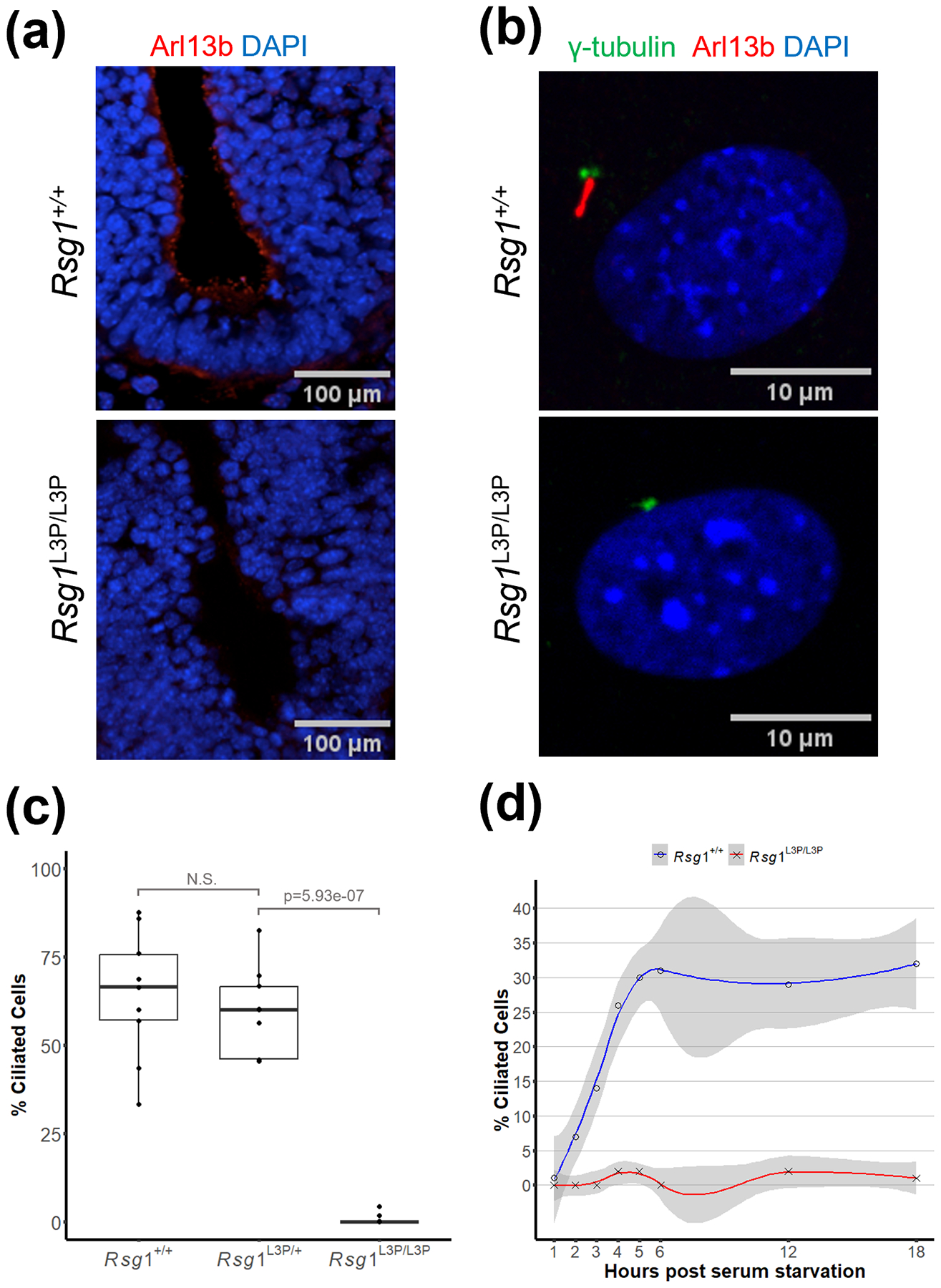
Cilia play a key role in the regulation of signaling pathways required for embryonic development, including the proper formation of the neural tube, the precursor to the brain and spinal cord. Forward genetic screens were used to generate mouse lines that display neural tube defects (NTD) and secondary phenotypes useful in interrogating function. We describe here the L3P mutant line that displays phenotypes of disrupted Sonic hedgehog signaling and affects the initiation of cilia formation. A point mutation was mapped in the L3P line to the gene Rsg1, which encodes a GTPase-like protein. The mutation lies within the GTP-binding pocket and disrupts the highly conserved G1 domain. The mutant protein and other centrosomal and IFT proteins still localize appropriately to the basal body of cilia, suggesting that RSG1 GTPase activity is not required for basal body maturation but is needed for a downstream step in axonemal elongation.

Keywords: RSG1 or CPLANE2; cilia; gene mapping; mouse; neural tube defects.


Conflict of interest statement

Conflict of interest disclosure: The authors declare no competing interests.

Figures

Figure 1:
Figure 1:. L3P mutant mouse embryos display exencephaly, polydactyly, eye defects, and disrupted SHH signaling
(a) E12.5 Rsg1L3P/L3P mutant embryos show exencephaly and eye defects. (b) E12.5 limbs stained with alcian blue to show cartilaginous skeleton. L3P mutant embryos display polydactyly. Asterisks highlight digit condensations. (c) Shh is expressed relatively normally in L3P limb buds at E10.5 or E11.5 by RNA in situ hybridization but the downstream target Ptch1 is decreased in Rsg1L3P/L3P :Ptch1+/LacZ limb buds as detected by Xgal staining. (d) Shh is present in the notochord but only at low levels in the floor plate of Rsg1L3P/L3P E10.5 embryo neural tubes compared to wildtype. This results in the expression of ISL1/2, LHX3, and PAX6 more ventrally than normal. Yellow bracket indicates domain of expression, blue bracket indicates ventral region where the protein is not present. Stainings for 1c and 1d were performed on a minimum on three embryos of each genotype, representative images are shown.
Figure 2:
Figure 2:. Rsg1L3P/L3P mutant cells lack primary cilia
(a) Rsg1L3P/L3P mutant E9.5 embryos lack primary cilia in the lumen of the neural tube, when compared to the wildtype. The presence of primary cilia is visualized by antibody against Arl13b which localizes to the cilia axoneme. Ventral floor plate is at bottom of panel. Immunostaining was performed on a minimum on three embryos of each genotype, representative images are shown. (b) L3P mutant mouse embryonic fibroblasts (MEFs) also lack primary cilia when compared to wildtype. Gamma-tubulin visualizes the location of the basal body and cilia are highlighted by Arl13b. (c) Quantification of MEFs stained as in panel b. MEFs within a 170μm2 field of view were evaluated for the presence of cilia. There were on average 27 cells per image with total cell counts n= 248 for WT; n = 223 for L3P/+; n = 254 for L3P/L3P. (d) Serum starvation fails to induce ciliogenesis in L3P mutant MEFs. Two biological replicates of wildtype and mutant embryos were used for the timed series of ciliogenesis.
Figure 3:
Figure 3:. Rsg1L3P/L3P mutant cells retain ability to recruit centrosomal proteins
Cultured MEFs isolated from wildtype and Rsg1L3PL3P embryos display co-localization of the basal body and (a) Ninein, (b) CEP164, and (c) IFT88. In all images the basal body is marked by γ-tubulin and the primary cilia is visualized by an antibody against Arl13b which localizes to the cilia axoneme. Immunostaining was performed on MEFs from a minimum of three embryos of each genotype and at least 100 cells per embryo visualized, representative images are shown. Scale bars in basal body zoom panels are 1μm. Scale bars in panels that depict the entire cell are 10μm.
Figure 4:
Figure 4:. Rsg1L3P mutation affects the GTP binding domain but does not affect RSG1 localization to the basal body
(a) Protein data bank model of RSG1 (7Q3E) with the GTPase G1 domain shown in blue, bound GTP in red, and the T69I mutation in green. The V169D mutation shown in purple lies outside of the GTP binding domain. (b) Cultured MEFs isolated from wildtype and Rsg1L3P/L3P embryos display co-localization of the basal body and RSG1. (c) There is no statistical difference between colocalization of RSG1 and γ-tubulin in Rsg1L3P/L3P MEFs compared to wildtype. Each datapoint represents quantification from a field of view of cells as shown in 4b with an average of 22 cells per image.

References

    1. Adler PN, & Wallingford JB (2017). From Planar Cell Polarity to Ciliogenesis and Back: The Curious Tale of the PPE and CPLANE proteins. Trends in Cell Biology, 27(5), 379–390. 10.1016/j.tcb.2016.12.001 - DOI - PMC - PubMed
    1. Agbu SO, Liang Y, Liu A, & Anderson KV (2018). The small GTPase RSG1 controls a final step in primary cilia initiation. The Journal of Cell Biology, 217(1), 413–427. 10.1083/jcb.201604048 - DOI - PMC - PubMed
    1. D’Angelo A, & Franco B (2009). The dynamic cilium in human diseases. PathoGenetics, 2, 3. 10.1186/1755-8417-2-3 - DOI - PMC - PubMed
    1. Feig LA (1999). Tools of the trade: Use of dominant-inhibitory mutants of Ras-family GTPases. Nature Cell Biology, 1(2), E25–E27. 10.1038/10018 - DOI - PubMed
    1. Goodrich LV, Milenković L, Higgins KM, & Scott MP (1997). Altered Neural Cell Fates and Medulloblastoma in Mouse patched Mutants. Science, 277(5329), 1109–1113. 10.1126/science.277.5329.1109 - DOI - PubMed

Publication types

Substances

LinkOut - more resources