. 2024 Jul 31;120(9):1037-1050.
doi: 10.1093/cvr/cvae098.

Inhibition of TBL1 cleavage alleviates doxorubicin-induced cardiomyocytes death by regulating the Wnt/β-catenin signal pathway

Affiliations

Inhibition of TBL1 cleavage alleviates doxorubicin-induced cardiomyocytes death by regulating the Wnt/β-catenin signal pathway

Sun-Ho Lee et al. Cardiovasc Res. .

Abstract

Aims: Doxorubicin (DOX) is a widely used anthracycline anticancer agent; however, its irreversible effects on the heart can result in DOX-induced cardiotoxicity (DICT) after cancer treatment. Unfortunately, the pathophysiology of DICT has not yet been fully elucidated, and there are no effective strategies for its prevention or treatment. In this investigation, the novel role of transducin beta-like protein 1 (TBL1) in developing and regulating DICT was explored.

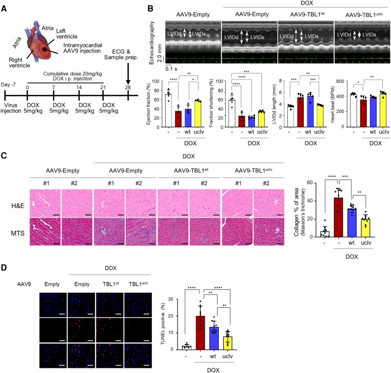
Methods and results: We observed a reduction in TBL1 protein expression levels as well as cleavage events in the transplanted cardiac tissues of patients diagnosed with Dilated Cardiomyopathy and DICT. It was revealed that DOX selectively induces TBL1 cleavage at caspase-3 preferred sites-D125, D136, and D215. Interestingly, overexpression of the uncleaved TBL1 mutant (TBL1uclv) variant reduced apoptosis, effectively preventing DOX-induced cell death. We confirmed that cleaved TBL1 cannot form a complex with β-catenin. As a result, Wnt reporter activity and Wnt target gene expression collectively indicate a decrease in Wnt/β-catenin signalling, leading to DICT progression. Furthermore, the cleaved TBL1 triggered DOX-induced abnormal electrophysiological features and disrupted calcium homeostasis. However, these effects were improved in TBL1uclv-overexpressing human-induced pluripotent stem cell-derived cardiomyocytes. Finally, in a DICT mouse model, TBL1uclv overexpression inhibited the DICT-induced reduction of cardiac contractility and collagen accumulation, ultimately protecting cardiomyocytes from cell death.

Conclusion: Our findings reveal that the inhibition of TBL1 cleavage not only mitigates apoptosis but also enhances cardiomyocyte function, even in the context of DOX administration. Consequently, this study's results suggest that inhibiting TBL1 cleavage may be a novel strategy to ameliorate DICT.

Keywords: Cardiotoxicity; Doxorubicin; Human induced pluripotent stem cell derived cardiomyocytes; Transducin beta-like protein 1; Wnt/β-catenin signal pathway.


Conflict of interest statement

Conflict of interests: none declared.

Figures

Graphical Abstract
Graphical Abstract
Figure 1
Figure 1
TBL1 expression is low in cardiac tissues from patients with DCM. (A) TBL1 expression in cardiac tissues from patients with idiopathic DCM (n = 6) compared to normal cardiac tissues (n = 15). TBL1 expression is low in the cardiac tissues of patients with DCM. Slides were stained with an antibody against an N-terminal TBL1 epitope using IHC. Representative images are shown. Scale bar = 50 µm (left panel). The DAB (3,3′-diaminobenzidine) staining intensity score was calculated using the IHC profiler plugin in ImageJ software (right panel). ****P < 0.0001 (Student's t-test) (B) Data from publicly available datasets on TBL1 expression levels in cardiac tissues from patients with DCM and normal cardiac tissues. GSE57338, n = 313 (normal n = 136, iCMP n = 95, iDCMP n = 82); GSE42955 n = 29 (normal n = 5, iCMP n = 12, DCMP n = 12). Quantification of TBL1 mRNA levels by microarray (two probes; ILMN_1248994 and ILMN_2624451) in the GSE57337 and by RNA sequencing in GSE42955. n.s., no significance.
Figure 2
Figure 2
TBL1 is cleaved at D125, D136, and D215 in a caspase-3-dependent manner and consequently destabilized through ubiquitin-dependent degradation following doxorubicin treatment in H9c2 cells. (A) TBL1 cleavage in heart tissues sampled from patients with idiopathic DCM and DICT. Proteins were immunoblotted with the indicated antibodies. The arrows indicate cleaved-TBL1. (B) Endogenous TBL1 was cleaved in a time-dependent manner following DOX treatment. H9c2 cells were treated with 2 µM DOX, and total proteins were extracted and immunoblotted with the indicated antibodies. SE, short exposure; LE, long exposure. (C) TBL1 cleaved at D125, D136, and D215 in response to DOX. Wild-type TBL1 (TBL1wt), TBL1D125A, TBL1D136A, TBL1D215A, or mutations at three sites (TBL1uclv) were overexpressed in H9c2 cells which were then harvested and lysed to extract the protein. The total protein was used for immunoblot assays. The arrow marked as ‘Clv’ indicate cleaved TBL1 bands. SE, short exposure; LE, long exposure. (D) TBL1uclv abrogates its cleavage in response to DOX. H9c2 cells were transiently transfected with a double-tagged TBL1 (Flag-TBL1-Myc) plasmid and treated with DOX. Permeabilized cells were incubated with antibodies against Flag and Myc, and PLA probes added. The cell nuclei were counterstained with DAPI. Dot signals represent RCA products from immuno-RCA reactions detected using fluorescently labelled complementary oligonucleotides. Representative images of four independent experiments are shown. Scale bar = 50 µm (upper panel). The number of RCA products per cell from these images is shown in the bar graph. The values are presented as the mean ± SD from three independent experiments (lower panel). *P < 0.05 and ****P < 0.0001 (Student’s t-test). (E) TBL1 cleavage was blocked following the treatment of caspase inhibitors. H9c2 cells were exposed to either pan-caspase inhibitor (Z-VAD) or caspase 3/7 specific inhibitor (Z-DEVD) with or without 2 µM DOX for 24 h. The cells were lysed, and protein extracts were used for immunoblot assays with the indicated antibodies. (F) In the DOX response, TBL1 is cleaved in a caspase-3-dependent manner. siCaspase-3 or siCaspase-7 was transiently transfected, and subsequently, H9c2 cells were treated with 2 µM DOX for 24 h. Protein extracts from the cells were immunoblotted with the indicated antibodies. (G) MG132 treatment induces the accumulation of TBL1 cleavage. Endogenous TBL1 was cleaved in a time-dependent manner following DOX treatment. The cells were treated with 1 µM DOX for the indicated time and 10 µM MG132 for 12 h. Cells were incubated for the indicated time, and whole-cell lysates were immunoblotted using the indicated antibodies. Arrows indicate cleaved TBL1. (H) TBL1 ubiquitination increases in response to DOX in a time-dependent manner. H9c2 cells were transfected HA-tagged Ub plasmid and treated with 10 µM MG132 with or without 1 µM DOX for the indicated time. Whole-cell lysates were immunoprecipitated with an anti-TBL1(C) antibody and immunoblotted with the indicated antibodies. (I) The TBLuclv mutation abolishes DOX-induced TBL1 ubiquitination. H9c2 cells were transfected with HA-Ub and the indicated Myc-tagged TBL1 plasmids and then treated with 10 µM MG132 with or without 1 µM DOX for 24 h. Whole-cell lysates were immunoprecipitated with anti-Myc antibody and immunoblotted.
Figure 3
Figure 3
Depletion of TBL1 abrogates the DOX-induced TBL1 cleavage and p53-dependent apoptotic cell death. (A) Endogenous TBL1 was cleaved in a time-dependent manner following DOX treatment. The cells were treated with 2 µM DOX for the indicated time. Protein from the H9c2 cells was extracted and immunoblotted with the indicated antibodies. (B) Depletion of TBL1 abrogates the DOX-induced TBL1 cleavage and p53-dependent apoptotic cell death. DOX-induced TBL1 cleavage regulated the expression of apoptotic proteins. H9c2 cells were treated with 2 µM DOX for the indicated times. Cells were harvested, and total protein extracted. Whole-cell lysates were immunoblotted with the indicated antibodies. (C) TBL1 Knockdown increased the expression of apoptotic proteins in response to the DOX treatment. Either siControl (siCONT) or siTBL1 was transiently transfected in H9c2 cells and treated with 2 µM DOX for the indicated times. Whole-cell lysates were used for immunoblotting assays with the indicated antibodies. (D) Abrogation of TBL1 cleavage inhibits apoptotic cell death of rat cardiomyocytes in response to DOX. Either TBL1wt or TBL1uclv construct was transfected into H9c2 cells, and the cells were exposed to 2 µM DOX for 24 h. Annexin V-positive cells were assessed by flow cytometry. A representative image of three independent experiments is shown. (E) Representative image of TUNEL-positive apoptotic cells vs. DAPI. Total cells positive for Empty, wild-type TBL1, and uncleaved-TBL mutant overexpression were detected by fluorescent microscopy. Cells were exposed to 2 µM DOX for 24 h. Representative images of three independent experiments are demonstrated (right panel). Scale bar = 20 µm. The histogram depicts the quantification of apoptosis (in percentage; the total number of TUNEL-positive cells vs. DAPI-positive cells) for TBL1wt and TBL1uclv. The values are presented as the mean ± SD from three independent experiments. n.s., no significance. ****P < 0.0001 (Student's t-test).
Figure 4
Figure 4
DOX-induced TBL1 cleavage abrogates Wnt/β-catenin signal pathway activation. (A) TBL1 knockdown decreased mRNA expression of the Wnt target genes. H9c2 cells were transfected with either siControl or siTBL1 and exposed to 1 µM DOX for 24 h. cDNA was synthesized from mRNA, and the levels of Axin2 and c-Myc were analyzed using real-time PCR. (B) Exogenous overexpressed TBL1wt was dissociated from β-catenin following the DOX treatment. The indicated plasmid was transfected with or without 1 µM DOX for 24 h in H9c2 cells. Whole-cell lysates were immunoprecipitated with a β-catenin antibody and sequentially immunoblotted with the indicated antibodies. (C) In situ PLA assay demonstrating the dissociation of cleaved TBL1wt from β-catenin in response to DOX. Flag-TBL1wt or -TBL1uclv plasmid was overexpressed with or without 2 µM DOX for 24 h in H9c2 cells. Permeabilized cells were incubated with antibodies against Flag and β-catenin, and the PLA probes were added. The cell nuclei were counterstained with DAPI. Positive signals were analyzed using confocal microscopy. RCA dots indicate the TBL1-β-catenin complex. Representative images of four independent experiments are demonstrated. Scale bar = 50 µm (left panel). The numbers of RCA products per cell from those images are shown in the bar graph (right panel). (D) DOX-induced TBL1 cleavage mediates its abnormal localization. The cleaved TBL1wt was relocalized into the cytosol in response to the DOX treatment. Flag-TBL1wt-Myc or Flag-TBL1uclv-Myc plasmid was transfected into H9c2 cells with or without 1 µM DOX for 24 h. Immunofluorescence analysis was performed as described in the Materials and Methods section. Representative images of three independent experiments are shown. Scale bar = 50 µm. (E) DOX induces β-catenin nuclear translocation. Cells were treated with 2 µM DOX for the indicated time. H9c2 cells were analyzed using immunofluorescence staining. Representative images of four independent experiments are demonstrated. Scale bar = 50 µm. H9c2 cells with or without 2 µM DOX for 24 h were fractionized into the cytosol and nucleus and used for western blot assays (lower panel). (F) DOX-induced TBL1 cleavage abrogates its occupancy in the promoter region of Wnt target genes. H9c2 cells were transiently transfected with either the TBL1wt or TBL1uclv plasmid with or without 1 µM DOX for 24 h. Chromatin immunoprecipitation assays were performed with the indicated antibodies. Precipitated samples were analyzed using real-time PCR. (G) Overexpression of TBL1uclv enhances β-catenin-mediated transcription activity under DOX-exposed conditions. H9c2 cells were co-transfected with TOP/FOPFLASH reporter and the indicated TBL1 plasmid for 24 h. Whole-cell lysates were used for luciferase assays. The results are represented as the mean ± SD from the three independent experiments. n.s., no significance. *P < 0.05, **P < 0.01, ***P < 0.001, and ****P < 0.0001 (Student’s t-test).
Figure 5
Figure 5
Abrogation of TBL1 cleavage reverses DOX-induced abnormal physiological changes in human-induced pluripotent stem cell-derived cardiomyocytes (hiPSC-CMs). (A) iPSC-CMs were cleaved following DOX treatment. iPSC-CMs were treated with 2 µM DOX, and total proteins were extracted and immunoblotted with the indicated antibodies. (B) Representative image of TUNEL-positive apoptotic cells vs. DAPI. Ad-Empty, -TBL1wt, or Ad-TBL1uclv inoculated into iPSC-CMs and TUNEL-positive cells were detected using fluorescent microscopy. Cells were exposed to 2 µM DOX for 24 h. Representative images of three independent experiments are demonstrated (right panel). Scale bar = 20 µm. The histogram depicts the quantification of apoptosis (in percentage; the total number of TUNEL-positive cells vs. DAPI-positive cells) for TBL1wt and TBL1uclv. (C) Overexpression of Ad-TBL1uclv in hiPSC-CMs improves field potential in response to DOX. Field potential traces data recorded with the MEA exhibiting mean spontaneous beating trace. Representative trace images recorded with the MEA exhibiting a field potential trace in hiPSC-CMs with the overexpression of Ad-TBL1wt and Ad-TBL1uclv with or without DOX. The graph indicates beat period and spike amplitude. (D) Overexpression of Ad-TBL1uclv in hiPSC-CMs improves decreased spike amplitude in response to DOX. An active map through the total active lid count is shown. The degree of the waveform refers to the highs and lows of the spike amplitudes for iPSC-CMs, respectively. Representative images are presented after three independent experiments. (E) Overexpression of Ad-TBL1uclv in hiPSC-CMs improves decreased conduction velocity in response to DOX. Conduction plot showing propagation delay of hiPSC-CMs with Ad-Flag-TBL1wt or Ad-Flag-TBL1uclv, the two points that appear to be the starting points of the waveform represent the beginning and end of the conduction. Representative images are demonstrated after three independent experiments (left panel). The data with percent changes compared to that of the baseline were measured using MEA (right panel). (F) Overexpression of TBL1uclv reversed the disrupted electrophysiological activity in DOX-exposed iPSC-CMs. Ad-TBL1wt or -TBL1uclv was used to infect hiPSC-CMs and exposed to 1 µM DOX. The changes in the beat period, spike amplitude, and beat period irregularity with percent changes compared to that in the baseline were measured using an MEA assay. (G and H) Overexpression of TBL1uclv attenuates calcium decay in DOX-exposed iPSC-CMs. Representative tracing of spontaneous rhythmic calcium transience in hiPSC-CMs injected with Ad-TBL1wt and -TBL1uclv. A typical line scan (X-T mode) image of spontaneous calcium transients was obtained. The systolic calcium (F/F0), time to baseline 50% (s), calcium amplitude (F/F0), Tau (s), and frequency of calcium transient (per min) were measured. F/F0, fluorescence (F) normalized to baseline fluorescence (F0); s, seconds. The data with percent changes compared to that in the baseline were measured using MEA. The values are the mean ± SD from three independent experiments. * P < 0.05 and ** P < 0.01 (Student’s t-test).
Figure 6
Figure 6
Blocking TBL1 cleavage protects mouse cardiac function by ameliorating genotoxic damage and fibrosis in cardiomyocytes. (A) Mouse experimental protocol. The indicated viral particles were injected into the mouse cardiac using a sophisticated closed-chest echocardiography-guided intramyocardial (i.c.) injection method. After viral injection, 5 mg/kg of DOX was intraperitoneally (i.p.) injected four times (cumulative dose, 20 mg/kg). (B) Overexpression of AAV9-TBL1uclv protects mouse cardiac function against DICT. After a week from intramyocardial (i.c.) injection of the indicated viral particle and sequential intraperitoneal (i.p.) injection of DOX, mouse cardiac function was observed with echocardiography. Representative M-mode echocardiogram images were demonstrated. LVIDd and LVIDs are marked with a double arrow (upper panel). EF%, FS%, and LVIDd length were measured. The values are demonstrated as the mean ± SE from three independent experiments. *P < 0.05 and **P < 0.01 (Student’s t-test; n = 5). (C) Overexpression of TBL1uclv inhibits DOX-induced cardiac fibrosis in mice. After echocardiography, mice were sacrificed, and their cardiac tissues dissected. The extent of fibrosis was measured using haematoxylin and eosin (H&E) or Masson's trichrome staining (MTS). Two representative images of H&E or MTS staining are demonstrated (left panel). Scale bar = 50 µm. The percent of the collagen-stained area was calculated IHC profiler plugin in ImageJ software (right panel). The values are the mean ± SE from three independent experiments (n = 5). **P < 0.01, and ****P < 0.0001 (Student’s t-test). (D) Inhibition of TBL1 cleavage suppresses mouse cardiomyocyte death. After echocardiography, mice were sacrificed, and their cardiac tissues dissected. DNA damage in the paraffin-embedded cardiac tissues caused by DICT was determined using TUNEL assays. The values are represented as the mean ± SE from three independent experiments. *P < 0.05 and ****P < 0.0001 (Student’s t-test). Scale bar = 50 µm.
Figure 7
Figure 7
Schematic representation of the major findings from this study. Briefly, DOX-induced TBL1 cleavage at D125, D136, and D215 causes its ubiquitin-dependent degradation. Owing to TBL1 destabilization, it fails to form a complex with β-catenin in the nucleus and blocks β-catenin from occupying the Axin2 and c-myc promoters. Consequently, Wnt activity is decreased, and the death of cardiomyocytes is induced in response to DOX. Therefore, inhibition of TBL1 cleavage protects cardiomyocytes from DICT by controlling the series of signalling mechanisms and consequently maintains normal cardiac function. This image was created with BioRender.com.

References

    1. De Angelis A, Urbanek K, Cappetta D, Piegari E, Ciuffreda LP, Rivellino A, Russo R, Esposito G, Rossi F, Berrino L. Doxorubicin cardiotoxicity and target cells: a broader perspective. Cardiooncology 2016;2:2. - PMC - PubMed
    1. Lefrak EA, Pitha J, Rosenheim S, Gottlieb JA. A clinicopathologic analysis of Adriamycin cardiotoxicity. Cancer 1973;32:302–314. - PubMed
    1. Zamorano JL, Lancellotti P, Rodriguez Munoz D, Aboyans V, Asteggiano R, Galderisi M, Habib G, Lenihan DJ, Lip GYH, Lyon AR, Lopez Fernandez T, Mohty D, Piepoli MF, Tamargo J, Torbicki A, Suter TM; Group ESCSD . 2016 ESC position paper on cancer treatments and cardiovascular toxicity developed under the auspices of the ESC committee for practice guidelines: the task force for cancer treatments and cardiovascular toxicity of the European Society of Cardiology (ESC). Eur Heart J 2016;37:2768–2801. - PubMed
    1. Henriksen PA. Anthracycline cardiotoxicity: an update on mechanisms, monitoring and prevention. Heart 2018;104:971–977. - PubMed
    1. Haupt LP, Rebs S, Maurer W, Hubscher D, Tiburcy M, Pabel S, Maus A, Kohne S, Tappu R, Haas J, Li Y, Sasse A, Santos CCX, Dressel R, Wojnowski L, Bunt G, Mobius W, Shah AM, Meder B, Wollnik B, Sossalla S, Hasenfuss G, Streckfuss-Bomeke K. Doxorubicin induces cardiotoxicity in a pluripotent stem cell model of aggressive B cell lymphoma cancer patients. Basic Res Cardiol 2022;117:13. - PMC - PubMed

Publication types

MeSH terms