. 2024 Aug 8;73(9):1543-1553.
doi: 10.1136/gutjnl-2024-332033.

Novel IL-4/HB-EGF-dependent crosstalk between eosinophils and macrophages controls liver regeneration after ischaemia and reperfusion injury

Affiliations

Novel IL-4/HB-EGF-dependent crosstalk between eosinophils and macrophages controls liver regeneration after ischaemia and reperfusion injury

Yang Yang et al. Gut. .

Abstract

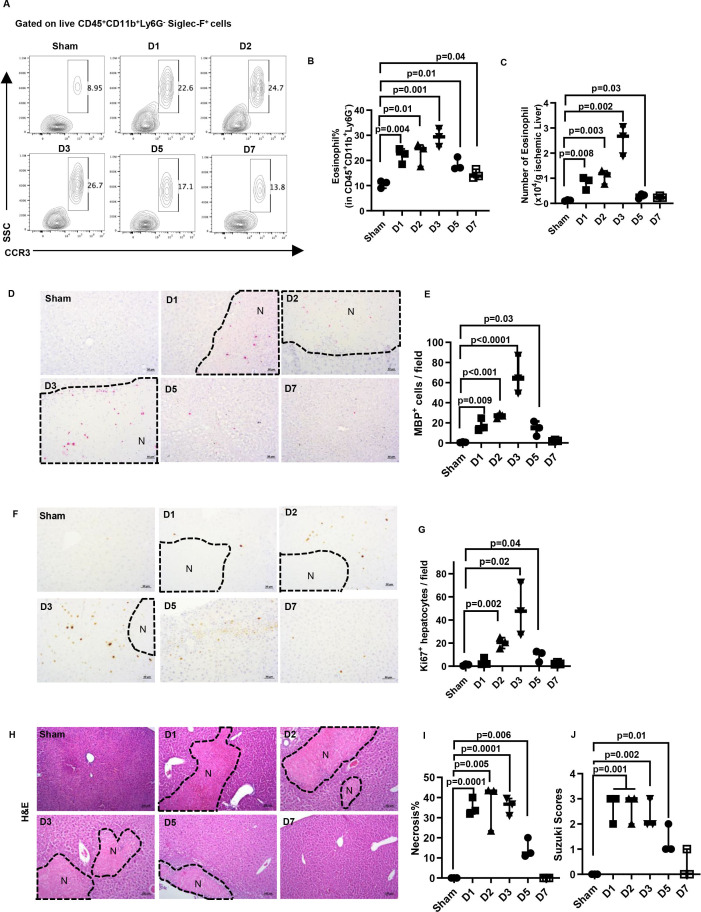
Objective: Previous studies indicate that eosinophils are recruited into the allograft following orthotopic liver transplantation and protect from ischaemia reperfusion (IR) injury. In the current studies, we aim to explore whether their protective function could outlast during liver repair.

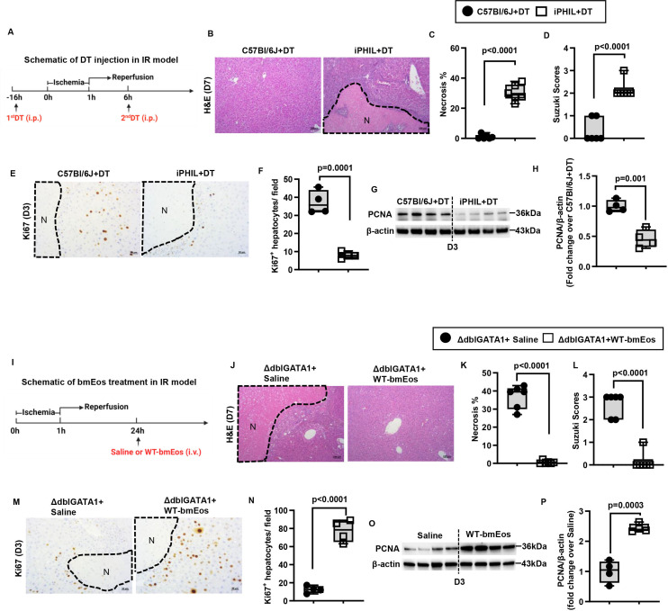
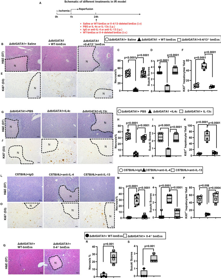
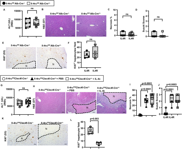
Design: Eosinophil-deficient mice and adoptive transfer of bone marrow-derived eosinophils (bmEos) were employed to investigate the effects of eosinophils on tissue repair and regeneration after hepatic IR injury. Aside from exogenous cytokine or neutralising antibody treatments, mechanistic studies made use of a panel of mouse models of eosinophil-specific IL-4/IL-13-deletion, cell-specific IL-4rα-deletion in liver macrophages and hepatocytes and macrophage-specific deletion of heparin-binding epidermal growth factor-like growth factor (hb-egf).

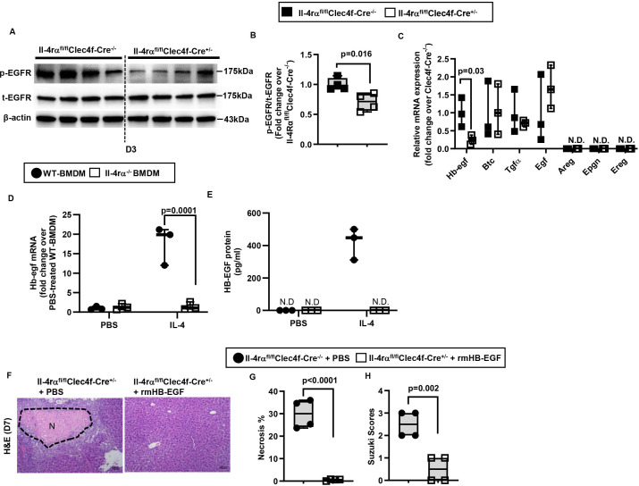
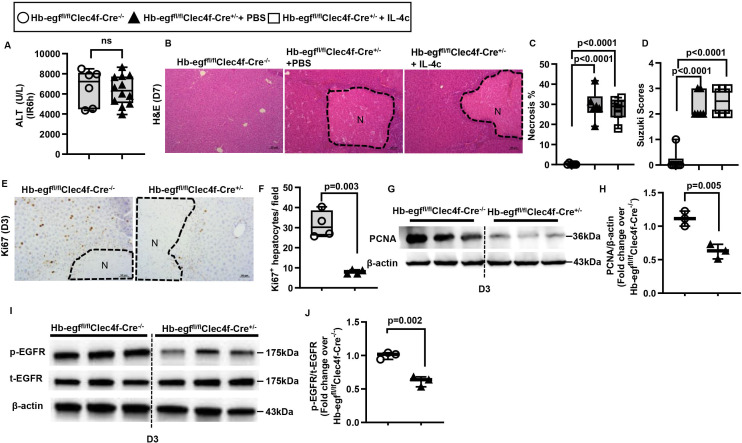
Result: We observed that eosinophils persisted over a week following hepatic IR injury. Their peak accumulation coincided with that of hepatocyte proliferation. Functional studies showed that eosinophil deficiency was associated with a dramatic delay in liver repair, which was normalised by the adoptive transfer of bmEos. Mechanistic studies demonstrated that eosinophil-derived IL-4, but not IL-13, was critically involved in the reparative function of these cells. The data further revealed a selective role of macrophage-dependent IL-4 signalling in liver regeneration. Eosinophil-derived IL-4 stimulated macrophages to produce HB-EGF. Moreover, macrophage-specific hb-egf deletion impaired hepatocyte regeneration after IR injury.

Conclusion: Together, these studies uncovered an indispensable role of eosinophils in liver repair after acute injury and identified a novel crosstalk between eosinophils and macrophages through the IL-4/HB-EGF axis.

Keywords: growth factors; immunology in hepatology; ischaemia-reperfusion; liver regeneration; macrophages.


Conflict of interest statement

Competing interests: None declared.

Figures

Figure 1
Figure 1
The kinetics of hepatic eosinophil accumulation and the time course of liver repair after IR injury in WT mice. Male C57Bl/6J mice were subjected to hepatic ischemia (60 mins) followed by reperfusion and sacrificed at various times (n=3/group). Sham mice underwent the same surgery without vascular blockage and were sacrificed on day 7. (A-C) The percentage and numbers of eosinophils (CD45+CD11b+Ly6G-Siglec-F+CCR3+cells) in the liver were measured by flow cytometry and quantified. (D, E) IHC staining for eosinophils by anti-mouse major basic protein (MBP) antibody and the numbers of MBP+ cells quantified. (F, G) Proliferating hepatocytes were stained by anti-Ki67 antibody, and the numbers of positive cells were quantified. (H, I) Liver necrosis (N, outlined areas) was evaluated and quantified. (J) Liver pathology was assessed by using the Suzuki’s scoring system. Two-tailed unpaired Student’s t-test with Welch’s correction was performed in B, C, E, G, I and J.
Figure 2
Figure 2
Eosinophils promote liver repair after IR injury. (A-H) Male iPHIL mice and WT littermates were subjected to hepatic IR surgery. Mice were administered (i.p.) the first dose of DT at 16h prior to IR surgery and the second dose at 6h after surgery. Mice were sacrificed on day 3 (n=4/group) or day 7 (n=6/group) after IR surgery. (B, C) Liver necrosis (N, outlined areas) was evaluated and quantified on day 7 after IR surgery. (D) Liver pathology was determined by the Suzuki score on day 7 after IR surgery. (E, F) Liver tissue sections were stained for Ki67 to quantify proliferating hepatocytes on day 3 after IR surgery. (G, H) PCNA protein expression was detected by Western blotting and quantified. (I-P) Male ΔdblGATA-1 mice were subjected to hepatic IR surgery. After 24h, half of the mice were i.v. injected with bmEos (10x106) and the other half injected with saline as control. Mice were sacrificed on day 3 (n=4/group) or day 7 after IR surgery (n=6/group). (J, K) Liver necrosis (N, outlined area) was evaluated and quantified on day 7 after IR surgery. (L) Liver pathology was determined by the Suzuki score on day 7 after IR surgery. (M, N) Proliferative hepatocytes were stained for Ki67 and quantified on day 3 after IR surgery. (O, P) PCNA protein expression was detected by Western blotting and quantified. Two-tailed unpaired Student’s t-test with Welch’s correction was performed in C, D, F, H, K, L, N and P.
Figure 3
Figure 3
Eosinophil-derived IL-4 plays a critical role in liver repair after IR injury. (A-F) Male ΔdblGATA-1 were subjected to hepatic IR surgery. After 24h, the mice were divided into three groups and i.v. injected with saline, WT-bmEos (10x106) or Il-4/13-deleted bmEos (10x106). Mice were sacrificed on day 3 (n=4/group) or day 7 (n=6/group) after IR surgery. (B, C) Liver necrosis (N, outlined areas) was evaluated and quantified on day 7 after IR surgery. (D) Liver pathology was assessed by using the Suzuki’s scoring system on day 7 after IR surgery. (E, F) Proliferative hepatocytes were stained for Ki67 and quantified on day 3 after IR surgery. (A, G-K) Male ΔdblGATA-1 were subjected to hepatic IR surgery. After 24h, the mice were divided into three groups and i.p. injected with PBS, IL-4c (5 μg recombinant IL-4 complexed to 25 μg anti-IL-4 antibody) or IL-13c (5 µg of recombinant IL-13 complexed to 25 µg anti-IL-13 antibody). Mice were sacrificed on day 3 (n=4/group) or day 7 (n=6/group). (G, H) Liver necrosis (N, outlined areas) was evaluated and quantified on day 7 after IR surgery. (I) Liver pathology was assessed by using the Suzuki’s scoring system quantified on day 7 after IR surgery. (J, K) Proliferative hepatocytes were stained for Ki67 and quantified on day 3. (A, L-P) Male C57Bl/6J mice were subjected to hepatic IR surgery. On day 1 and day 3 after surgery, the mice were i.p. injected with anti-IL-4 antibody (10 μg/mouse) or anti-IL-13 antibody (10 μg/mouse). Control mice were injected with IgG. Mice were sacrificed on day 3 (n=4/group) or day 7 (n=6/group) after IR surgery. (L, M) Liver necrosis (N, outlined areas) was evaluated and quantified on day 7 after IR surgery. (N) Liver pathology was assessed by using the Suzuki’s scoring system on day 7 after IR surgery. (O, P) Proliferative hepatocytes were stained for Ki67 and quantified on day 3. (A, Q-S) Male ΔdblGATA-1 mice were subjected to hepatic IR surgery. After 24h, half of the mice were i.v. injected with WT-bmEos (10x106) and the other half injected with Il-4-deleted bmEos (10x106). Mice were sacrificed on day 7 after IR surgery (n=4/group). (Q, R) Liver necrosis (N, outlined areas) was evaluated and quantified on day 7 after IR surgery. (S) Liver pathology was assessed by using the Suzuki’s scoring system on day 7 after IR surgery. Two-tailed unpaired Student’s t-test with Welch’s correction was performed in R and S. One-way ANOVA was performed in C, D, F, H, I, K, M, N and P.
Figure 4
Figure 4
IL-4Rα signaling in liver macrophages, rather than hepatocytes, promotes liver repair after IR injury. (A-F) Male Il-4rαfl/flAlb-Cre+/- mice and WT littermates (Male Il-4rαfl/flAlb-Cre-/-) were subjected to hepatic IR surgery and sacrificed after 3 days (n=4/group) or 7 days (n=6/group). (G-L) Male Il-4rα fl/flClec4f-Cre+/- mice and WT littermates (Il-4rαfl/flClec4f-Cre-/-) were subjected to hepatic IR surgery. After 24h, the Il-4rαfl/flClec4f-Cre+/- mice were divided into two groups and injected (i.v.) with PBS or IL-4c (n=6/group) and and sacrificed after 3 days (n=4/group) or 7 days (n=6/group) after IR surgery. (A, G) Serum ALT levels 6h after IR surgery. (B, C, H, I) Liver necrosis (N, outlined areas) was evaluated and quantified on day 7 after IR surgery. (D, J) Liver pathology was assessed by using the Suzuki’s scoring system on day 7 after IR surgery. (E, F, K, L) Proliferative hepatocytes were stained for Ki67 and quantified on day 3 after IR surgery. Two-tailed unpaired Student’s t-test with Welch’s correction was performed in A, C, D, F, G and L. One-way ANOVA was performed in I and J.
Figure 5
Figure 5
IL-4/IL-4Rα signaling in hepatic macrophage induces HB-EGF expression. (A-C) Male Il-4rαfl/flClec4f-Cre+/- mice and WT littermates (Male Il-4rαfl/flClec4f-Cre-/-) were subjected to hepatic IR surgery and sacrificed after 3 days. Liver tissues were collected and hepatic macrophages were purified by magnetic-associated cell sorting (MACS) using anti-F4/80 antibody. (A, B) phospho(p)-EGFR and total (t)-EGFR protein levels in the liver tissue were detected by Western blotting and quantified (n=4/group). (C) mRNA levels of all EGFR ligands expressed by hepatic macrophages were measured by q-PCR (n=3/group). (D, E) WT-BMDM and Il-4rα-deleted BMDM were cultured with IL-4 for 6h or 24h. Control groups were treated with PBS (n=3/group). mRNA and protein levels of HB-EGF were measured at 6h by q-PCR and 24h by ELISA, respectively. (F-H) rmHB-EGF was i.p. injected to male Il-4rαfl/flClec4f-Cre+/- mice on day 1 and day 3 after liver IR surgery. Control mice were injected with PBS. All mice were sacrificed on day 7 after IR surgery (n=4/group). (F, G) Liver necrosis (N, outlined areas) was evaluated and quantified on day 7. (H) Liver pathology was assessed by using the Suzuki’s scoring system. Two-tailed unpaired Student’s t-test with Welch’s correction was performed in B, C, G and H. One-way ANOVA was performed in D and E.
Figure 6
Figure 6
Liver repair after IR injury is impaired when hb-egf is deleted in hepatic macrophages. Male Hb-egffl/flClec4f-Cre+/-mice and WT littermates (Male Hb-egffl/flClec4f-Cre-/-) were subjected to hepatic IR surgery. After 24h, Hb-egffl/flClec4f-Cre+/- mice were divided into two groups and injected (i.p.) with PBS or IL-4c (n=6/group). (A) Serum ALT levels at 6h after IR surgery. (B, C) Liver necrosis (N, outlined areas) was evaluated and quantified on day 7 after IR surgery. (D) Liver pathology was assessed by using the Suzuki’s scoring system on day 7 after IR surgery. (E, F) Proliferative hepatocytes were stained for Ki67 and quantified on day 3 after IR surgery (n=4/group). (G, H) PCNA protein expression was detected by Western blotting and quantified. (I, J) p-EGFR and t-EGFR protein expression levels were detected by Western blotting and quantified. Two-tailed unpaired Student’s t-test with Welch’s correction was performed in A, F, H and J. One-way ANOVA was performed in C and D.

References

    1. Dutkowski P, Linecker M, DeOliveira ML, et al. . Challenges to liver transplantation and strategies to improve outcomes. Gastroenterology 2015;148:307–23. 10.1053/j.gastro.2014.08.045 - DOI - PubMed
    1. Wertheim JA, Petrowsky H, Saab S, et al. . Major challenges limiting liver transplantation in the United States. Am J Transplant 2011;11:1773–84. 10.1111/j.1600-6143.2011.03587.x - DOI - PMC - PubMed
    1. Fellström B, Aküyrek LM, Backman U, et al. . Postischemic reperfusion injury and allograft arteriosclerosis. Transplant Proc 1998;30:4278–80. 10.1016/s0041-1345(98)01412-2 - DOI - PubMed
    1. Howard TK, Klintmalm GB, Cofer JB, et al. . The influence of preservation injury on rejection in the hepatic transplant recipient. Transplantation 1990;49:103–7. 10.1097/00007890-199001000-00023 - DOI - PubMed
    1. Busuttil RW, Tanaka K. The utility of marginal donors in liver transplantation. Liver Transpl 2003;9:651–63. 10.1053/jlts.2003.50105 - DOI - PubMed

LinkOut - more resources