. 2024 Nov 18;95(12):1112-1122.
doi: 10.1136/jnnp-2023-331979.

Neurophysiological features of STN LFP underlying sleep fragmentation in Parkinson's disease

Affiliations

Neurophysiological features of STN LFP underlying sleep fragmentation in Parkinson's disease

Guokun Zhang et al. J Neurol Neurosurg Psychiatry. .

Abstract

Background: Sleep fragmentation is a persistent problem throughout the course of Parkinson's disease (PD). However, the related neurophysiological patterns and the underlying mechanisms remained unclear.

Method: We recorded subthalamic nucleus (STN) local field potentials (LFPs) using deep brain stimulation (DBS) with real-time wireless recording capacity from 13 patients with PD undergoing a one-night polysomnography recording, 1 month after DBS surgery before initial programming and when the patients were off-medication. The STN LFP features that characterised different sleep stages, correlated with arousal and sleep fragmentation index, and preceded stage transitions during N2 and REM sleep were analysed.

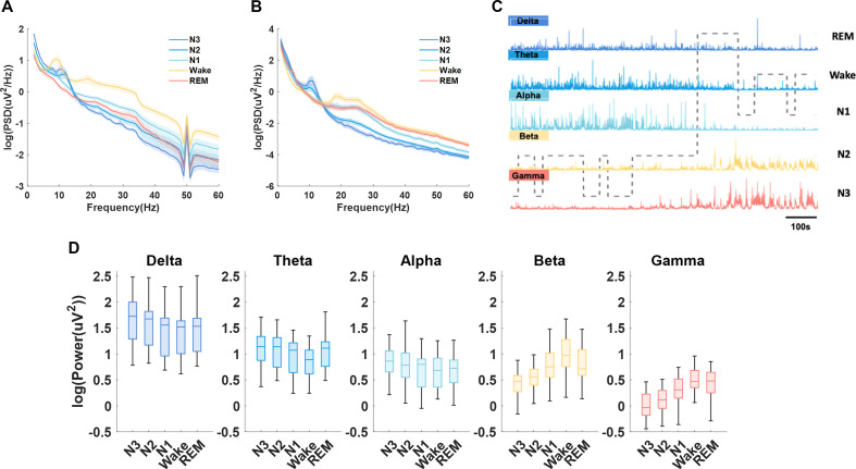
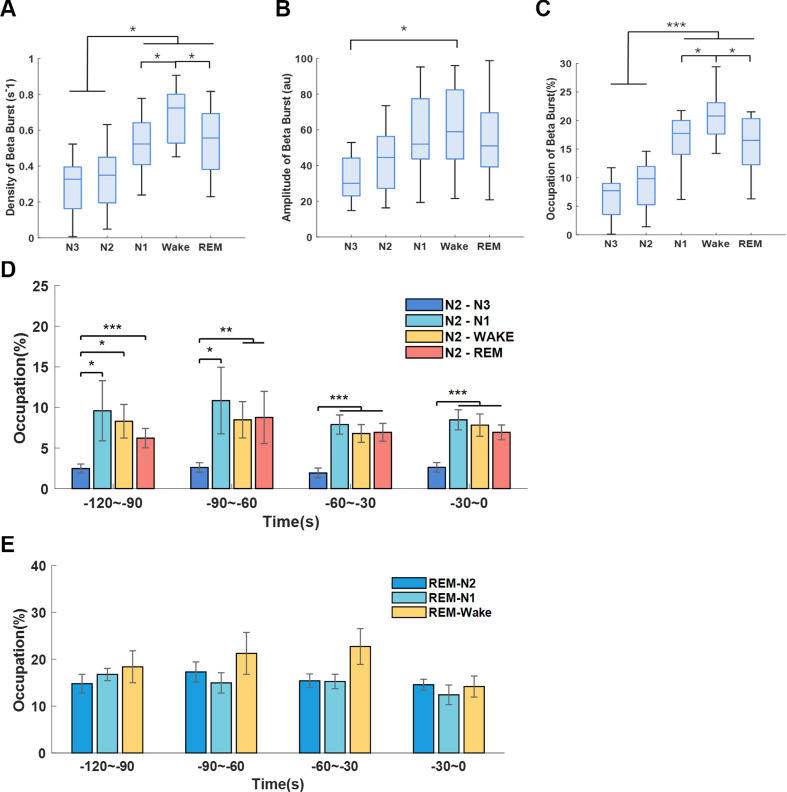
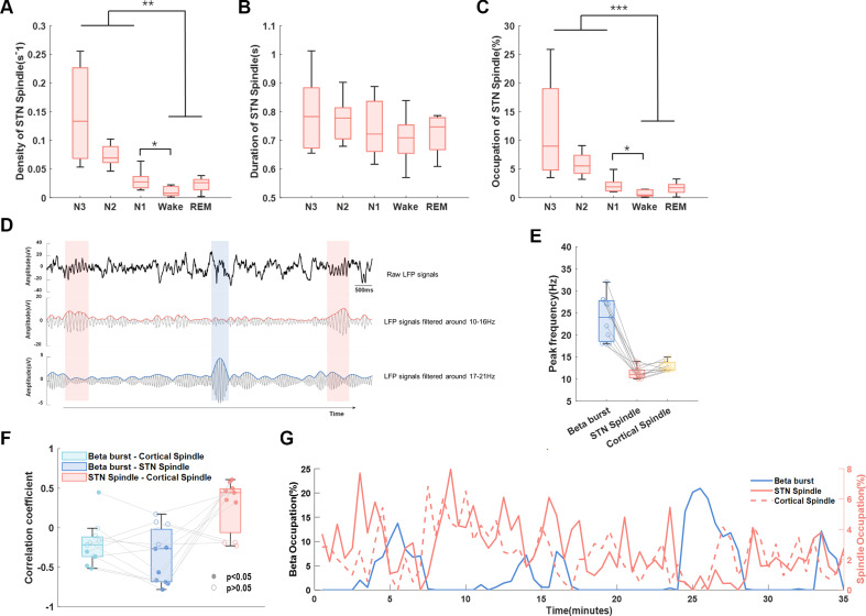
Results: Both beta and low gamma oscillations in non-rapid eye movement (NREM) sleep increased with the severity of sleep disturbance (arousal index (ArI)-betaNREM: r=0.9, p=0.0001, sleep fragmentation index (SFI)-betaNREM: r=0.6, p=0.0301; SFI-gammaNREM: r=0.6, p=0.0324). We next examined the low-to-high power ratio (LHPR), which was the power ratio of theta oscillations to beta and low gamma oscillations, and found it to be an indicator of sleep fragmentation (ArI-LHPRNREM: r=-0.8, p=0.0053; ArI-LHPRREM: r=-0.6, p=0.0373; SFI-LHPRNREM: r=-0.7, p=0.0204; SFI-LHPRREM: r=-0.6, p=0.0428). In addition, long beta bursts (>0.25 s) during NREM stage 2 were found preceding the completion of transition to stages with more cortical activities (towards Wake/N1/REM compared with towards N3 (p<0.01)) and negatively correlated with STN spindles, which were detected in STN LFPs with peak frequency distinguishable from long beta bursts (STN spindle: 11.5 Hz, STN long beta bursts: 23.8 Hz), in occupation during NREM sleep (β=-0.24, p<0.001).

Conclusion: Features of STN LFPs help explain neurophysiological mechanisms underlying sleep fragmentations in PD, which can inform new intervention for sleep dysfunction.

Trial registration number: NCT02937727.

Keywords: PARKINSON'S DISEASE; SLEEP.


Conflict of interest statement

Competing interests: Luming Li and Hongwei Hao serve on the scientific advisory board for Beijing Pins Medical Co., Ltd and are listed as inventors in issued patents and patent applications on the deep brain stimulator used in this work.

Figures

Figure 1
Figure 1. Lead localisation, schematic of recording platform and waveform examples of electroencephalogram (EEG) and subthalamic nucleus (STN) local field potential (LFP). (A) Locations of electrodes from all 12 patients, for whom the LFPs were analysed, were reconstructed by lead DBS and viewed from superior to inferior and posterior to anterior. (B) The polysomnography system consisted of six channels of EEG, submental electromyogram (EMG), electrooculogram (EOG) and ECG recordings. LFPs were recorded through the chronically implanted sensible DBS system and transmitted wirelessly to a PC with radio-frequency (RF) modules. (C, D) Example waveforms of five sleep stages of EEG (C) and STN LFP signal (D) obtained from a single subject.
Figure 2
Figure 2. Sleep parameters and clinical correlation. (A) Comparison of sleep stage percentage between Parkinson patients (grey bars) and health control (white bars) within the similar age group. The grey bar illustrated the mean±SD of sleep stage percentage from 13 patients. (B) Average sleep stage transition probabilities across sleep stages. The pie charts in the first row presented the probabilities of sleep transition from one sleep stage to another different sleep stages; the second raw presented the probabilities of each sleep stage transitioning to another sleep stage in the following epoch. (C) Correlations between sleep efficiency and Minimum Mental State Examination (MMSE). (D) Correlations between sleep efficiency and Unified PD Rating Scale motor score (UPDRS III). (E) Correlations between N3 percentage and UPDRS III. (F) Correlations between N2–N3 transition probability and UPDRS III. (G) Correlations between N3–N3 transition probability and UPDRS III. (H) Correlations between N2–N2 transition probability and UPDRS III. In (C–H), each dot represents data from one participant; the red solid line and grey shading indicate the linear fitting and 95% CIs.
Figure 3
Figure 3. Sleep-stage-dependent characteristics of subthalamic nucleus (STN) local field potential (LFP) and electroencephalogram (EEG). (A) Averaged power spectrum densities (PSDs) (mean±SEM) of different sleep stages from C3 and C4 channels of EEG. (B) Averaged PSDs (mean±SEM) of different sleep stages from LFP. The PSD results were averaged for all hemispheres. (C) Illustration of how delta, theta, alpha, beta and gamma oscillations in STN LFPs changes with time and across different sleep stages in one exemplar patient. (D) Box-and-whisker plots depicting changes in the power of delta, theta, alpha, beta and gamma frequencies during different sleep stages. The box edges represented the first quartile to the third quartile, with a vertical line drawing through the box at the median.
Figure 4
Figure 4. Correlation between sleep fragmentation severity and subthalamic nucleus (STN) local field potential (LFP) oscillations. (A–C) Correlations between arousal index and average power of beta, gamma and theta oscillations during NREM sleep. (D, E) Correlations between sleep fragmentation index and average power of beta, gamma and theta oscillations during NREM sleep. In (A–F), each dot represents data from one participant (the LFP features were averaged across the two hemispheres for each participant); the grey solid line and red shading indicate the linear fittings and 95% CIs were shown.
Figure 5
Figure 5. Analysis of low-to-high power ratio (LHPR) during N2 and REM transition process. (A) Comparison of LHPR preceding N2–N3, N2–N1, N2–Wake and N2–REM transitions. (B) Comparison of LHPR preceding REM–N2, REM–N1 and REM–Wake transitions. In both panels A and panel B, the bar graphs illustrated the mean±SEM of LHPR for all hemispheres in non-overlapping 30 s epochs, from 120 s to 0 s before completion of the transitions. **P<0.01; ***p<0.001; (C, D) LHPR changes with time before different transitions from N2 and REM, respectively. In (C) and (D), time 0 indicates the change of sleep stage label, the solid lines and shades represent the mean±SEM across hemispheres. (E, F) Correlations between sleep fragmentation index/arousal index and the average LHPR of NREM sleep. (G, H) Correlations between sleep fragmentation index/arousal index and average LHPR of REM sleep. In (E–H), each dot represents data from one participant (LFP features were averaged across the two hemispheres for each participant); the grey solid line and red shading indicate the linear fittings and 95% CIs were shown.
Figure 6
Figure 6. Long beta burst of subthalamic nucleus (STN) local field potential (LFP) during sleep. (A–C) The density (number of events per second), duration and occupations of long beta bursts (>0.25 s) during different sleep stages. The box edges represent the first quartile to the third quartile, with a vertical line drawn through the box showing the median of all hemispheres. *P<0.05; ***p<0.001. (D) Comparison of long beta bursts occupations between N2–N3, N2–N1, N2–Wake and N2–REM transitions. (e) Comparison of long beta bursts occupations between REM–N2, REM–N1, REM–Wake transitions. In (D) and (E), the bar graphs illustrate the mean±SEM of long beta bursts occupations for all hemispheres in non-overlapping 30 s epochs, from 120 s to 0 s before completion of the transitions. No statistical significance was shown.
Figure 7
Figure 7. Interaction between long beta burst and sleep spindles. (A–C) The density, duration and occupations of sleep spindles of subthalamic nucleus (STN) local field potential (LFP) in different sleep stages. The box edges represent the first quartile to the third quartile, with a vertical line drawn through the box showing the median of all hemispheres. **P<0.01; ***p<0.001. (D) Example of detected sleep spindles and long beta bursts of STN LFP during NREM sleep in one hemisphere. The detected beta burst and spindles were marked by blue and red shadow, respectively. These three subplots showed the raw LFP signal (top), the 10–16 Hz bandpass filtered signal and its envelope (middle), and the 17–21 Hz bandpass filtered signal and its envelope in sequence (bottom). (E) The peak frequency of beta and sleep spindles in both STN and cortex was distinguishable from each other. (F) The Spearman correlation coefficients between the occupation of STN beta burst and cortical spindle, STN beta bursts and STN spindle, as well as STN spindles and cortical spindles, respectively, for hemispheres in which both prominent beta and spindles were detected. The filled dots represent the coefficients with a p value less than 0.05 while the unfilled dots represent coefficients with a p value greater than 0.05. (G) A typical example of the occupation of long beta bursts, STN spindles and cortical spindles changes over time during NREM sleep from a single subject. The occupations of spindles and beta bursts were averaged over each 120 s window and concatenated during NREM sleep.

References

    1. Oerlemans WGH, de Weerd AW. The prevalence of sleep disorders in patients with Parkinson’s disease. A self-reported, community-based survey. Sleep Med. 2002;3:147–9. doi: 10.1016/s1389-9457(01)00127-7. - DOI - PubMed
    1. Mantovani S, Smith SS, Gordon R, et al. An overview of sleep and circadian dysfunction in Parkinson’s disease. J Sleep Res. 2018;27:e12673. doi: 10.1111/jsr.12673. - DOI - PubMed
    1. Chung S, Bohnen NI, Albin RL, et al. Insomnia and Sleepiness in Parkinson disease: associations with symptoms and Comorbidities. J Clin Sleep Med. 2013;9:1131–7. doi: 10.5664/jcsm.3150. - DOI - PMC - PubMed
    1. Stavitsky K, Neargarder S, Bogdanova Y, et al. The impact of sleep quality on cognitive functioning in Parkinson’s disease. J Int Neuropsychol Soc. 2012;18:108–17. doi: 10.1017/S1355617711001482. - DOI - PMC - PubMed
    1. Rutten S, Vriend C, van der Werf YD, et al. The Bidirectional longitudinal relationship between insomnia, depression and anxiety in patients with early-stage, medication-Naïve Parkinson’s disease. Parkinsonism Relat Disord. 2017;39:31–6. doi: 10.1016/j.parkreldis.2017.01.015. - DOI - PMC - PubMed

Associated data