. 2024 Sep;31(9):1404-1412.
doi: 10.1038/s41594-024-01288-5. Epub 2024 May 9.

Computational design of non-porous pH-responsive antibody nanoparticles

Affiliations

Computational design of non-porous pH-responsive antibody nanoparticles

Erin C Yang et al. Nat Struct Mol Biol. 2024 Sep.

Abstract

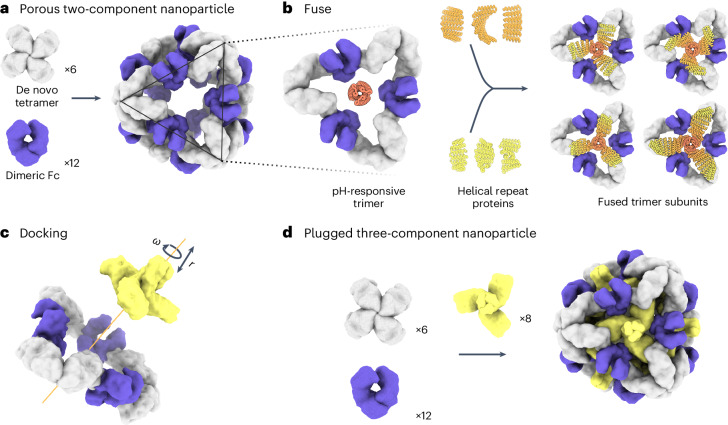
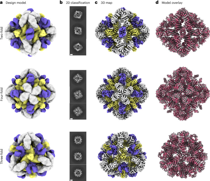
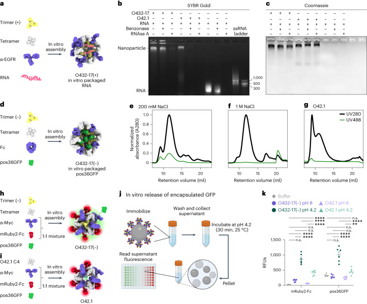
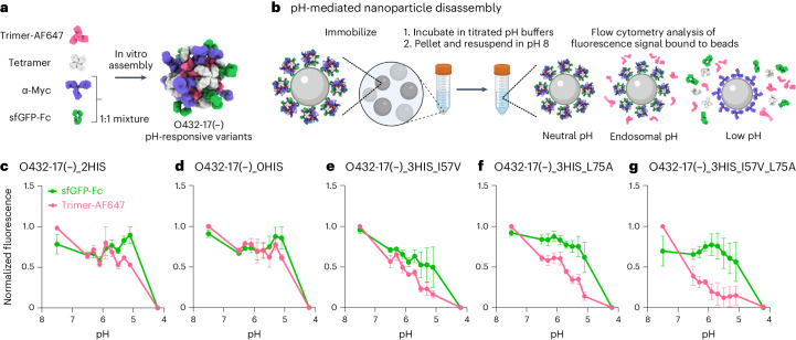
Programming protein nanomaterials to respond to changes in environmental conditions is a current challenge for protein design and is important for targeted delivery of biologics. Here we describe the design of octahedral non-porous nanoparticles with a targeting antibody on the two-fold symmetry axis, a designed trimer programmed to disassemble below a tunable pH transition point on the three-fold axis, and a designed tetramer on the four-fold symmetry axis. Designed non-covalent interfaces guide cooperative nanoparticle assembly from independently purified components, and a cryo-EM density map closely matches the computational design model. The designed nanoparticles can package protein and nucleic acid payloads, are endocytosed following antibody-mediated targeting of cell surface receptors, and undergo tunable pH-dependent disassembly at pH values ranging between 5.9 and 6.7. The ability to incorporate almost any antibody into a non-porous pH-dependent nanoparticle opens up new routes to antibody-directed targeted delivery.


Conflict of interest statement

A provisional patent application has been filed (63/493,252) on the plugged antibody nanoparticle sequences by the University of Washington, listing D.B., E.C.Y., N.P.K., R.D., J.L., W.S., G.U., and J.F. as inventors. The other authors declare no competing interests.

Figures

Fig. 1
Fig. 1. Design of symmetry-matched plugs to fill empty symmetry axes in protein nanoparticles.
a, Six de novo tetramers (gray) and 12 dimeric Fc domains (purple) assemble into a porous octahedral O42 nanoparticle. The tetramers are aligned along the four-fold symmetry axis, and the Fc domains along the two-fold symmetry axis. b, Combinations of helical repeat proteins are fused to each other and to the pH trimer subunit at regions of high backbone overlap between pairs of helices to generate fused trimer subunits large enough to fully occupy the void along the three-fold axis in the original nanoparticle. c, The three-fold symmetry axes of the resulting pH-dependent trimeric fusions and the nanoparticle are aligned. Favorable docked arrangements are then generated by sampling rotations and translations along this axis. d, The resulting docked three-component nanoparticles have eight new trimeric plug subunits (yellow) that occupy the three-fold symmetry axes of the octahedral architecture. UCSF ChimeraX 1.6 (ref. ) and the PyMOL Molecular Graphics System version 2.5 (Schrödinger) were used to create ad.
Fig. 2
Fig. 2. Efficient assembly of three-component nanoparticles from independently purified components for both Fc and IgG.
a, Design models with Fc or IgG (purple), designed nanoparticle-forming tetramers (gray) and pH-dependent plug-forming trimers (yellow). b, Overlay of representative SEC traces of the full assembly formed by mixing designed tetramers, trimers and Fc or IgG (black) with those of the single components in gray (tetramer), yellow (trimeric plug) or purple (Fc or IgG). c, Representative DLS of fractions collected from the O432 assembly peak, showing average hydrodynamic diameters of 34 nm (polydispersity index (PDI), 0.05) and 40 nm (PDI, 0.07) for the O432 assemblies with Fc and full-length IgG, respectively. d, Negatively stained electron micrographs with reference-free 2D class averages along each axis of symmetry in inset; electron microscopy images were collected prior to SEC purification. Scale bars, 100 nm and 10 nm for the micrograph and 2D averages, respectively. The PyMOL Molecular Graphics System version 2.5 (Schrödinger) was used to create a. Source data
Fig. 3
Fig. 3. Cryo-EM analysis of 72-subunit nanoparticles composed of three structural components.
a, Cryo-EM characterization of the O432 assembly with Fc before SEC purification. Computational design models viewed along each axis of symmetry of the octahedral architecture are shown. b, Representative 2D class averages along each axis of symmetry. Scale bars, 10 nm. c, 3D EM density map with 7 Å resolution, reconstructed from the collected dataset. d, Overlay of the design model (gray) and the model refined into the 3D EM density map (pink), showing high agreement. UCSF ChimeraX 1.6 (ref. ) was used to create a.
Fig. 4
Fig. 4. Plugged antibody nanoparticles electrostatically package protein and nucleic acid cargoes.
a, Designed positively charged trimer variants were assembled by incorporating the designed tetramer, pegRNA and antibody to EGFR (α-EGFR) to form O432-17(+) nucleocapsids. b,c, O432-17(+) nucleocapsids and O42.1 assembled in vitro with RNA were treated with Benzonase or RNAse A for 1 h, electrophoresed on non-denaturing 0.8% agarose gels and stained with SYBR Gold (b; nucleic acid) and Coomassie (c; protein). d, Designed negatively charged trimer variants were assembled with designed tetramer, pos36GFP and human Fc to form O432-17(–) nanoparticles with in vitro-packaged pos36GFP. e,f, SEC chromatograms of in vitro packaging reactions involving O432-17(–) and pos36GFP were performed in either 200 mM NaCl (e) or 1 M NaCl (f). g, As a comparison, SEC chromatograms of in vitro-assembled O42.1 with pos36GFP in 200 mM NaCl showed no co-migration of pos36GFP. Absorbance was monitored at 280 nm (black) and 488 nm (green). h, O432-17(–) nanoparticles for in vitro release of pos36GFP were assembled with negatively charged trimer, designed tetramer, pos36GFP and a 1:1 mixture of a Myc-targeted monoclonal antibody (α-Myc) and mRuby2-Fc. i, As a comparison, O42.1 was assembled in vitro with O42.1 C4, pos36GFP and a 1:1 mixture of antibody to Myc and mRuby2-Fc. j, Experimental design for in vitro release of encapsulated pos36GFP in acidic conditions. Assembled nanoparticles were immobilized on Myc-peptide-coated S. cerevisiae yeast cells. Cells and supernatant were collected by centrifugation and resuspended and incubated in acidic conditions. The supernatant containing released components and cargo was buffer exchanged to pH 8, and fluorescence intensity was analyzed. k, Fluorescence intensity of the supernatant of O432-17(–) and O42.1 assembled with pos36GFP and mRuby2-Fc before and after acidic incubation. mRuby2-Fc fluorescence was used as an indicator of nanoparticle assembly; a positive fluorescence signal indicates nanoparticle disassembly. Positive pos36GFP fluorescence indicates release of pos36GFP cargo. *P ≤ 0.05, **P ≤ 0.01, ***P ≤ 0.001, ****P < 0.0001, two-way ANOVA with Tukey’s correction for multiple comparisons. n.s., not significant. Data are presented as mean values ± s.e.m. and measured over three independent samples in duplicate (Supplementary Table 6). UCSF ChimeraX 1.6 (ref. ) and the PyMOL Molecular Graphics System version 2.5 (Schrödinger) were used to create a, d, h and i. j was created using BioRender. GraphPad Prism version 9.3.1 (GraphPad Software) was used to create k. RFUs, relative fluorescence units. Source data
Fig. 5
Fig. 5. Plugged antibody nanoparticles exhibit tunable dissociation in response to acidification.
a, O432-17 nanoparticles were assembled with AF647-conjugated trimeric plug variants, designed tetramer, sfGFP-Fc and α-Myc antibody. b, O432-17 nanoparticles were immobilized on Myc-peptide-coated beads and incubated in titrated pH buffers. Beads were washed by centrifugation and resuspended in 25 mM Tris and 500 mM NaCl, pH 8, and the remaining trimer and sfGFP-Fc fluorescence on the beads was analyzed by flow cytometry. cg, AF647 and sfGFP fluorescence levels were normalized to the minimum and maximum values across the titration and analyzed as a function of the pH for each nanoparticle variant (O432-17(–)_2HIS (c), O432-17(–)_0HIS (d), O432-17(–)_3HIS_I57V (e), O432-17(–)_3HIS_L75A (f), O432-17(–)_3HIS_I57V_L75A (g)). Data are normalized to the minimum and maximum fluorescence per titration and presented as mean ± s.e.m. for three biologically independent replicates. UCSF ChimeraX 1.6 (ref. ) and the PyMOL Molecular Graphics System version 2.5 (Schrödinger) were used to create a; b was created using BioRender; GraphPad Prism version 9.3.1 (GraphPad Software) was used to create cg. Source data
Extended Data Fig. 1
Extended Data Fig. 1. Reducing SDS–PAGE screening gels of co-expressed trimer-tetramer variants 1–45.
Tetramer protein band is marked (right), designs where the designed trimers and tetramers co-eluted are marked with pink stars next to the trimer band. Gel samples were derived from the same experiment and electrophoresis was processed in parallel.
Extended Data Fig. 2
Extended Data Fig. 2. Example experimental screen of O432 three−component nanoparticles.
a, Clarified lysates of 16 designs where the designed trimer co-eluted with the tetramer were supplemented with a purified sfGFP-Fc fusion protein, purified by IMAC, and subject to reducing SDS-PAGE. Trimer protein bands selected for subsequent characterization are marked with pink stars. b, Non-denaturing native PAGE gel electrophoresis of clarified lysates from 16 designs, supplemented with purified sfGFP-Fc fusion protein and purified by IMAC. All designs were compared against the previously designed, two-component antibody nanoparticle (O42.1), O42.1 tetrameric component (O42.1 C4) and sfGFP-Fc (Divine et al.). Gel samples were derived from the same experiment and electrophoresis was processed in parallel.
Extended Data Fig. 3
Extended Data Fig. 3. Large-scale assembly competency screen of O432 three-component nanoparticles.
a, Subcloned components from putative three-component assemblies were purified separately. Peaks marked with a pink star were collected for stoichiometric in vitro assembly of three-component O432 nanoparticles. b, Elution profiles of tetrameric components were compared to the original O42.1 tetramer (O42.1 C4). c, Material permitting, stoichiometric in vitro assembly of trimer, tetramer, and Fc were purified by SEC, d, Elution profiles of resulting assemblies were compared to the SEC elution profile of previously designed O42.1 (Divine et al.). e, Non-reducing SDS-PAGE of the void (V) and shoulder (S) peaks from SEC of O432-17 with purified trimer, tetramer, and Fc components as controls. f, Non-reducing SDS-PAGE of the void (V) and shoulder (S) peaks from SEC purification of O432-23, O432-7, and O432-34 in vitro assemblies. g, 6× designed tetramers form protein-protein interfaces with both 12× Fc and 8× designed trimers. A schematic depicts a hypothetical nanoparticle assembly from the designed tetramer with only the Fc (top) or only the trimer (bottom). h, Representative SEC traces on the Superose 6 10/300 GL of assembly reactions containing only the designed tetramer and Fc (pink), compared to the full three-component assembly (black) and the individual components (gray and purple). i, Representative SEC traces on the Superdex 200 10/300 GL of assembly reactions containing only the designed tetramer and trimer at concentrations of 50 µM, 100 µM, 200 µM, 300 µM, and 400 µM, compared to the individual components (gray and yellow). UCSF ChimeraX 1.6 and the PyMOL Molecular Graphics System, version 2.5 (Schrödinger) was used to create g. Source data
Extended Data Fig. 4
Extended Data Fig. 4. Cryo-EM processing shows newly designed trimeric plug occupying all 3-fold symmetry axes of the nanoparticle octahedral architecture.
a, Representative micrograph of cryo-EM sample. Scale bar, 100 nm. b, Reference-free two-dimensional class averages. Scale bar, 10 nm. c, Ab initio three-dimensional reconstruction without applied octahedral symmetry. d, 3D reconstructions generated following a heterogeneous refinement in the absence of applied octahedral symmetry. All four classes show trimeric plugs occupying all facets of the designed nanoparticle. e, Gold-standard Fourier shell correlation curves for the O432-Fc EM density map with octahedral symmetry applied. f, Close up of plug interface between design model (light gray and yellow) and model built from the 3D reconstruction (dark gray). UCSF ChimeraX 1.6 and the PyMOL Molecular Graphics System, version 2.5 (Schrödinger) was used to create f.
Extended Data Fig. 5
Extended Data Fig. 5. Comparison of design models of O42.1 and O432-17 with models fit into Cryo-EM density.
a, The three-component O432-17 design model (light gray, purple, and yellow) overlaid on the two-component O42.1 density-refined model (dark gray) shows an RMSD of 1.9 Å. b, The O432-17 design model (light gray, purple, and yellow) deviates from its density-refined model (dark gray) by 1.6 Å. c, The O42.1 design model deviates from its density-refined model (dark gray) by 4.2 Å. The PyMOL Molecular Graphics System, version 2.5 (Schrödinger) was used to create a-c.
Extended Data Fig. 6
Extended Data Fig. 6. O432-17 nanoparticles form assemblies in the presence of protein and nucleic acid cargo.
a, Representative negative stain electron micrograph of O432-17(+) in the presence of RNA. Scale bar, 100 nm. b, Reference-free two-dimensional class averages showing multiple views of O432-17(+) nanoparticles. Scale bar, 10 nm. c, Representative negative-stain electron micrograph of O432-17(-) in the presence of pos36GFP. Scale bar, 100 nm. d, Reference-free two-dimensional class averages showing multiple views of O432-17(-) nanoparticles. Scale bar, 10 nm.
Extended Data Fig. 7
Extended Data Fig. 7. Assembly competency of O432-17(+) trimer variants.
a, SEC elution profile of the trimer component on a Superdex 200 GL 10/300 chromatography column. b, SEC elution profile of the full O432-17 assembly containing each trimer variant assembled with tetramer and Fc and purified on a Superose 6 GL 10/300 chromatography column. c, DLS profile of the resulting assembly as normalized intensity. d, Representative nsEM and reference-free two-dimensional class averages of each assembly variant. Scale bar 100 nm and 10 nm, respectively. Source data
Extended Data Fig. 8
Extended Data Fig. 8. Flow cytometry analysis of O432-17 nanoparticle variants.
a, Forward and side scattering intensity isolated all beads. b, Isolated beads were selected for singlet scattering. c, Singlet beads were gated for both AF647 and sfGFP-Fc signal with reference to negative controls: beads only (left), beads incubated with sfGFP-Fc (middle), and beads incubated with AF647-labeled trimeric plug (right). d, The mean fluorescence intensity of beads positive for AF647-labeled trimer and sfGFP-Fc signal was taken at each pH. This gating strategy was applied across all trimeric plug variants: 0HIS, 2HIS, 3HIS_I57V, 3HIS_L75A, 3HIS_I57V_L75A. e-f, Mean fluorescence intensity of O432-17(+) nanoparticles was measured as a function of pH for the trimeric plug variants and sfGFP-Fc. Histidine-containing O432-17(+) variants: O432-17(+)_3HIS_I57V, O432-17(+)_3HIS_L75A, O432-17(+)_3HIS_I57V_L75A); negative control: O432-17(+)_0HIS. Apparent pKas for O432-17(+)_3HIS_I57V, O432-17(+)_3HIS_L75A, and O432-17(+)_3HIS_I57V_L75A: pH 6.1 (AF647); pH 5.8 (sfGFP). O432-17(+)_0HIS apparent pKa: pH 5.1 (AF647); pH 5.0 (sfGFP). Data are presented as mean values +/- SEM over 3 biologically independent replicates. FlowJo v10.8.1(BD Biosciences) was used to create a, b, c, and d. GraphPad Prism version 9.3.1 (GraphPad Software) was used to create e and f. Source data
Extended Data Fig. 9
Extended Data Fig. 9. Assembly competency of O432-17(-) trimer variants.
a, SEC elution profile of the trimer component on a Superdex 200 GL 10/300 chromatography column. b, SEC elution profile of the full O432-17 assembly containing each trimer variant assembled with tetramer and Fc and purified on a Superose 6 GL 10/300 chromatography column. c, DLS profile of the resulting assembly as normalized intensity. d, Representative nsEM and reference-free two-dimensional class averages of each assembly variant. Scale bar 100 nm and 10 nm, respectively. Source data
Extended Data Fig. 10
Extended Data Fig. 10. Quantification of targeted receptor-mediated uptake of O432 nanoparticles.
a, Targeted and non-targeted variants of O432-17(-) nanoparticles were assembled in vitro with AF647-conjugated trimeric plug, designed tetramer, and either premixed mRuby2-Fc and α-EGFR mAb (top) or mRuby2-Fc alone (bottom). b, Cellular uptake of O432-17-CTX and O432-17-Fc nanoparticles was measured by the AF647 and mRuby2 fluorescence within the cell area after 3 hours of incubation in A431 cells. Single confocal plane images; grayscale panels for lysosomal membranes (green), mRuby2-Fc (red), and Trimer-AF647 (gray). Scale bar, 10 µm. c, Percentage of A431 cells containing nanoparticles. Statistics: *P = 0.0306, two-tailed unpaired t-test. Data are presented as mean values +/- SEM over 3 images per condition. d, Integrated intensity per cell area among A431 cells containing nanoparticles. Statistics: P = 0.6025, ns = not significant, two-tailed, unpaired T-test. Data are presented as mean values +/- SEM across all cells in 3 images per condition. e, Targeted receptor-mediated uptake of O432-17-CTX nanoparticles in WT and EGFR KO HeLa cells after 3 hours. Scale bar, 10 µm. f, Percentage of HeLa WT and HeLa EGFR KO cells containing O432-17-CTX nanoparticles with and without serum. Statistics: *P = 0.0116, ***P = 0.001, ****P < 0.0001, ns (P = 0.9349) = not significant, One-way ANOVA with Tukey’s correction for multiple comparisons. Data are presented as mean values +/- SEM over 3 images per condition. g, Integrated intensity per cell area among HeLa WT and HeLa EGFR KO cells containing O432-17-CTX nanoparticles with and without serum. Statistics: *P = 0.0359 (HeLa WT without serum vs. HeLa EGFR KO without serum), *P = 0.0161 (HeLa EGFR KO with serum vs. HeLa EGFR KO without serum), P = 0.9598 (HeLa WT with serum vs. HeLa WT without serum), P = 0.9298 (HeLa WT with serum vs. HeLa EGFR KO with serum), One-way ANOVA with Tukey’s correction for multiple comparisons. Data are presented as mean values +/- SEM across all cells in 3 images per condition; ImageJ v2 was used to create b,e; GraphPad Prism version 9.3.1 (GraphPad Software) was used to create c-d, f-g. Source data

Update of

References

    1. Edwardson, T. G. W., Mori, T. & Hilvert, D. Rational engineering of a designed protein cage for siRNA delivery. J. Am. Chem. Soc.140, 10439–10442 (2018). 10.1021/jacs.8b06442 - DOI - PubMed
    1. Mitchell, M. J. et al. Engineering precision nanoparticles for drug delivery. Nat. Rev. Drug Discov.20, 101–124 (2021). 10.1038/s41573-020-0090-8 - DOI - PMC - PubMed
    1. Banskota, S. et al. Engineered virus-like particles for efficient in vivo delivery of therapeutic proteins. Cell185, 250–265 (2022). 10.1016/j.cell.2021.12.021 - DOI - PMC - PubMed
    1. Wang, D., Tai, P. W. L. & Gao, G. Adeno-associated virus vector as a platform for gene therapy delivery. Nat. Rev. Drug Discov.18, 358–378 (2019). 10.1038/s41573-019-0012-9 - DOI - PMC - PubMed
    1. Hou, X., Zaks, T., Langer, R. & Dong, Y. Lipid nanoparticles for mRNA delivery. Nat. Rev. Mater.6, 1078–1094 (2021). 10.1038/s41578-021-00358-0 - DOI - PMC - PubMed

LinkOut - more resources