. 2024 May 9;21(1):40.
doi: 10.1186/s12987-024-00542-8.

Reduced cerebrospinal fluid motion in patients with Parkinson's disease revealed by magnetic resonance imaging with low b-value diffusion weighted imaging

Affiliations

Reduced cerebrospinal fluid motion in patients with Parkinson's disease revealed by magnetic resonance imaging with low b-value diffusion weighted imaging

Gabriela Pierobon Mays et al. Fluids Barriers CNS. .

Abstract

Background: Parkinson's disease is characterized by dopamine-responsive symptoms as well as aggregation of α-synuclein protofibrils. New diagnostic methods assess α-synuclein aggregation characteristics from cerebrospinal fluid (CSF) and recent pathophysiologic mechanisms suggest that CSF circulation disruptions may precipitate α-synuclein retention. Here, diffusion-weighted MRI with low-to-intermediate diffusion-weightings was applied to test the hypothesis that CSF motion is reduced in Parkinson's disease relative to healthy participants.

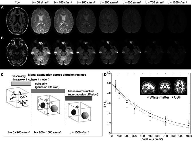
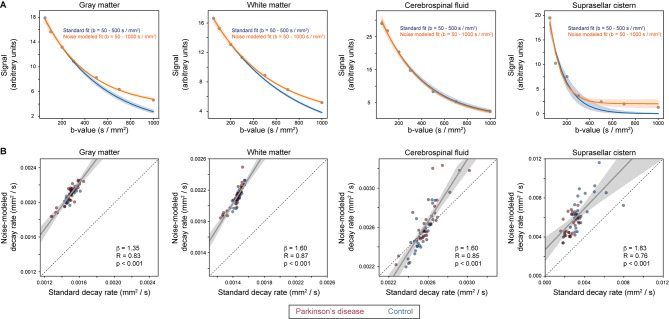
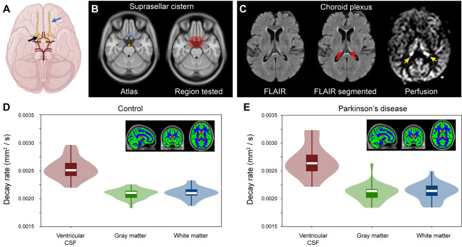
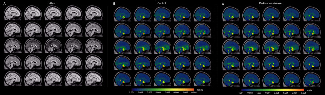
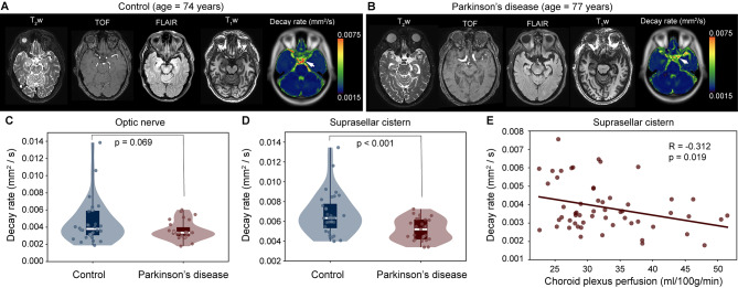
Methods: Multi-shell diffusion weighted MRI (spatial resolution = 1.8 × 1.8 × 4.0 mm) with low-to-intermediate diffusion weightings (b-values = 0, 50, 100, 200, 300, 700, and 1000 s/mm2) was applied over the approximate kinetic range of suprasellar cistern fluid motion at 3 Tesla in Parkinson's disease (n = 27; age = 66 ± 6.7 years) and non-Parkinson's control (n = 32; age = 68 ± 8.9 years) participants. Wilcoxon rank-sum tests were applied to test the primary hypothesis that the noise floor-corrected decay rate of CSF signal as a function of b-value, which reflects increasing fluid motion, is reduced within the suprasellar cistern of persons with versus without Parkinson's disease and inversely relates to choroid plexus activity assessed from perfusion-weighted MRI (significance-criteria: p < 0.05).

Results: Consistent with the primary hypothesis, CSF decay rates were higher in healthy (D = 0.00673 ± 0.00213 mm2/s) relative to Parkinson's disease (D = 0.00517 ± 0.00110 mm2/s) participants. This finding was preserved after controlling for age and sex and was observed in the posterior region of the suprasellar cistern (p < 0.001). An inverse correlation between choroid plexus perfusion and decay rate in the voxels within the suprasellar cistern (Spearman's-r=-0.312; p = 0.019) was observed.

Conclusions: Multi-shell diffusion MRI was applied to identify reduced CSF motion at the level of the suprasellar cistern in adults with versus without Parkinson's disease; the strengths and limitations of this methodology are discussed in the context of the growing literature on CSF flow.

Keywords: Cerebrospinal fluid; Choroid plexus; DWI; Glymphatic; Parkinson’s; Suprasellar cistern; α-synuclein.


Conflict of interest statement

No authors declare any relevant conflicts or disclosures regarding the work presented in this manuscript. Manus J. Donahue receives research related support from the National Institutes of Health (NINDS, NCI, NIA, NCCIH, NINR, and NHLBI), Philips Healthcare and is a paid consultant for Graphite Bio, Pfizer Inc, Global Blood Therapeutics, and LymphaTouch. He is a paid advisory board member for Novartis and bluebird bio and receives research funding from the National Institutes of Health and Pfizer Inc. Manus J. Donahue is also the CEO of Biosight Inc which operates as a clinical research organization and provides healthcare technology vendor services. Ciaran Considine?s financial disclosures include those associated with his private consulting practice, NeuropsyConsulting LLC, i.e., Forensic Consulting with Park Dietz & Associates, clinical-research advisory panel member with MDisrupt, external advisory board for HD Genetics. He also receives academic-research grant support from the NIH, NIA, and DoD, and acts as PI on an investigator-initiated clinical study funded by Acadia. Daniel O. Claassen has received research support from the NIH/NINDS/NIA/NICHD/NCCIH, Department of Defense, Griffin Family Foundation, and Huntington Disease Society of America; he has received pharmaceutical grant support from AbbVie, Alterity, Acadia, Biogen, BMS, Cerecour, Eli Lilly, Genentech-Roche, Lundbeck, Jazz Pharmaceuticals, Neurocrine, Teva Neuroscience, Wave Life Sciences, UniQure, and Vaccinex. He has received personal fees for consulting from Acadia, Alterity, Adamas, Anexon, Ceruvel, Lundbeck, Neurocrine, Spark, Uniqure, and Teva Neuroscience.

Figures

Fig. 1
Fig. 1
Multi-shell diffusion weighted imaging (DWI) using low-to-intermediate b-values. (A) Two slices at the level of the lateral ventricles (A) and suprasellar cistern (B) for a representative participant (age = 77 years; sex = male) are shown. (C) The approximate regime of physiological sensitivity for increasing b-values, whereby low b-values < 200 s/mm2 have known sensitivity to vascular structures and intravoxel incoherent motion, intermediate b-values of approxiamtely 200–1500 s/mm2 are sensitive to cellularity in the regime of gaussian diffusion, and high b-values above 1500 s/mm2 are most sensitive to non-gaussian diffusion and tissue microstructure assessments. (D) Example decay curves as a function of low-to-intermediate b-value in the transition range of intravoxel incoherent motion and gaussian diffusion demonstrate differences in cerebrospinal fluid and tissue. Error bars across the region shown in the insert are depicted as one-sided for clarity
Fig. 2
Fig. 2
Fitting procedures. (A) Signal decay, along with exponential fitting, across a range of regions and tissue structures. Given higher motion for neurofluids relative to gray and white matter, fitting and confidence intervals are shown in blue for the lower b-values (e.g., 5–500 s/mm2) without the noise floor modeled as well as separately in orange for all b-values by including the noise floor term (e.g., Eq. 3). Across all tissue types it can be appreciated that by modeling the noise floor, the goodness of fit improves, as expected, for the range of b-values. Quantitative fitting statistics are summarized in Table 2. (B) The quantitative relationship for decay rates when the noise floor is fit versus when it is excluded. Solid lines depict best fit lines whereas dashed lines depict lines of unity. In all subsequent analyses, the fitting procedure that incorporated the noise floor modeling was used
Fig. 3
Fig. 3
Localization and quantification procedures. (A) Suprasellar cistern location, which co-localizes with major cranial nerves including the olfactory (blue arrow) and optic (black arrow) nerves, along with circle of Willis (red vasculature). (B) The region of interest encompassing the suprasellar cistern from which spatial statistical testing was performed is overlaid on the standard 1 mm brain atlas; note that the region deliberately extended beyond the average suprasellar cistern boundaries to accommodate potential morphological variations. (C) Choroid plexus at the level of the lateral ventricles visible on FLuid Attenuated Inverstion Recovery (FLAIR) MRI, along with example of segmentation; the perfusion map is shown to right, which highlights (yellow arrows) the high choroid plexus perfusion signal which is comparable to gray matter perfusion signal. Given the high variability in choroid plexus anatomy, structures were segmented in native space for each participant using previously-reported machine learning routines (see Methods). (D-E) Across major tissue types (shown as inserts), decay rates are significantly elevated in CSF relative to other tissue types (p < 0.001), a well as in gray matter relative to white matter (p < 0.001). The plots show boxplots overlaid on violin plots. See Table 1 for quantitative values. No significant difference was observed between control and Parkinson’s disease participants for any tissue type
Fig. 4
Fig. 4
Group-averaged orthogonal maps of decay rates. Orthogonal representations are shown, for the level of the (A) T1-weighted atlas, (B) control participants, and (C) Parkinson’s disease (PD) participants. Reduced suprasellar flow is observed in PD relative to control participants. The changes are largely localized to the posterior region of the suprasellar cistern and pre-chiasmatic optic nerve. Sagittal representations across midline are shown separately in Fig. 5
Fig. 5
Fig. 5
Group-averaged sagittal maps of decay rates. Group-averaged maps are shown sagittal across midline. Note that the scale has been changed slightly from that in Fig. 4 to show contrast of additional structures. The most prominent changes are appreciated in the posterior aspect of the suprasellar cistern and spaces along major neurofluid routes (e.g., fourth ventricle, cerebral aqueduct, and cisterns) with general similarity across cortex and other subarachnoid spaces
Fig. 6
Fig. 6
Case and group findings. Case example of an age- and sex-matched control (A) and Parkinson’s disease (PD) (B) participant showing anatomical imaging at the level of the suprasellar cistern. (C-D) Below, group level results are shown. Violin plots show the distribution of decay rates in control vs. PD participants, both in the suprasellar cistern and at the approximate location of the pre-chiasmatic optic nerve. The decay rates were significantly different in the suprasellar cistern (p < 0.001). (E) The decay rate was observed to inversely correlate with the choroid plexus perfusion in the suprasellar cistern (p = 0.019)

Update of

Similar articles

Cited by

References

    1. Bloem BR, Okun MS, Klein C. Parkinson’s disease. Lancet. 2021;397(10291):2284–303. doi: 10.1016/S0140-6736(21)00218-X. - DOI - PubMed
    1. McKnight CD, Trujillo P, Lopez AM, et al. Diffusion along perivascular spaces reveals evidence supportive of glymphatic function impairment in Parkinson disease. Parkinsonism Relat Disord. 2021;89:98–104. doi: 10.1016/j.parkreldis.2021.06.004. - DOI - PMC - PubMed
    1. Salman MM, Kitchen P, Iliff JJ, Bill RM. Aquaporin 4 and glymphatic flow have central roles in brain fluid homeostasis. Nat Rev Neurosci. 2021;22(10):650–1. doi: 10.1038/s41583-021-00514-z. - DOI - PubMed
    1. McKnight CD, Rouleau RM, Donahue MJ, Claassen DO. The regulation of cerebral spinal fluid Flow and its relevance to the Glymphatic System. Curr Neurol Neurosci Rep. 2020;20(12):58. doi: 10.1007/s11910-020-01077-9. - DOI - PMC - PubMed
    1. Ringstad G, Valnes LM, Dale AM et al. Brain-wide glymphatic enhancement and clearance in humans assessed with MRI. JCI Insight 2018;3(13). - PMC - PubMed