Review
. 2024 May 9;22(1):235.
doi: 10.1186/s12951-024-02349-z.

Cucurbit[8]uril-based supramolecular theranostics

Affiliations
Review

Cucurbit[8]uril-based supramolecular theranostics

Dan Wu et al. J Nanobiotechnology. .

Abstract

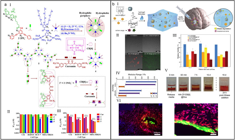
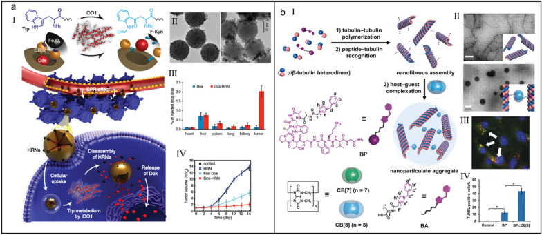
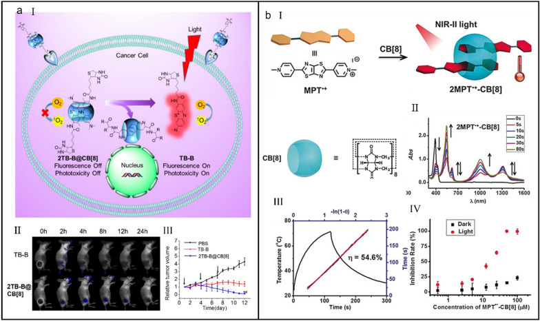
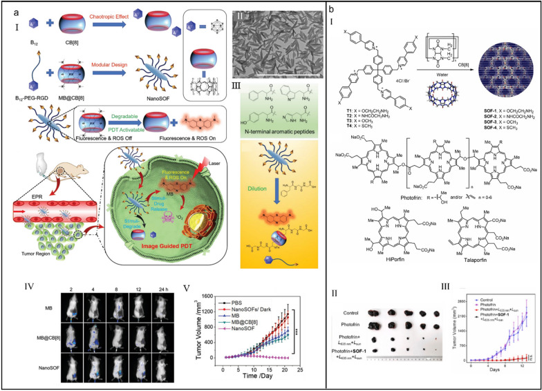
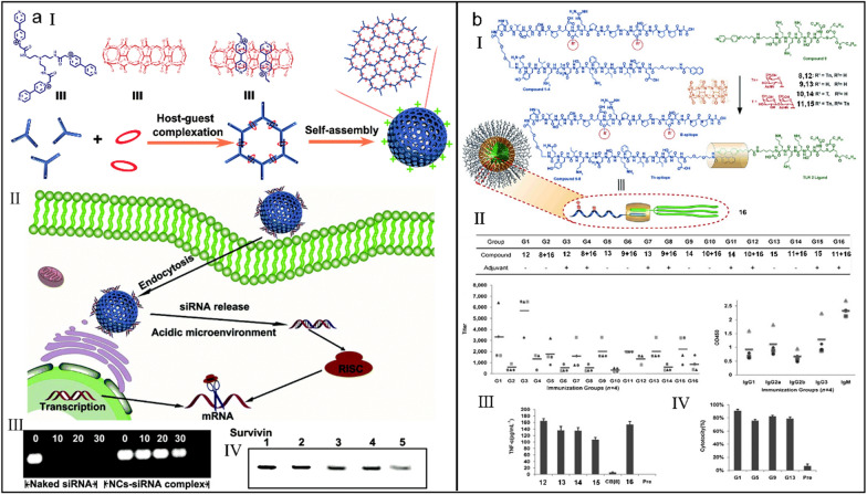
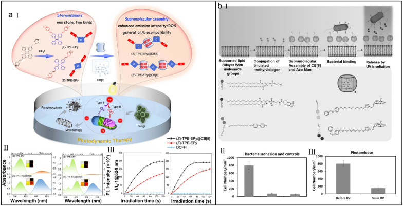
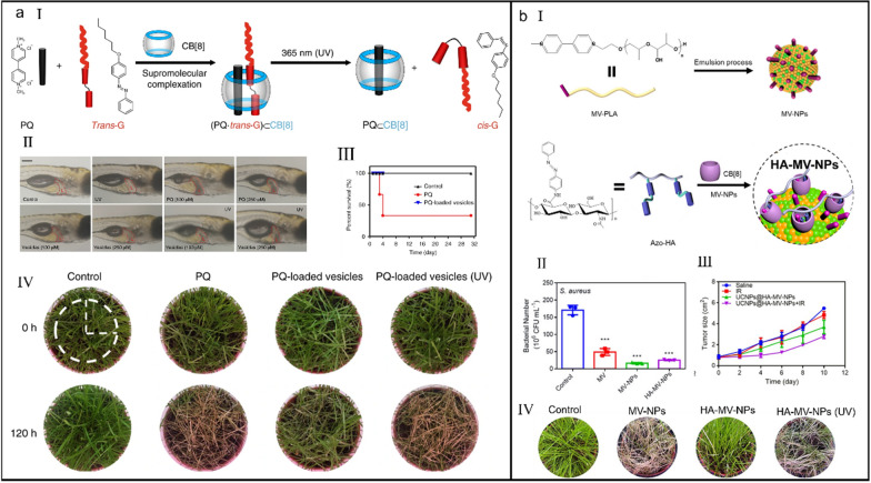
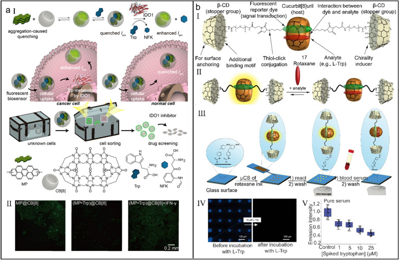
Different from most of the conventional platforms with dissatisfactory theranostic capabilities, supramolecular nanotheranostic systems have unparalleled advantages via the artful combination of supramolecular chemistry and nanotechnology. Benefiting from the tunable stimuli-responsiveness and compatible hierarchical organization, host-guest interactions have developed into the most popular mainstay for constructing supramolecular nanoplatforms. Characterized by the strong and diverse complexation property, cucurbit[8]uril (CB[8]) shows great potential as important building blocks for supramolecular theranostic systems. In this review, we summarize the recent progress of CB[8]-based supramolecular theranostics regarding the design, manufacture and theranostic mechanism. Meanwhile, the current limitations and corresponding reasonable solutions as well as the potential future development are also discussed.

Keywords: Cucurbit[8]uril; Host–guest reactions; Self-assembly; Supramolecular nanomedicine; Supramolecular theranostics.


Conflict of interest statement

The authors declare no competing interests.

Figures

Fig. 1
Fig. 1
(a) Two-stage enhanced NIR supramolecular assemblies for cell imaging. (I) Illustration of the self-assemble process of NIR supramolecular assemblies. (II) Fluorescence emission spectroscopy of ENDT, ENDT/SC4AD, ENDT/CB[8] and ENDT/CB[8]/SC4AD. (III) Illustration of the sled n:n binding motif. (IV) Fluorescence photographs of ENDT, ENDT/CB[8] and ENDT/CB[8]/SC4AD. (V) Confocal laser scanning microscopy (CLSM) images of A549 cells treated with ENDT/CB[8]/SC4AD and LysoTracker Blue. Reproduced with permission [78]. Copyright 2018 Wiley–VCH Verlag GmbH & Co. KGaA, Weinheim (b) NIR supramolecular assemblies for two-photon targeting imaging. (I) Illustration of the formation of NIR supramolecular nanoparticles and cartoons of each component. Assembly schematic diagram and SEM images of TPE-2SP/CB[8] (II) and TPE-2SP/CB[8]/HA-CD (III). (IV) CLSM images of A549 cells treated with TPE-2SP/CB[8]/HA-CD and Mito-Tracker Green. Reproduced with permission [90]. Copyright 2021 Wiley–VCH GmbH
Fig. 2
Fig. 2
(a) Room-temperature phosphorescence emissive supramolecular assembly excited by visible-light. (I) X-ray diffraction single-crystal structure of supramolecular assembly (TBP)2·CB[8]2. (II) Phosphorescent emission spectra of TBP with gradual addition of CB[8]. (III) Photographs of hydrogels with different ratios of TBP and CB[8] under daylight or UV light. (IV) CLSM images of Hela cells incubated with TBP and·CB[8] (1:1). Reproduced with permission [98]. Copyright 2019 Wiley–VCH Verlag GmbH & Co. KGaA, Weinheim. (b) Supramolecular phosphorescence-capturing assembly for NIR lysosome imaging. (I) Illustration of the establishment of RTP-capturing system featured with a delayed NIR emission. (II) Phosphorescent emission spectra of G with gradual addition of CB[8]. (III) Phosphorescent emission spectra of G⊂CB[8]. Inset: The time-resolved phosphorescence decay plot of G⊂CB[8] at 530 nm. Phosphorescent emission spectra of G⊂CB[8]@SC4AH/NiR (IV) and G⊂CB[8]@SC4AH/NiB (V) at different ratios of donor and acceptor. Reproduced with permission [99]. Copyright 2021 Wiley–VCH GmbH
Fig. 3
Fig. 3
(a) Phosphorescent biaxial pseudorotaxane for selectively imaging tumor cells. (I) Illustration of the supramolecular assembly and disassembly of biaxial pseudorotaxane. (II) Photoluminescence spectra of BPTN in the presence of different concentrations of CB[8]. (III) Photoluminescence spectra of BPTNC⊂CB[8] and BPTNCCB[8]⊂SSP[4]. (IV) Photoluminescence spectra of BPTNC⊂CB[8], BPTNCCB[8]⊂SSP[4], BPTNCCB[8]⊂SSP[4] + GSH and BPTNCCB[8]⊂SSP[4] + weak acid. Reproduced with permission [106]. Copyright 2022 The Author(s) Published by the Royal Society of Chemistry. (b) Ultralong phosphorescence supramolecular polymer for tumor cell imaging. (I) Illustration of the construction of CBs/HA-BrBP supramolecular polymers. (II) The proposed mechanism of ultralong phosphorescence of supramolecular polymer. (III) CLSM images of A549 cells treated with CB[8]/HA-BrBP. Reproduced with permission [110]. Copyright 2020 The Author(s)
Fig. 4
Fig. 4
(a) CB[8]-based supramolecular nanomedicine for tumor therapy. (I) Chemical structures of different building blocks and the preparation of supramolecular nanomedicine. (II) Illustration of the imaging-guided selective drug release. (III) Pharmacokinetics of free DOX and DOX-loaded SNPs. (IV) Tumor volume change of mice with different treatments. Reproduced with permission [120]. Copyright 2017 American Chemical Society. (b) Supramolecular DOX-dimer for selective drug release. (I) Chemical structures of different building blocks and the construction of supramolecular dimeric prodrug. Cell viability of BEL 7402 cells (II) and LO2 cells (III) after different treatments. Reproduced with permission [126]. Copyright 2019 Chinese Chemical Society and Institute of Materia Medica, Chinese Academy of Medical Sciences. Published by Elsevier B.V. All rights reserved
Fig. 5
Fig. 5
(a) Orthogonal organoplatinum(II) metallacycle for tumor therapy. (I) Schematic illustration of the self-assembly of supramolecular system. (II) IC50 value of 2', 4' and 5' measured on different cell lines. (III) IC50 value of 1, 4 and 5 measured on different cell lines. Reproduced with permission [132]. Copyright 2018 Published under the PNAS license. (b) A CB[8]-based hydrogel delivery vehicle for GB therapy. (I) Illustration of the preparation of supramolecular hydrogel and its cure mechanism. (II) Fluorescence images of GB cells after different treatments. (III) Cell viability of different cells after different treatments. (IV) The moduli comparation between tissue and supramolecular hydrogel. (V) The stability study of supramolecular hydrogel. (VI) The immumohistochemical staining of GB tissue reflecting the tissue penetrability of supramolecular hydrogel delivery vehicle. Reproduced with permission [139]. Copyright 2018 Published by Elsevier Ltd
Fig. 6
Fig. 6
(a) Trp/CB[8]-mediated hybrid nanoparticles for targeted drug delivery in IDO1-overexpressed tumor cells. (I) Illustration of the targeted release mechanism of hybrid supramolecular nanoparticles. (II) Transmission electron microscope (TEM) images of hybrid nanoparticles (left) and their collapse upon exposure to IDO1 (right). (III) Biodistribution of DOX in major organs and tumors at 24 h post-injection of free DOX and hybrid supramolecular nanoparticles. (IV) Tumor volume change of mice during treatment. Reproduced with permission [146]. Copyright 2019 WILEY–VCH Verlag GmbH & Co. KGaA, Weinheim. (b) CB[8]-mediated microtubule aggregation for enhancing cell apoptosis. (I) Illustration of BP⊂CB[8]-mediated targeted microtubular aggregation. (II) TEM images of free MTs (up) and BP@MTs (down). (III) CLSM image of A549 cells treated with BP⊂CB[8]. (IV) The percentage of TUNEL-positive cells in tumor tissue of mice after different treatments. Reproduced with permission [152]. Copyright 2019 Wiley–VCH Verlag GmbH & Co. KGaA, Weinheim
Fig. 7
Fig. 7
(a) CB[8]-regulated aPS for imaging-guided PDT. (I) Illustration of the mechanism of aPS-mediated imaging-guided PDT. (II) In vivo imaging of mice intravenously administrated with TB-B and 2TB-B@CB[8]. (III) Tumor volume change of mice during treatments. Reproduced with permission [164]. Copyright 2016 American Chemical Society. (b) A CB[8]-based supramolecular radical dimer with a high NIR-II photothermal conversion efficiency. (I) Illustration of the self-assembly of 2MPT•+-CB[8]. (II) UV/Vis–NIR spectra of 2MPT•+-CB[8] with different irradiation time. (III) Heating and cooling cycle of 2MPT•+-CB[8] and the calculated photothermal conversion efficiency. (IV) Inhibition rate plots of HepG2 cells after 2MPT•+-CB[8] induced PTT. Reproduced with permission [171]. Copyright 2019 Wiley–VCH Verlag GmbH & Co. KGaA, Weinheim
Fig. 8
Fig. 8
(a) CB[8]-regulated supramolecular organic frameworks for imaging-guided PDT. (I) Construction of CB[8]-regulated supramolecular organic frameworks and their application for imaging-guided PDT. (II) TEM image of the supramolecular organic framework. (III) The chemical structures of N-terminal aromatic peptides (up) and the illustration of dilution effect and N-terminal aromatic peptides-co-triggered degradation of supramolecular organic frameworks (down). (IV) In vivo fluorescence images of mice with different treatments. (V) Tumor volume change of mice with different treatments. Reproduced with permission [175]. Copyright 2020 Wiley–VCH GmbH. (b) Supramolecular organic frameworks applied to improve the safety of clinical porphyrin photosensitizers without breaking their antitumor efficacy. (I) Illustration of the formation of supramolecular organic frameworks. (II) Photos of excised tumor tissues of mice with different treatments. (III) Tumor volume change of mice with different treatments.. Reproduced with permission [181]. Copyright 2022 Elsevier Ltd. All rights reserved
Fig. 9
Fig. 9
(a) Photoresponsive supramolecular complexes for efficiently regulating DNA. (I) Chemical structures of 6 and 7 and the optically controlled configuration interconversion process of supramolecular complexes. (II) Atomic force microscope (AFM) images of pBR322 DNA (left) and DNA condensation induced by trans-7⊂CB[8] (right). Reproduced with permission [187]. Copyright 2014 The Author(s). (b) Rodlike supramolecular nanoassemblies for effective delivery of ncRNAs. (I) The synthesis process of supramolecular nanoassemblies and their application for co-delivering pc3.0-MEG3 and pc3.0-miR-101. (II) AFM image of ncRNAs-loaded supramolecular nanoassemblies. (III) Tumor-suppressive effect of ncRNAs-loaded supramolecular nanoassemblies. Reproduced with permission [194]. Copyright 2017 WILEY–VCH Verlag GmbH & Co. KGaA, Weinheim
Fig. 10
Fig. 10
(a) Supramolecular polymer nanocapsules for effective siRNA delivery. (I) Illustration of the construction of supramolecular polymer nanocapsules. (II) Illustration of the intracellular siRNA delivery by supramolecular polymer nanocapsules. (III) Biostability test of siRNA with or without supramolecular polymer nanocapsules. (IV) Western blot analysis of intracellular survivin protein after different treatments (1: control; 2: scramble siRNA; 3: lipofectamine 2000-siRNA complex; 4: NC-siRNA complex (50 nM); 5: NC-siRNA complex (100 nM). Reproduced with permission [200]. Copyright 2019 The Royal Society of Chemistry. (b) A noncovalent strategy to construct chemically synthesized vaccines. (I) Illustration of the construction of synthesized vaccines. (II) ELISA anti-MUC1 IgG antibody titers (left) and analyses (right) after different immunizations. (III) The secretion of TNF-α cytokine by dendritic cells after different stimulations. (IV) Cytotoxicity assay of MCF-7 cells after different immunizations. Reproduced with permission [206]. Copyright 2014 Wiley–VCH Verlag GmbH & Co. KGaA, Weinheim
Fig. 11
Fig. 11
(a) Supramolecular engineering of AIE photosensitizers for fungal killing. (I) Chemical structures of stereoisomers and corresponding supramolecular assemblies and the illustration of their sterilization mechanism via PDT. (II) Absorption and emission spectra of stereoisomers. (III) ROS generation assessment of stereoisomers and corresponding supramolecular assemblies. Reproduced with permission [214]. Copyright 2022, The Author(s). (b) CB[8]-mediated photoswitchable adhesion and release of bacteria on SLBs. (I) Chemical structures of different components and the illustration of the mechanism of bacteria adhesion and release. (II) The number of bacteria immobilized on supramolecular SLBs. (III) The number of residual bacteria immobilized on supramolecular SLBs. Reproduced with permission [218]. Copyright 2015 Wiley–VCH Verlag GmbH & Co. KGaA, Weinheim
Fig. 12
Fig. 12
(a) Photo-responsive supramolecular vesicles for user-friendly herbicide. (I) Illustration of the CB[8]-mediated supramolecular complexation and photo-driven, reversible complexation and decomplexation. (II) Liver tissue observation of zebrafish after different treatments. (III) Survival curves of mice after different treatments. (IV) Weed control efficacy of different treatment methods. Reproduced with permission [223]. Copyright 2018 The Author(s). (b) DIA of supramolecular toxic nanoparticles for multifunctional applications. (I) Illustration of the preparation of MV-NPs and HA-MV-NPs. (II) The comparation of bacteriostasis rate after different treatments. (III) Tumor volume change of mice after different treatments. (IV) Weed control efficacy of different treatment methods. Reproduced with permission [224]. Copyright 2020 American Chemical Society
Fig. 13
Fig. 13
(a) An off −on supramolecular fluorescent biosensor for monitoring IDO1 activity in living cells. (I) Illustration of the detection mechanism of supramolecular fluorescent biosensor. (II) Fluorescence images of HepG2 cells with different treatments. Reproduced with permission [233]. Copyright 2019 American Chemical Society. (b) CB[8]-based rotaxane chemosensor for optical detection of Trp in biological samples. (I) Design principle of supramolecular rotaxane 17. (II) Illustration of the analyte binding by rotaxane 17. (III) Illustration of the fluorescence imaging of Trp in blood serum by rotaxane 17-immobilizated glass surfaces. (IV) Fluorescence images of a microarray before and after treatment with Trp. (V) Emission intensity change of a sensor chip after treatment with different serums. Reproduced with permission [238]. Copyright 2023 The Author(s)
Fig. 14
Fig. 14
(a) A supramolecular fluorescent probe for determination of norfloxacin. (I) Schematic illustration of the self-assembly of supramolecular fluorescent probe. (II) The fluorescence emission change of DBXPY@CB[8] after addition of different drugs. (III) Fluorescence photographs of DBXPY@CB[8] after addition of various drugs, pesticides and amino acids. (IV) Schematic illustration of the detection process of supramolecular fluorescent probe. Reproduced with permission [239]. Copyright 2023 Elsevier B.V. All rights reserved. (b) Supramolecular phosphorescent probe for determination of dodine. (I) Chemical structures of different components and the schematic illustration of the detection mechanism of supramolecular phosphorescent probe. (II) Phosphorescent emission change of CB[8]-BPCOOH after addition of different pesticides. (III) Phosphorescent photographs of CB[8]-BPCOOH-based solid film in the presence of different pesticides. (IV) Phosphorescent photographs of CB[8]-BPCOOH-based indicator paper in the presence of different concentrations of dodine. Reproduced with permission [240]. Copyright 2022 American Chemical Society

Similar articles

Cited by

References

    1. Song N, Lou X-Y, Ma L, Gao H, Yang Y-W. Supramolecular nanotheranostics based on pillarenes. Theranostics. 2019;9(11):3075–3093. doi: 10.7150/thno.31858. - DOI - PMC - PubMed
    1. Gravel J, Schmitzer AR. Imidazolium and benzimidazolium-containing compounds: from simple toxic salts to highly bioactive drugs. Org Biomol Chem. 2017;15(5):1051–1071. doi: 10.1039/C6OB02293F. - DOI - PubMed
    1. Karimi M, Zangabad PS, Mehdizadeh F, Malekzad H, Ghasemi A, Bahrami S, Zare H, Moghoofei M, Hekmatmanesh A, Hamblin MR. Nanocaged platforms: modification, drug delivery and nanotoxicity. Opening synthetic cages to release the tiger. Nanoscale. 2017;9(4):1356–1392. doi: 10.1039/C6NR07315H. - DOI - PMC - PubMed
    1. Wu D, Zhang Z, Li X, Zhu T, Wang J, Hu Q. Supramolecular theranostic nanomedicine for in situ self-boosting cancer photochemotherapy. Biomacromol. 2023;24(2):1022–1031. doi: 10.1021/acs.biomac.2c01469. - DOI - PubMed
    1. Wu D, Zhang Z, Li X, Han J, Hu Q, Yu Y, Mao Z. Cucurbit[10]uril-based supramolecular radicals: powerful arms to kill facultative anaerobic bacteria. J Control Release. 2023;354:626–634. doi: 10.1016/j.jconrel.2023.01.040. - DOI - PubMed

LinkOut - more resources