. 2024 Jul;11(28):e2401327.
doi: 10.1002/advs.202401327. Epub 2024 May 9.

Supramolecular Nanofibers Ameliorate Bleomycin-Induced Pulmonary Fibrosis by Restoring Autophagy

Affiliations

Supramolecular Nanofibers Ameliorate Bleomycin-Induced Pulmonary Fibrosis by Restoring Autophagy

Debin Zheng et al. Adv Sci (Weinh). 2024 Jul.

Abstract

Idiopathic pulmonary fibrosis (IPF) is a progressive and ultimately fatal interstitial lung disease, with limited therapeutic options available. Impaired autophagy resulting from aberrant TRB3/p62 protein-protein interactions (PPIs) contributes to the progression of IPF. Restoration of autophagy by modulating the TRB3/p62 PPIs has rarely been reported for the treatment of IPF. Herein, peptide nanofibers are developed that specifically bind to TRB3 protein and explored their potential as a therapeutic approach for IPF. By conjugating with the self-assembling fragment (Ac-GFFY), a TRB3-binding peptide motif A2 allows for the formation of nanofibers with a stable α-helix secondary structure. The resulting peptide (Ac-GFFY-A2) nanofibers exhibit specific high-affinity binding to TRB3 protein in saline buffer and better capacity of cellular uptake to A2 peptide. Furthermore, the TRB3-targeting peptide nanofibers efficiently interfere with the aberrant TRB3/p62 PPIs in activated fibroblasts and fibrotic lung tissue of mice, thereby restoring autophagy dysfunction. The TRB3-targeting peptide nanofibers inhibit myofibroblast differentiation, collagen production, and fibroblast migration in vitro is demonstrated, as well as bleomycin-induced pulmonary fibrosis in vivo. This study provides a supramolecular method to modulate PPIs and highlights a promising strategy for treating IPF diseases by restoring autophagy.

Keywords: autophagy; peptide nanofiber; protein‐protein interactions; pulmonary fibrosis; self‐assembly.


Conflict of interest statement

The authors declare no conflict of interest.

Figures

Figure 1
Figure 1
Enhanced TRB3/p62 PPIs and autophagy suppression in pulmonary fibrosis. a) TRB3 mRNA expression was upregulated in IPF fibroblasts from the GDS4580, two‐tailed Student's t‐test ** p<0.01. b) Western bolt analysis of TRB3, p62 and Col 1 from MRC‐5 cells treated with different concentrations of TGF‐β1 for 24 h. c) Western bolt analysis for TRB3, p62, and Col 1 of lung tissue from normal mice and bleomycin‐injured mice. d) Representative immunoblots from (b) and the ratio of the indicated protein to β‐actin (n = 3, two‐way ANOVA). e) Representative immunoblots from (c) and the ratio of the indicated protein to β‐actin (n = 4, two‐way ANOVA), * p<0.05, ** p<0.01, *** p<0.001, **** p<0.0001. f) Immunostaining images depict expression and co‐localization of the cargo protein p62 and TRB3 in MRC‐5 cells treated with 0 or 10 ng mL−1 TGF‐β1 for 24 h, the nucleus stained by DAPI, bar represents 7.5 µm. g) MRC‐5 cells were infected with mRFP‐GFP‐LC3 adenovirus. After 24 h, the cells were treated with 0 or 10 ng/mL TGF‐β1 for 24 h, and the autophagic flux rate was detected with Live Cell Imaging Microscopy, bar represents 10 µm.
Figure 2
Figure 2
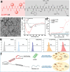
Peptide nanofibers specifically bind to the TRB3 protein. a) Chemical structure of the self‐assembly peptide Ac‐GFFY‐A2 (A2 is TRB3 targeting fragment). b) TEM images of Ac‐GFFY‐A2 (0.5 wt%) peptide nanofibers after the heat‐cooling process, bar represents 100 nm. c) The CD spectrum both of Ac‐GFFY‐A2 (0.5 wt%) and A2 (0.5 wt%) in saline buffer. d) The MST analysis of Ac‐GFFY‐A2 or A2 binding to TRB3 protein, BSA protein as negative control group. e) Flow cytometry analysis for cellular uptake of NBD labeled different peptides (100 µm) after incubation with MRC‐5 cells for 4 h. f) Schematic explaining the supramolecular self‐assembly strategy regulated the secondary conformation of A2 peptide to boost cellular uptake and protein binding affinity.
Figure 3
Figure 3
TRB3‐targeting peptide (Ac‐GFFY‐A2) nanofibers disrupt TRB3/p62 PPIs and restore autophagy. a) Co‐IP analysis for TRB3/p62 PPIs in MRC‐5 cells treated with TGF‐β1 (10 ng/mL), saline, A2 (50 µm) or Ac‐GFFY‐A2 (50 µm), respectively. + represents treated,–represents untreated. b) CLSM images depict the co‐localization of the protein p62 and TRB3 in MRC‐5 cells treated with different methods. c) After MRC‐5 cells were infected with mRFP‐GFP‐LC3 adenovirus, the cells were treated with different methods, and the autophagic flux rate was detected with Live Cell Imaging Microscopy, bar represents 10 µm. d) Western bolt analysis for autophagic marker protein LC3‐ II/LC3‐ I and p62 in MRC‐5 cells treated with TGF‐β1 (10 ng/mL), saline, A2 (50 µm) or Ac‐GFFY‐A2 (50 µm), respectively. + represents treated,–represents untreated.
Figure 4
Figure 4
TRB3‐targeting peptide nanofibers inhibit TGF‐β1‐induced fibroblast activation, ECM production, and fibroblast migration in vitro. a) Western bolt analysis for α‐SMA, Col 1, and FN in cell extracts (Cell) and Col 1 and FN (fibronectin) in the medium supernatant (SN). + represents treated,–represents untreated. b) Representative immunoblots from (a) and the gray value ratio of the indicated protein to β‐actin (n = 3, two‐way ANOVA), * p<0.05, ** p<0.01. c) Migration and invasion assay of MRC‐5 cells treated with saline, A2 (50 µm) or Ac‐GFFY‐A2 (50 µm) in the absence or presence of TGF‐β1 at 24 h, stained with 0.5% crystal violet, bar represents 50 µm. d) Wound‐healing assay of MRC‐5 cells treated with saline, A2, or Ac‐GFFY‐A2 in the absence or presence of TGF‐β1 at indicated times. e) The rate of wound closure was determined at 48 h, and the rate of migration and invasion was quantified using the OD value at 540 nm. All experiments were conducted in triplicate (n = 3, one‐way ANOVA), * p<0.05, ** p<0.01.
Figure 5
Figure 5
TRB3‐targeting peptide selectively accumulates in the lung tissue. a) NIR fluorescence imaging of the bio‐distribution of sulfo‐Cy5 labeled peptide conjugates (i.v., 0.3 mg per mouse) in the wild male C57BL/6 mice (0 day) and 2U kg−1 bleomycin‐treated male C57BL/6 mice (14 day) in vivo. b) Quantitative fluorescence analyses of (a) at the corresponding time in the lung area of mice. c) NIR fluorescence images of excised lung tissue post injection at 8 h. d) Quantitative fluorescence analyses of (c) for corresponding tissue ex vivo. One‐way ANOVA, mean± SD, ** p < 0.01, ns represent no significance.
Figure 6
Figure 6
TRB3‐targeting peptide nanofibers ameliorate bleomycin‐induced pulmonary fibrosis in mice. a) Schematic representation of the therapeutic timeline of Ac‐GFFY‐A2 (10 mg/kg) or A2 (no‐self‐assembling peptide) to mice with established fibrosis model following bleomycin‐induced lung injury for 7 days, mice intratracheally injected with saline were used as a sham control, therapeutic formulation administrated with i.v. injection. b) Percentages of survived mice during a 21‐day period, (n = 16, Log‐rank (Mental‐Cox) test). c) The curve of average mice body weight, (saline + saline group: n = 6, BLM + saline group: n = 12, BLM + A2 group: n = 8, BLM + Ac‐GFYY‐A2 group: n = 12). d) Representative images of lung sections stained with hematoxylin and eosin (H&E), Masson's trichrome as well as immunohistochemical analysis for α‐SMA and Collagen 1, bar represents 100 µm. e) Lung fibrotic score analysis shows percentages of fibrotic areas in lung sections, one‐way ANOVA. f Hydroxyproline contents in lung tissue. One‐way ANOVA, **** p<0.0001, *** p<0.001, ** p<0.01.
Figure 7
Figure 7
Disrupting the TRB3/p62 PPIs restores the autophagy and inhibits the progression of pulmonary fibrosis. a) Co‐IP analysis for TRB3/p62 interaction in the lung tissues. b) Western bolt analysis for the α‐SMA, FN, LC3‐II/LC3‐I, and p62 in the lung tissues. c) Grayscale analysis of the ratio of the indicated protein to β‐actin in lung tissue, (n = 3, two‐way ANOVA). The quantitative polymerase chain reaction (q‐PCR) analysis for mRNA level in lung tissues of d) α‐SMA, e) Col 1, f) FN, one‐way ANOVA, ** p<0.01, * p<0.05, ns represents no significance.
Scheme 1
Scheme 1
Proposed mechanism of the autophagy‐associated lung fibrosis progression or the Ac‐GFFY‐A2 peptide nanofibers against lung fibrosis in vivo.

References

    1. a) Lederer D. J., Martinez F. J., N. Engl. J. Med. 2018, 378, 1811; - PubMed
    2. b) Sgalla G., Iovene B., Calvello M., Ori M., Varone F., Richeldi L., Respir. Res. 2018, 19, 32. - PMC - PubMed
    1. a) King T. E., Pardo A., Selman M., Lancet 2011, 378, 1949; - PubMed
    2. b) Honda K., Saraya T., Ishii H., J. Clin. Med. 2023, 12, 3564. - PMC - PubMed
    1. Harari S., Davi M., Biffi A., Caminati A., Ghirardini A., Lovato V., Cricelli C., Lapi F., Intern. Emerg. Med. 2020, 15, 437. - PubMed
    1. a) Bazdyrev E., Rusina P., Panova M., Novikov F., Grishagin I., Nebolsin V., Pharmaceuticals 2021, 14, 807; - PMC - PubMed
    2. b) George P. M., Wells A. U., Jenkins R. G., Lancet Respir. Med. 2020, 8, 807. - PubMed
    1. Glass D. S., Grossfeld D., Renna H. A., Agarwala P., Spiegler P., Kasselman L. J., Glass A. D., DeLeon J., Reiss A. B., Respir. Investig. 2020, 58, 320. - PubMed

MeSH terms

LinkOut - more resources