Case Reports
. 2024 Apr 9;16(4):e57922.
doi: 10.7759/cureus.57922. eCollection 2024 Apr.

Management of Recurrent Giant Cell Tumor of Distal Tibia With Intramedullary Hindfoot Fusion Nail and Trabecular Metal Implant Construct

Affiliations
Case Reports

Management of Recurrent Giant Cell Tumor of Distal Tibia With Intramedullary Hindfoot Fusion Nail and Trabecular Metal Implant Construct

Kylie T Callan et al. Cureus. .

Abstract

Reconstruction options for giant cell tumors (GCTs) of bone are limited and challenging due to the amount of structural compromise and the high recurrence rates. This is especially true for GCTs of the foot and ankle, as the area is vital for weight bearing and function. The typical treatment for GCTs is currently excision, curettage, and cementation, although that is not always effective. A 36-year-old otherwise healthy female presented with an original diagnosis of a large aneurysmal bone cyst (ABC) of the distal tibia that had recurred despite two previous attempts at treatment with resection and cementation. She was treated with surgical resection of the lesion, reconstruction, and ankle and subtalar joint arthrodesis with a tibiotalocalcaneal intramedullary nail in combination with a trabecular metal cone. The final pathology of the intraoperative samples was consistent with GCT. Postoperatively, she recovered well, and her imaging was consistent with a successful fusion. This case report provides evidence that tibiotalocalcaneal fusion with a unique combination of hindfoot nail and trabecular metal cone construct in a single procedure is a successful option for the treatment of large, recurrent GCT lesions in the distal tibia.

Keywords: aneurysmal bone cyst; ankle arthritis; distal tibia; foot and ankle reconstruction; giant-cell tumor of bone; tibiotalocalcaneal nail; tumor recurrence.


Conflict of interest statement

The authors have declared financial relationships, which are detailed in the next section.

Figures

Figure 1
Figure 1. Initial x-rays
AP (A) and lateral (B) x-ray views of the left ankle of the patient when she initially presented to the clinic. The lesion is marked with arrows.
Figure 2
Figure 2. Prior MRI
Sagittal (A), coronal (B), and axial (C) MRI views of the left ankle of the patient prior to her previous surgeries. The lesion is marked with arrows.
Figure 3
Figure 3. Repeat MRI
Sagittal (A), coronal (B), and axial (C) MRI views of the left ankle of the patient when she returned to the clinic several months later. The lesion is marked with arrows.
Figure 4
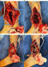
Figure 4. Intraoperative photos – initial approach and dissection
(A) A marking pen was used to draw out the intended incision that would be used to isolate the lesion (marked with the two purple arrows). The lower dotted line was used for the ankle fusion portion of the procedure. (B) The initial dissection down towards the lesion. The blue vessel loop was used to isolate and protect the superficial peroneal nerve.
Figure 5
Figure 5. Intraoperative photos – evacuation and treatment of the lesion
(A) The lesion was isolated and opened. An arrow indicates the inside of the lesion. (B) Samples of the serosanguinous core of the lesion (indicated with an arrow) were sent for intraoperative frozen pathology. (C) The lesion after being burned out and irrigated with peroxide. An arrow indicates the inside of the lesion. (D) The lesion after being treated with argon. An arrow indicates the inner surface of the lesion.
Figure 6
Figure 6. Intraoperative photos – K-wire fixation
K-wires (indicated with arrows) were used to hold provisional fixation of the ankle.
Figure 7
Figure 7. Intraoperative photos – metal cone implants and passage of nail
(A) An arrow indicates the trabecular metal cone placed inside the hollowed-out lesion in the tibia. (B) A reamer was used to ream out the path of the eventual tibiotalocalcaneal nail. (C) The 11.5x250mm straight hindfoot fusion nail was passed through the calcaneus and talus and into the tibia, through the trabecular metal cone. The arrow indicates where the nail is passing through the cone.
Figure 8
Figure 8. Intraoperative photos – bone graft and bone cement coverage and closure
(A) Autograft, allograft, and demineralized bone matrix (marked with an arrow) were used to fill in the residual defect surrounding the trabecular metal cone and nail. (B) Bone cement with tobramycin (marked with an arrow) was used to recreate the cortices of the tibia prior to closure.
Figure 9
Figure 9. Postoperative x-rays
AP (A) and lateral (B) x-ray views of the left ankle with trabecular metal cones and tibiotalocalcaneal nail in place six months postoperatively. The tibiotalar fusion site is marked with arrows in (B).

References

    1. Giant cell tumor of bone. Yip KM, Leung PC, Kumta SM. Clin Orthop Relat Res. 1996:60–64. - PubMed
    1. Giant cell tumour around the foot and ankle. Kamath S, Jane M, Reid R. Foot Ankle Surg. 2006;12:99–102.
    1. Distal tibial giant cell tumour treated with curettage and stabilisation with an Ilizarov frame. Cribb GL, Cool P, Hill SO, Mangham DC. Foot Ankle Surg. 2009;15:28–32. - PubMed
    1. An unusual case of giant cell tumor of the distal tibia. Mohapatra AR, Choudhury P, Patel PS, Malhotra RS, Patil AB. J Orthop Case Rep. 2018;8:29–31. - PMC - PubMed
    1. Giant cell tumor of lower end of tibia. Bami M, Nayak AR, Kulkarni S, Kulkarni A, Gupta R. Case Rep Orthop. 2013;2013:429615. - PMC - PubMed

Publication types

LinkOut - more resources