Case Reports
. 2024 Apr 8;16(4):e57880.
doi: 10.7759/cureus.57880. eCollection 2024 Apr.

Chameleonic Chloroma: A Case of Myeloid Sarcoma Presenting as a Pancreatic Head Mass

Affiliations
Case Reports

Chameleonic Chloroma: A Case of Myeloid Sarcoma Presenting as a Pancreatic Head Mass

Benjamin F Cardenas et al. Cureus. .

Abstract

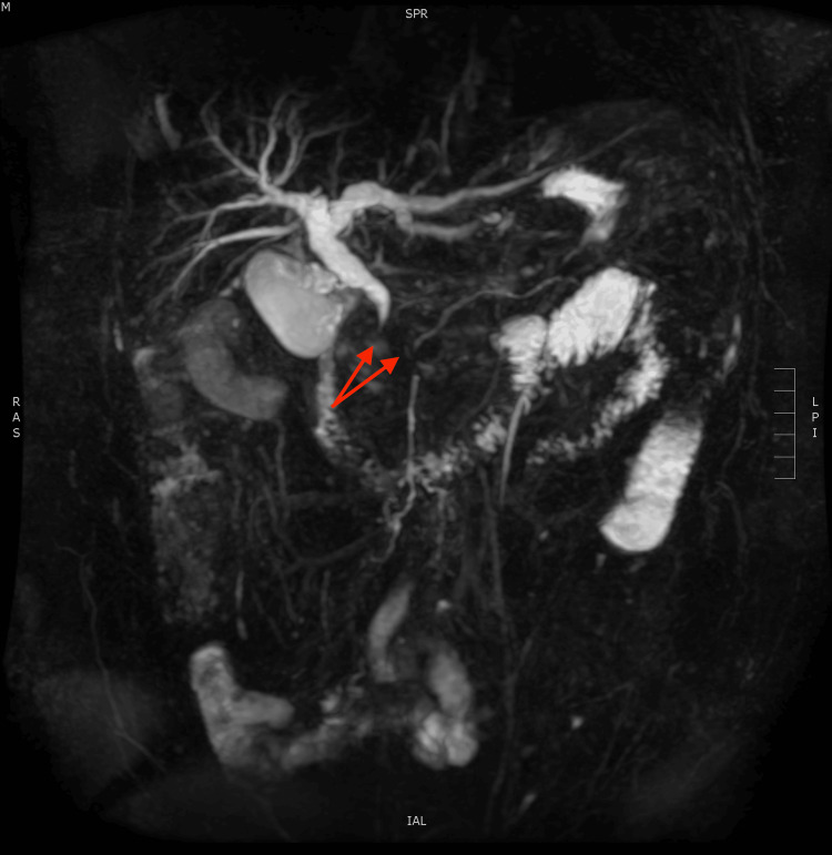
We report a case of pancreatic myeloid sarcoma (MS), an extremely rare manifestation of acute myeloid leukemia (AML), in a 35-year-old male who presented with epigastric pain and watery stools. Initial diagnostic testing was inconclusive; however, following an extensive evaluation, endoscopic biopsies suggested AML, which was confirmed by a bone marrow biopsy. Given that few cases are documented in the literature, pancreatic MS without a preexisting hematologic malignancy poses a significant diagnostic challenge.

Keywords: acute myeloid leukemia (aml); chloroma; granulocytic sarcoma; myeloid sarcoma; pancreatic cancer.


Conflict of interest statement

The authors have declared that no competing interests exist.

Figures

Figure 1
Figure 1. Magnetic resonance cholangiopancreatography (coronal view) demonstrating a 4.8 x 4.0 x 3.7 cm homogeneous hypo-enhancing mass (red arrow) in the pancreatic head region and common bile duct dilation.
Figure 2
Figure 2. 3D reconstruction of the pancreaticobiliary system demonstrating abrupt cessation of the common bile duct and pancreatic duct, or double duct sign (red arrows).
Figure 3
Figure 3. Abdominal computed tomography (axial view) with oral and IV contrast re-demonstrating homogeneous mass, 5.6 cm in the longest dimension, in the region of the pancreatic head circumferentially surrounding the common bile duct stent.
Figure 4
Figure 4. Contrast artifact versus food products versus later discovered gastric mass can be seen in the lesser gastric curvature (white arrow); notably, this was not commented on in the original radiography report. The red arrow shows the pancreatic head mass.
Figure 5
Figure 5. Endoscopic image of the retrograde view of a large, smooth mass along the lesser gastric curvature. Gross bleeding is present following an excisional biopsy of the mass.
Figure 6
Figure 6. Positron emission tomography (coronal view) demonstrating abnormally elevated F-FDG uptake in the pancreatic head (white arrow) and gastrum (red arrow).

References

    1. Myeloid sarcoma derived from the gastrointestinal tract: a case report and review of the literature. Yu T, Xu G, Xu X, Yang J, Ding L. Oncol Lett. 2016;11:4155–4159. - PMC - PubMed
    1. Clinical characteristics, treatment, and prognosis of 118 cases of myeloid sarcoma. Zhao H, Dong Z, Wan D, et al. Sci Rep. 2022;12:6752. - PMC - PubMed
    1. Pancreatic myeloid sarcoma. Al-Obaidi A, Parker NA, Hussein Agha Y, Alqam H, Page S. Cureus. 2020;12:0. - PMC - PubMed
    1. Isolated pancreatic myeloid sarcoma: a potential mimicker of pancreatic adenocarcinoma. Soni A, Jindal S, Narang V, Singh A, Paul D, Kaur H. https://journals.lww.com/ijpm/pages/articleviewer.aspx?year=2022&issue=6.... Indian J Pathol Microbiol. 2022;65:676–678. - PubMed
    1. Isolated myeloid sarcoma in the pancreas and orbit: a case report and review of literature. Zhu T, Xi XY, Dong HJ. World J Clin Cases. 2018;6:477–482. - PMC - PubMed

Publication types

LinkOut - more resources