. 2024 Apr 25:15:1380065.
doi: 10.3389/fimmu.2024.1380065. eCollection 2024.

Targeting PD-L1 in solid cancer with myeloid cells expressing a CAR-like immune receptor

Affiliations

Targeting PD-L1 in solid cancer with myeloid cells expressing a CAR-like immune receptor

Kayla Myers Chen et al. Front Immunol. .

Abstract

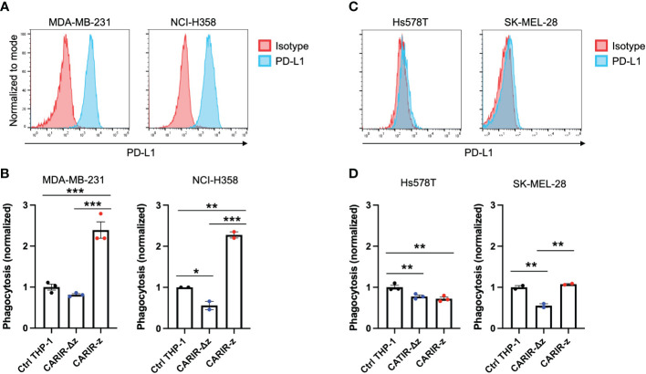
Introduction: Solid cancers Myeloid cells are prevalent in solid cancers, but they frequently exhibit an anti-inflammatory pro-tumor phenotype that contribute to the immunosuppressive tumor microenvironment (TME), which hinders the effectiveness of cancer immunotherapies. Myeloid cells' natural ability of tumor trafficking makes engineered myeloid cell therapy an intriguing approach to tackle the challenges posed by solid cancers, including tumor infiltration, tumor cell heterogenicity and the immunosuppressive TME. One such engineering approach is to target the checkpoint molecule PD-L1, which is often upregulated by solid cancers to evade immune responses.

Method: Here we devised an adoptive cell therapy strategy based on myeloid cells expressing a Chimeric Antigen Receptor (CAR)-like immune receptor (CARIR). The extracellular domain of CARIR is derived from the natural inhibitory receptor PD-1, while the intracellular domain(s) are derived from CD40 and/or CD3ζ. To assess the efficacy of CARIR-engineered myeloid cells, we conducted proof-of-principle experiments using co-culture and flow cytometry-based phagocytosis assays in vitro. Additionally, we employed a fully immune-competent syngeneic tumor mouse model to evaluate the strategy's effectiveness in vivo.

Result: Co-culturing CARIR-expressing human monocytic THP-1 cells with PD-L1 expressing target cells lead to upregulation of the costimulatory molecule CD86 along with expression of proinflammatory cytokines TNF-1α and IL-1β. Moreover, CARIR expression significantly enhanced phagocytosis of multiple PD-L1 expressing cancer cell lines in vitro. Similar outcomes were observed with CARIR-expressing human primary macrophages. In experiments conducted in syngeneic BALB/c mice bearing 4T1 mammary tumors, infusing murine myeloid cells that express a murine version of CARIR significantly slowed tumor growth and prolonged survival.

Conclusion: Taken together, these results demonstrate that adoptive transfer of PD-1 CARIR-engineered myeloid cells represents a promising strategy for treating PD-L1 positive solid cancers.

Keywords: PD-1; PD-L1; adoptive cell therapy; chimeric antigen receptor (CAR); macrophages; phagocytosis; solid cancer.


Conflict of interest statement

Authors KC, DG, BG, SV, MM, A-HZ, and PA are employees and/or shareholders of Vita Therapeutics, Inc. A-HZ, PA, KC, and DG are inventors of pending patents involving the generation and use of CARIR-modified myeloid cells for treating cancer.

Figures

Figure 1
Figure 1
Functional expression of CARIR in human monocytic THP-1 cells. (A) Schematic illustration of lentiviral vector-encoded CARIR transgenes, with (CARIR-z) or without an intracellular signaling domain derived from human CD3ζ (CARIR-Δz). PD1 (extracellular portion of programmed cell death protein 1), TM (transmembrane domain), 2A (2A cleavage peptide), tEGFR (truncated extracellular portion of the Epidermal Growth Factor Receptor containing domain III and IV). (B) CARIR expression in transduced human monocytic THP-1 cells based on flow staining of human PD-1. THP-1 cells were transduced with lentiviral vector encoding PD-1 CARIR at MOI of 3, and flow analysis was conducted 2 days following the lentiviral transduction. (C) The histogram shows the upregulation of co-stimulatory molecule CD86 in CARIR-z transduced THP-1 macrophages following co-culture with RM-1hPD-L1 cells. Non-transduced (non-modified) or CARIR transduced THP-1 cells were stimulated with 1ng/ml PMA for 24 hours, followed co-culture for 3 days as indicated. The CD86 expression was analyzed by flow cytometry, and the cells were gated on THP-1 cells based on the characteristic FSC/SSC parameters of the cells. (D) Cytokines levels measured by ELISA in the culture supernatant of the experiment as described in (C) above. (E) Representative flow cytometry dot plots depicting the phagocytosis events appeared in the Q2 quadrant, which is double positive for CellTrace CFSE labeled target cells and CellTrace Violet labeled THP-1 macrophages. Non-modified or CARIR transduced THP-1 cells were treated with 5ng/ml PMA for 24 hours to differentiate the cells toward macrophages, followed by co-culture with RM-1 or RM-1hPD-L1 at E:T ratio of 5:1 for 3 hours. Both the effectors and the target cells were fluorescent dye labeled before setting up the co-culture. The cells were gated on FSC/SSC, singlets, and live THP-1 macrophages (CellTrace Violet+). (F) Bar graph summarizes the % phagocytosis during the 3-hour co-culture, in experiment as described for (F). (G) BAR graph shows the inhibition on phagocytosis in the presence of anti-PD-1 blockade antibody or cytochalasin D in experiment as described for (F). The data were expressed as mean ± SEM. **p < 0.01 and ***p < 0.001, by non-paired student t test with 2-tailed distribution. Data shown are representatives of at least 3 independent repeats (B, C, E).
Figure 2
Figure 2
CARIR expression in THP-1 macrophages enabled significant cytotoxicity against PD-L1+ target cells. Non-modified- (Ctrl), CARIR-Δz, or CARIR-z-engineered THP-1 effector cells were co-cultured in the presence of 5ng/ml PMA for 3 days with RM-1hPD-L1 or WT-RM-1 target cells at effector to target ratio of 5:1. Following the co-culture, the cells were stained with APC anti-human CD11b, Brilliant Violet 605 anti-human PD-L1, and Zombie NIR viability dye. The number of the remaining live CD11b- target cells following the co-culture was quantified by flow cytometry with the use of absolute counting beads. (A) Representative contour plots of triplicate experiments show the percentage change for PD-L1+, PD-L1int, and PD-L1- RM-1hPD-L1 tumor cells following the co-culture. (B) BAR graphs summarize the percentage data shown on (A). (C) BAR graphs summarize the absolute cell count data shown on (A). (D) Representative contour plots of triplicate experiments show the percentage change of RM-1 tumor cells (PD-L1-) following the co-culture. (E) BAR graph summarizes the percentage data shown on (D). (F) BAR graph summarizes the absolute cell count data shown on (D). Tumor cells were gated on non-beads, live, singlets, and CD11b-. Data were presented as mean ± SEM. *p < 0.05, **p < 0.01, ***p < 0.001, and ****p < 0.0001 by one-way ANOVA.
Figure 3
Figure 3
CARIR expression in primary human macrophages enhanced phagocytosis against human PD-L1+ target cells. (A) Schematic illustrations show different CARIR constructs varying in the intracellular signaling domain(s). (B) Flow dot plots show the efficiency of CARIR transduction in human HSCs based on staining for PD-1. (C) Representative flow plots showing the % phagocytosis of CARIR expressing primary human macrophage against RM1 or RM1hPD-L1 target cells. For generating CARIR modified human macrophages to serve as the effector cells, human CD34+ hematopoietic stem cells (HSCs) were engineered to express CARIR through lentiviral transduction, and then differentiated into macrophages. Fluorescent dye labeled effector cells and target cells were co-cultured for 3 hours before flow analysis for the % phagocytosis. The cells were gated on FSC/SSC, singlets, live, and PD-1+Violet+ macrophages. The CellTrace Violet and CFSE double positive population represent macrophages that have phagocytosed target cells. Data shown are representatives of triplicate experiments (B, C). (D) The bar graph summarizes the flow data shown in (C) **p < 0.01, ***p < 0.001, and ****p < 0.0001 by unpaired student t test with two tailed distributions.
Figure 4
Figure 4
CARIR expression in human THP-1 macrophages increased phagocytosis against PD-L1+ human tumor cells. (A, C) Histogram shows the detection of cell surface PD-L1 expression by flow cytometry in cultured human tumor cell lines. Data shown are representatives of 3 independent experiments. (B, D) Phagocytosis activity against PD-L1+ tumor cell lines MDA-MB-231 and NCI-H358 (B), or PD-L1- tumor cell lines Hs578T and SK-MEL-28 (D). Human monocytic THP-1 cells were either nonmodified (Ctrl THP-1) or engineered to express either CARIR-Δz or CARIR-z through lentiviral transduction, and then differentiated toward macrophages by culturing in the presence of 1 ng/ml PMA for 24 hours. CellTrace Violet labeled THP-1 effectors were co-cultured with CellTrace Yellow labeled indicated tumor cells for 4 hours, followed by flow cytometry analysis of the % phagocytosis. Cells were gated on live, singlets, and violet+ cells. The events that were double positive for CellTrace Violet and CellTrace Yellow were considered as phagocytic events. The phagocytosis activity was normalized to the non-modified THP-1 condition. The data was presented as mean ± SEM. *p < 0.05, **p < 0.01, and ***p < 0.00 by one-way ANOVA.
Figure 5
Figure 5
Adoptive transfer of CARIR modified myeloid cells (CARIR-M) slows 4T1 tumor growth and prolongs survival. (A) Schematic timeline for the animal experiment. Syngeneic Balb/c mice (8 mice/group) were subcutaneously implanted with 5 × 10⁴ 4T1 breast cancer cells on day -7. Starting on day 0, the mice were injected through the tail vein with 3 weekly doses of 10 × 106 either CARIR-M or control non-modified myeloid cells (WT-M). An additional control group of mice were treated with PBS. Body weight (B), tumor volume (C), and probability of survival (D) were measured 2-3 times per week. Data are presented as mean ± SEM. **p < 0.01: CARIR-M vs WT-IMC or PBS by type II ANOVA. #p < 0.05 (CARIR-M vs PBS) and p < 0.01(CARIR-M vs WT-M) by Gehan- Breslow-Wilcoxon test.

References

    1. U.S. Department of Health and Human Services. Center for Disease Control and Prevention. National Cancer Institute . U.S. Cancer Statistics Data Visualizations Tool, based on 2020 submission data (1999-2018). Available online at: https://gis.cdc.gov/Cancer/USCS/#/AtAGlance/.
    1. Morotti M, Albukhari A, Alsaadi A, Artibani M, Brenton JD, Curbishley SM, et al. . Promises and challenges of adoptive T-cell therapies for solid tumours. Br J Cancer. (2021) 124:1759–76. doi: 10.1038/s41416-021-01353-6 - DOI - PMC - PubMed
    1. Klichinsky M, Ruella M, Shestova O, Lu XM, Best A, Zeeman M, et al. . Human chimeric antigen receptor macrophages for cancer immunotherapy. Nat Biotechnol. (2020) 38:947–53. doi: 10.1038/s41587-020-0462-y - DOI - PMC - PubMed
    1. Bauml J, Barton D, Ronczka A, Cushing D, Klichinsky M, Dees EC. A phase 1, first in human (FIH) study of adenovirally transduced autologous macrophages engineered to contain an anti-HER2 chimeric antigen receptor (CAR) in subjects with HER2 overexpressing solid tumors. Cytotherapy. (2021) 23. doi: 10.1016/s1465324921004205 - DOI
    1. Vitale I, Manic G, Coussens LM, Kroemer G, Galluzzi L. Macrophages and metabolism in the tumor microenvironment. Cell Metab. (2019) 30:36–50. doi: 10.1016/j.cmet.2019.06.001 - DOI - PubMed

Publication types