Case Reports
. 2024 May 7:12:2050313X241252589.
doi: 10.1177/2050313X241252589. eCollection 2024.

Successful management of coronary complications during percutaneous intervention: A case report

Affiliations
Case Reports

Successful management of coronary complications during percutaneous intervention: A case report

Ghulam Abbas Shaikh et al. SAGE Open Med Case Rep. .

Abstract

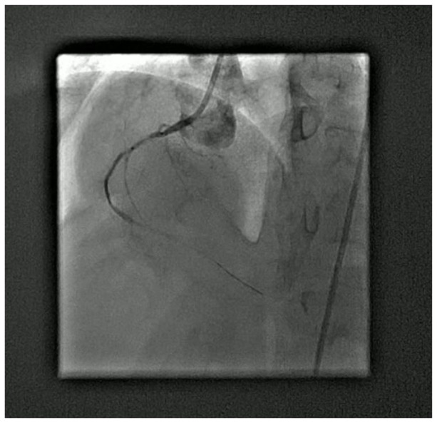
This case report delineates the complex management of a 65-year-old female with established diabetes, hypertension, and ischemic heart disease, who presented with refractory angina despite comprehensive medical management. Coronary angiography identified significant pathology in the right coronary artery alongside a previously placed, functioning stent in the left anterior descending artery. The intervention was complicated by the occurrence of a type B coronary artery dissection and a type III coronary perforation during an attempt to extract a stent. Immediate remedial measures, including balloon inflation and the placement of drug-eluting stents, were undertaken. The patient underwent a transient episode of collapse, from which she was successfully resuscitated. The concluding angiographic assessment confirmed the effective dilation of the lesion with no remaining dissection or perforation. This case accentuates the infrequent yet critical complications that can arise during percutaneous coronary intervention.

Keywords: Coronary artery complications; case report; coronary artery dissection; coronary artery perforation; percutaneous intervention.


Conflict of interest statement

The authors declared no potential conflicts of interest with respect to the research, authorship, and/or publication of this article.

Figures

Figure 1.
Figure 1.
Electrocardiogram (ECG) of the patient showing ST-T changes. The figure illustrates the ECG of the patient, revealing significant ST-T changes. These changes are indicative of myocardial ischemia and provide important diagnostic information regarding the patient’s cardiac condition.
Figure 2.
Figure 2.
Coronary angiogram of the right coronary artery (RCA). This figure showcases the coronary angiogram of the patient’s RCA. The angiogram reveals a severely diseased segment of the RCA, highlighting the need for intervention. The figure provides a visual representation of the anatomical features and extent of the disease in the RCA, aiding in the decision-making process for further treatment strategies.
Figure 3.
Figure 3.
Type B coronary artery dissection (CICAD). This figure presents a visual recording of a type B CICAD during the percutaneous intervention. The figure clearly depicts the presence of parallel tracts or multiple lumens separated by a radiolucent area following contrast injection. This type of dissection indicates a significant tear or break in the coronary artery wall, posing a serious risk to the patient’s cardiovascular health.
Figure 4.
Figure 4.
Type III coronary artery perforation (CAP). This figure captures the occurrence of a type III CAP during the percutaneous intervention. The figure provides a clear visualization of the extravasation of contrast medium into the surrounding spaces, such as the coronary sinus or the heart chamber, indicating a severe and potentially life-threatening complication. The figure highlights the urgent nature of addressing the perforation to prevent further complications, such as cardiac tamponade or myocardial infarction.
Figure 5.
Figure 5.
Successful resolution of coronary complications. This figure showcases the successful resolution of the coronary complications encountered during the percutaneous intervention. After the management of coronary artery dissection and perforation, the figure demonstrates the final angiographic results, revealing good lesion dilation and the absence of residual dissection or perforation. The figure provides a visual confirmation of the successful outcome achieved through prompt and effective interventions. This footage serves a valuable educational resource, demonstrating the importance of timely recognition and appropriate management in overcoming complex coronary complications during percutaneous interventions.

References

    1. Sanchis-Gomar F, Perez-Quilis C, Leischik R, et al.. Epi-demiology of coronary heart disease and acute coronary syndrome. Ann Transl Med 2016; 4(13): 256. - PMC - PubMed
    1. Chhabra L, Zain MA, Siddiqui WJ. Angioplasty. In: StatPearls. Treasure Island (FL): StatPearls Publishing, 2023.
    1. Giri J, Halaby R. Coronary stents: from revolution, to evolution, to pursuit of perfection. JACC Cardiovasc Interv 2021; 14(22): 2474–2476. - PubMed
    1. Stone PH, Libby P, Boden WE. Fundamental pathobiology of coronary atherosclerosis and clinical implications for chronic ischemic Heart Disease Management—The plaque hypothesis: a narrative review. JAMA Cardiol 2022; 7(12): 1234–1245. - PMC - PubMed
    1. Valtorta NK, Kanaan M, Gilbody S, et al.. Loneliness and social isolation as risk factors for coronary heart disease and stroke: systematic review and meta-analysis of longitudinal observational studies. Heart 2016; 102(13): 1009–1016. - PMC - PubMed

Publication types

LinkOut - more resources