. 2024 Apr 30;13(9):771.
doi: 10.3390/cells13090771.

Small-Molecule Inhibitors of TIPE3 Protein Identified through Deep Learning Suppress Cancer Cell Growth In Vitro

Affiliations

Small-Molecule Inhibitors of TIPE3 Protein Identified through Deep Learning Suppress Cancer Cell Growth In Vitro

Xiaodie Chen et al. Cells. .

Abstract

Tumor necrosis factor-α-induced protein 8-like 3 (TNFAIP8L3 or TIPE3) functions as a transfer protein for lipid second messengers. TIPE3 is highly upregulated in several human cancers and has been established to significantly promote tumor cell proliferation, migration, and invasion and inhibit the apoptosis of cancer cells. Thus, inhibiting the function of TIPE3 is expected to be an effective strategy against cancer. The advancement of artificial intelligence (AI)-driven drug development has recently invigorated research in anti-cancer drug development. In this work, we incorporated DFCNN, Autodock Vina docking, DeepBindBC, MD, and metadynamics to efficiently identify inhibitors of TIPE3 from a ZINC compound dataset. Six potential candidates were selected for further experimental study to validate their anti-tumor activity. Among these, three small-molecule compounds (K784-8160, E745-0011, and 7238-1516) showed significant anti-tumor activity in vitro, leading to reduced tumor cell viability, proliferation, and migration and enhanced apoptotic tumor cell death. Notably, E745-0011 and 7238-1516 exhibited selective cytotoxicity toward tumor cells with high TIPE3 expression while having little or no effect on normal human cells or tumor cells with low TIPE3 expression. A molecular docking analysis further supported their interactions with TIPE3, highlighting hydrophobic interactions and their shared interaction residues and offering insights for designing more effective inhibitors. Taken together, this work demonstrates the feasibility of incorporating deep learning and MD simulations in virtual drug screening and provides inhibitors with significant potential for anti-cancer drug development against TIPE3-.

Keywords: TIPE3; anti-tumor; deep learning; small-molecule compounds.


Conflict of interest statement

The authors declare no conflicts of interest.

Figures
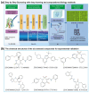
Figure 1
Figure 1
The virtual screening procedure integrates deep learning, docking, and force-field-based methods to identify highly reliable drug candidates for TIPE3. (a) A schematic diagram of the screening of TIPE3 inhibitors from the ZINC dataset. (b) The chemical structures of the six compounds selected for experimental validation.
Figure 2
Figure 2
The MD- and metadynamics simulation-related analysis. (a) RMSD values during a 40 ns simulation for the six selected inhibitors of TIPE3. (b) The calculated binding free energy landscape from the metadynamics simulation of the six selected compounds; the three labeled in red are the experimentally validated active compounds.
Figure 3
Figure 3
Cytotoxic activity of the six selected small-molecule compounds on cancer cell lines in vitro. (ad) HT-29 and LoVo cells were treated with different concentrations of K784-8160, E745-0011, 7238-1516, D393-0320, or a DMSO control for 48 h or 72 h. Cell viability was determined using a CCK-8 assay. (e,f) HT-29 and TE-1 cells were treated with different concentrations of STL125869, 711409088, or a DMSO control for 48 h or 72 h. Cell viability was determined using an MTT assay. To exclude the effect of DMSO on cell viability, we set up a DMSO control group for each indicated drug concentration (by adding an equal volume of DMSO as in the experimental group). Data were expressed as mean ± SEM values; n = 3. NS, not significant (p > 0.05); * p < 0.05, ** p < 0.01, *** p < 0.001, and **** p < 0.0001. Significance was determined using an unpaired Student’s t test. The experiments were performed at least three times with similar results.
Figure 4
Figure 4
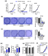
K784-8160 exhibits non-selective toxicity toward both tumor cells and normal human cells. (a) A cell growth analysis of human HT-29 and LoVo cells after either treatment with K784-8160 at a concentration of 20 μM or a DMSO control over the indicated times. (b,c) Numbers of colonies of HT-29 and LoVo cells after treatment with different concentrations of K784-8160 were detected by a colony formation assay. (d,e) The migration of HT-29 and LoVo treated with or without K784-8160 was determined using a wound healing assay. The wound closure rates of HT-29 and LoVo cells treated with or without K784-8160 for 96 h were calculated separately. The corresponding bar chart is shown on the right. (f) Jurkat cells were treated with K784-8160 at different doses for 48 h and 72 h. Cell viability was assessed by a CCK-8 assay. (g) Human T cells were treated with different doses of K784-8160 for 48 h and 72 h. Cell viability was determined using a CCK-8 assay. (h) A cell growth analysis of human T cells after either treatment with K784-8160 at a concentration of 20 μM or a DMSO control over the indicated times. Data were expressed as mean ± SEM values. NS, not significant (p > 0.05); * p < 0.05, ** p < 0.01, *** p < 0.001, and **** p < 0.0001. Significance was determined using an unpaired Student’s t test. The experiments were performed at least three times with similar results.
Figure 5
Figure 5
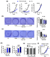
E745-0011 is effective against colon-cancer-derived cell lines in vitro while having no toxicity to normal human primary cells. (a) A cell growth analysis of HT-29, LoVo, and Jurkat cells after treatment with either 20 μΜ E745-0011 or a DMSO control over the indicated times. (b,c) Numbers of colonies of HT-29 and LoVo cells treated with different concentrations of E745-0011, as determined by a colony formation assay. (d) HT-29 and LoVo cells were treated with different concentrations of E745-0011 for 48 h. Apoptosis was measured by annexin V/propidium iodide staining and flow cytometry. (e) Human primary T cells were treated with different concentrations of E745-0011 for 72 h, and cell viability was determined using a CCK-8 assay. (f) A cell growth analysis of human T cells after treatment with either 20 μΜ E745-0011 or a DMSO control over the indicated times. Data were expressed as mean ± SEM values; n = 3. NS, not significant (p > 0.05); * p < 0.05, ** p < 0.01, *** p < 0.001, and **** p < 0.0001. In (ac,ef), significance was determined using an unpaired Student’s t test. Significance in (d) was determined by a one-way ANOVA. The experiments were performed at least three times with similar results.
Figure 6
Figure 6
7238-1516 is effective against colon-cancer-derived cell lines in vitro while having no toxicity to normal human primary cells. (a) A cell growth analysis of HT-29, LoVo, and Jurkat cells after treatment with either 20 μΜ 7238-1516 or a DMSO control over the indicated times. (b,c) Numbers of colonies of HT-29 and LoVo cells treated with different concentrations of 7238-1516, as determined by a colony formation assay. (d) HT-29 and LoVo cells were treated with 7238-1516 for 48 h and 24 h, respectively. Apoptosis was measured by flow cytometry. (e) Human T cells were treated with different concentrations of 7238-1516 for 72 h, and cell viability was determined using a CCK-8 assay. (f) A cell growth analysis of human T cells after treatment with either 20 μΜ 7238-1516 or a DMSO control over the indicated times. Data were expressed as mean ± SEM values; n = 3. NS, not significant (p > 0.05); * p < 0.05, ** p < 0.01, *** p < 0.001. In (ac,ef), significance was determined using an unpaired Student’s t test. Significance in (d) was determined by a one-way ANOVA. The experiments were performed at least three times with similar results.
Figure 7
Figure 7
Interaction analysis of TIPE3 with compounds identified from this study with docked conformations. (a) Compound ZINC000009238243 (7238-1516) binding to the TIPE3, displaying both 3D detailed interactions (middle) and 2D representation (right). (b) Interaction between ZINC000020558784 (E745-0011) and TIPE3. (c) Interaction between ZINC000006756082 (K784-8160) and TIPE3. Residues in all 3D visualizations were color-coded by rainbow in PyMOL. The 2D diagrams employ colors and symbols as standardized by the Schrödinger 2D interaction plots.

References

    1. Padmavathi G., Banik K., Monisha J., Bordoloi D., Shabnam B., Arfuso F., Sethi G., Fan L., Kunnumakkara A.B. Novel tumor necrosis factor-α induced protein eight (TNFAIP8/TIPE) family: Functions and downstream targets involved in cancer progression. Cancer Lett. 2018;432:260–271. doi: 10.1016/j.canlet.2018.06.017. - DOI - PubMed
    1. Goldsmith J.R., Fayngerts S., Chen Y.H. Regulation of inflammation and tumorigenesis by the TIPE family of phospholipid transfer proteins. Cell Mol. Immunol. 2017;14:482–487. doi: 10.1038/cmi.2017.4. - DOI - PMC - PubMed
    1. Lou Y., Liu S. The TIPE (TNFAIP8) family in inflammation, immunity, and cancer. Mol. Immunol. 2011;49:4–7. doi: 10.1016/j.molimm.2011.08.006. - DOI - PubMed
    1. Cui J., Hao C., Zhang W., Shao J., Zhang N., Zhang G., Liu S. Identical expression profiling of human and murine TIPE3 protein reveals links to its functions. J. Histochem. Cytochem. 2015;63:206–216. doi: 10.1369/0022155414564871. - DOI - PMC - PubMed
    1. Fayngerts S.A., Wu J., Oxley C.L., Liu X., Vourekas A., Cathopoulis T., Wang Z., Cui J., Liu S., Sun H., et al. TIPE3 is the transfer protein of lipid second messengers that promote cancer. Cancer Cell. 2014;26:465–478. doi: 10.1016/j.ccr.2014.07.025. - DOI - PMC - PubMed

Publication types

MeSH terms

Substances

LinkOut - more resources