Review
. 2024 May 10;166(1):209.
doi: 10.1007/s00701-024-06074-2.

Retrogasserian trigeminal radiofrequency-thermorhizotmoy for trigeminal neuralgia

Affiliations
Review

Retrogasserian trigeminal radiofrequency-thermorhizotmoy for trigeminal neuralgia

A Brinzeu et al. Acta Neurochir (Wien). .

Abstract

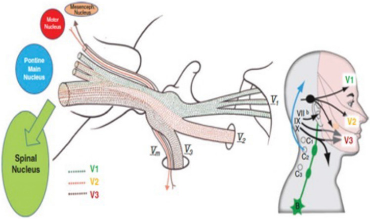
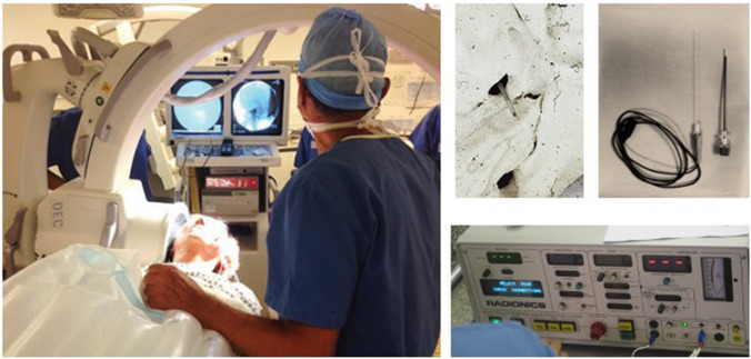
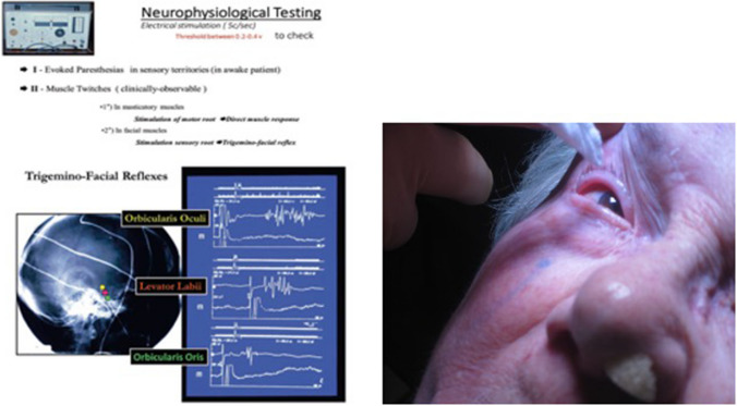
Based on a personal experience of 4200 surgeries, radiofrequency thermocoagulation is useful lesional treatment for those trigeminal neuralgias (TNs) not amenable to microvascular decompression (idiopathic or secondary TNs). Introduced through the foramen ovale, behind the trigemnial ganglion in the triangular plexus, the needle is navigated by radiology and neurophysiological testing to target the retrogasserian fibers corresponding to the trigger zone. Heating to 55-75 °C can achieve hypoesthesia without anaesthesia dolorosa if properly controlled. Depth of anaesthesia varies dynamically sedation for cannulation and lesioning, and awareness during neurophysiologic navigation. Proper technique ensures long-lasting results in more than 75% of patients.

Keywords: Hartel’s approach; Percutaneous; Rf-Thermocoagulation; Trigeminal neuralgia.


Conflict of interest statement

The authors declare that they have no conflicts of interest relevant to this work.

The authors declare they have no competing interests pertaining to this work.

Figures

Fig. 1
Fig. 1
Relevant anatomy of the trigeminal system from the periphery to the brain stem. The three sensory branches of the trigeminal nerve (V1 ophthalmic, V2 maxillary, V3 mandibular) are formed by sensory fibers from the face and cranial mucosae that converge to the sensory trigeminal ganglion. Behind it lies the triangular plexus through which the fibers pass but keep a somatotopic organization. Fibers from the V1 branch remain superomedial while those from the V3 branch are inferolateral. This overall organization is kept through the trigeminal root running within the cerebello-pontine angle cistern. The ganglion and the triangular plexus are in the trigeminal Meckel’s Cave and the plexus is surrounded by CSF (trigeminal cistern). At the trigeminal root entry zone the fibers change their organization from somatotopic to functional to reach their respective nuclei according to their modality: pain fibers towards the spinal trigeminal nucleus, tactile ones towards the main nucleus in the pons, proprioceptive fibers towards the mesencephalic nucleus. In the midpons is situated the motor trigeminal nucleus whose fibers exit the pons via a distinct root(let) exiting above the main root. This will then descend adjacent to V1 to the triangular plexus where it passes below the plexus diagonally from posterior to anterior and medial to lateral. It then joins the V3 peripheral branch exiting adjacent to it through the FO in which it is situated posterior and lateral
Fig. 2
Fig. 2
OR Setup for Rf-TR. The patient is supine with the head on a horseshoe head rest to clear the shoulders to place the plate of the C-Arm close to the face on the contralateral side to the pain. Also on this side is placed a unilateral nasal canula delivering oxygen. The C-Arm overarches the patient, and both the screen of the radiology machine and the anesthetist are placed headside. The surgeon directly faces the patient on the side of the pain with the instrument table and the RF machine lateral to him. The RF machine allows for complete control of the current for stimulation. Frequencies of 5 Hz are used to be able to see masticatory and trigemino-facial reflexes. Sensory threshold should be below 400 mV and is carefully controlled. The machine is then switched to radio frequencies to create the thermal lesion. The probe has a thermiresistance, the temperature should be kept below 75 degrees
Fig. 3
Fig. 3
Approach to the Foramen ovale (FO) through Hartel’s trajectory. Landmarks are clearly marked on the face. The entry point is situated 60 mm lateral to the middle of the interlabial line. A point 35 mm anterior to the anterior wall of the external acoustic canal in the orbito-meatal plane, on the inferior zygomatic edge. The pupil in neutral position or a point situated immediately below it on the inferior orbital rim. These three points determine the base of a triangular pyramid with the apex situated at the level of the FO. The needle is inserted through the cheek with the index finger inside the mouth to control the integrity of the buccal mucosa and guide the direction of the needle towards FO through the pterygo-maxillary fossa, and avoiding the lateral pterygoid process
Fig. 4
Fig. 4
Fluoroscopic control of the position of the needle. Imaging control should be performed at each step of the procedure. A strict lateral view is essential (this is checked by aligning the acoustic meati). A The FO is marked by the demilune. An important radiological landmarking the face is the posterior angle of the maxillary sinus (white star). The needle should draw a line between these two points. The tip of the needle should be placed in the triangular plexus. The landmark is the intersection of the clivus and the upper petrous ridge. B False trajectories. An excessive postero-lateral direction could puncture the internal jugular vein (IJV) at jugular foramen or the internal carotid artery at entrance into the petrous carotid canal. An excessive medial direction could enter the foramen lacerum and injure the internal carotid artery (ICA) at its C5 segment. An excessive anterior direction could penetrate the orbital apex through the inferior orbital fissure and injure the optic nerve (ON). The appropriate trajectory is yellow. C Projection of the peripheral (V1, V2 and V3) branches, ganglion (GG), and trigeminal plexus (TP) on a lateral skull X-Ray. The triangular plexus is situated at the intersection of the projection of the clivus and upper petrous ridge. D Respective projections for the probe for V1-V2-V3 lesions
Fig. 5
Fig. 5
Neurophysiological guidance. Stimulation is used to: 1 Evoke paresthesias in the sensory territory touched by the needle tip, for this a 5 Hz stimulation is used and communication with the patient allows for good sensory targeting. The threshold for sensory stimulation should be between 0.1-0.4 v (100-400 mV). Above 400 mV the needle is too far from the target fibers and should be replaced; 2 Evoke muscle twitches in the masticatory muscles in order to avoid the Vm branch. These masticatory responses are specific for Vm and if evoked at below 400 mV the needle should be replaced; 3 Evoke trigeminofacial reflexes. They have a localizing value concerning the position of the needle within the triangular plexus. Thus a location in V1 will give responses in the orbicularis oculi, V2 in levator labii, V3 in orbicularis oris; 4 The corneal reflex must be tested through the entire procedure to ensure that corneal hypoesthesia is avoided
Fig. 6
Fig. 6
Decision algorithm for surgical treatment of TN. Classical means by definition with a significant probability that the TN is due to an NVC [3]

References

    1. Alvernia JE, Sindou MP, Dang ND, Maley JH, Mertens P (2010) Percutaneous approach to the foramen ovale: an anatomical study of the extracranial trajectory with the incorrect trajectories to be avoided. Acta Neurochir (Wien) 152(6):1043–1053. 10.1007/s00701-010-0604-y - PubMed
    1. Bernard F, Mercier P, Sindou M (2019) Morphological and functional anatomy of the trigeminal triangular plexus as an anatomical entity: a systematic review. Surg Radiol Anat 41(6):625–637. 10.1007/s00276-019-02217-8 - PubMed
    1. Cruccu G, Finnerup NB, Jensen TS, Scholz J, Sindou M, Svensson P, Treede RD, Zakrzewska JM, Nurmikko T (2016) Trigeminal neuralgia: New classification and diagnostic grading for practice and research. Neurology. 87(2):220–8. 10.1212/WNL.0000000000002840 - PMC - PubMed
    1. Hartel F (1912) Die Leitungsanasthese und Injecktionbehandlung des Ganglioni gasseri und der Trigeminusstamme Arch Klin Chir 100:627–638
    1. Kirschner M (1933) Die Punktionstechnik und die Elektrokoagulation des Ganglion Gasseri. Arch Klin Chir. 176:581–620

MeSH terms