Review
. 2024 Oct 31;144(18):1855-1872.
doi: 10.1182/blood.2023021786.

A practical approach to the modern diagnosis and classification of T- and NK-cell lymphomas

Affiliations
Review

A practical approach to the modern diagnosis and classification of T- and NK-cell lymphomas

Laurence de Leval et al. Blood. .

Abstract

T- and natural killer (NK)-cell lymphomas are neoplasms derived from immature T cells (lymphoblastic lymphomas), or more commonly, from mature T and NK cells (peripheral T-cell lymphomas, PTCLs). PTCLs are rare but show marked biological and clinical diversity. They are usually aggressive and may present in lymph nodes, blood, bone marrow, or other organs. More than 30 T/NK-cell-derived neoplastic entities are recognized in the International Consensus Classification and the classification of the World Health Organization (fifth edition), both published in 2022, which integrate the most recent knowledge in hematology, immunology, pathology, and genetics. In both proposals, disease definition aims to integrate clinical features, etiology, implied cell of origin, morphology, phenotype, and genetic features into biologically and clinically relevant clinicopathologic entities. Cell derivation from innate immune cells or specific functional subsets of CD4+ T cells such as follicular helper T cells is a major determinant delineating entities. Accurate diagnosis of T/NK-cell lymphoma is essential for clinical management and mostly relies on tissue biopsies. Because the histological presentation may be heterogeneous and overlaps with that of many benign lymphoid proliferations and B-cell lymphomas, the diagnosis is often challenging. Disease location, morphology, and immunophenotyping remain the main features guiding the diagnosis, often complemented by genetic analysis including clonality and high-throughput sequencing mutational studies. This review provides a comprehensive overview of the classification and diagnosis of T-cell lymphoma in the context of current concepts and scientific knowledge.


Conflict of interest statement

Conflict-of-interest disclosure: L.d.L. had a consulting or advisory role for Abbvie, Lunaphore Technologies, Bayer, Blueprint Medicines, Novartis, and Roche (all institutional), and received travel grants from Roche. P.G. received research funding from Alderan, Innate Pharma, Takeda, and Sanofi; had a consulting or advisory role for Takeda and Gilead; and received travel grants from Roche. A.D. received research support from Roche and Astra-Zeneca.

Figures

None
Graphical abstract
Figure 1.
Figure 1.
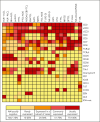
Immunophenotypic characteristics in common T and NK-cell neoplastic entities. Each column in the heat map represents a T/NK-cell neoplastic entity, and each row represents a diagnostic marker assessed by immunophenotyping of tissue or cell samples. The prevalence of cases positive for each marker is color-coded, as shown in the legend. ALCL, anaplastic large cell lymphoma; ALK, anaplastic lymphoma kinase; ATLL, adult T-cell leukemia/lymphoma; BIA-ALCL, breast implant–associated ALCL; EATL, enteropathy-associated T-cell lymphoma; HSTL, hepatosplenic T-cell lymphoma; ITCL-NOS, intestinal T-cell lymphoma, NOS; MEITL, monomorphic epitheliotropic intestinal T-cell lymphoma; MF, mycosis fungoides; NA, not available; NK-LGLL/CLPD-NK, NK-large granular lymphocytic leukemia/chronic LPD of NK cells; NK-LPD GI, indolent NK-cell LPD of the gastrointestinal tract; nodal EBV TCL, primary nodal EBV+ T/NK-cell lymphoma; pcALCL, primary cutaneous ALCL; PTCL-NOS, peripheral T-cell lymphoma, NOS; SPTCL, subcutaneous panniculitis-like T-cell lymphoma; SS, Sezary syndrome; TFHL, follicular helper T-cell lymphoma; T-LBL, T-lymphoblastic leukemia/lymphoma; T-LPD GI, indolent clonal T-cell LPD of the gastrointestinal tract; T-LGLL, T-cell large granular lymphocytic leukemia; T-PLL, T-cell prolymphocytic leukemia.
Figure 2.
Figure 2.
Mutational landscape of common T- and NK-cell neoplastic entities. Each column in the heat map represents a T/NK-cell neoplastic entity, and each row represents selected single nucleotide variants/indels and rearrangements (R), grouped according to functional annotations. The prevalence of mutations is color-coded, as shown in the legend. Genes with a mutational frequency of at least 10% observed in 1 or more mature T/NK-cell neoplasm were selected for display. Mutation frequencies 5%-10% were reported in some entities given their diagnostic or clinical value. ∗Refers to germ line mutation. BIA-ALCL, breast implant–associated ALCL; MF, mycosis fungoides; NK-LGLL, NK-large granular lymphocytic leukemia/chronic LPD of NK cells; pcALCL, primary cutaneous ALCL; SPTCL, subcutaneous panniculitis-like T-cell lymphoma; SS, Sezary syndrome; T-LBL, T-lymphoblastic leukemia/lymphoma; T-LGLL, T-cell large granular lymphocytic leukemia.
Figure 3.
Figure 3.
Diagnostic approach to CD30+ T-cell lymphoma. Cohesive growth of large cells including hallmark cells, with strong and homogeneous expression of CD30 at the membrane and in the cytoplasm with a paranuclear “Golgi-like” pattern, suggests anaplastic large cell lymphoma (ALCL). Demonstration of T-cell surface markers or expression of cytotoxic molecules is required to show T-cell lineage and distinguish from potential mimics, such as metastatic melanoma or carcinoma, or a variety of other hematologic neoplasms that may be CD30+, notably classic Hodgkin lymphoma. Clonality studies may be necessary or useful in some instances to show monoclonal TR gene rearrangements because immunohistochemistry results may not accurately represent cell lineages, and some cases of ALCL may be completely negative for T-cell markers (“null” phenotype). EBV testing is recommended in cases of presumed ALCL or CD30+ PTCLs, and HTLV-1 serology may be helpful in selected instances because EBV-associated NK- or T-cell lymphomas (extranodal NK/T-cell lymphoma, nasal type [ENKTCL]; primary nodal EBV+T/NK-cell lymphoma or aggressive NK-cell leukemia) and ATLL can present as tumors with anaplastic morphology and CD30 expression,, , and EBV is by definition negative in ALCL. Immunohistochemistry is the routinely used method to detect ectopic ALK expression reflecting ALK rearrangement. In selected cases, FISH analysis or other genetic assays may be useful to confirm an ALK rearrangement or specific ALK fusion transcripts. In the small cell and the lymphohistiocytic variants of ALK+ ALCL, the neoplastic cells tend to be smaller with less numerous hallmark cells and show more heterogeneous CD30 staining, therefore ALK testing should be generously applied to other PTCLs with various levels of CD30 expression, especially in pediatric cases where ALK+ ALCL is most prevalent. Differentiating ALK ALCL from CD30+ peripheral T-cell lymphoma, NOS (PTCL, NOS), may be difficult or subjective, and there is a number of cases whose classification remains uncertain., , In addition, other lymphomas such as enteropathy-associated T-cell lymphoma (EATL), or transformed mycosis fungoides (MF), may resemble ALCL and involve lymph nodes. Therefore, clinical history, topography of the lesion, and staging need to be integrated into the diagnosis. Only cases with strong CD30+ expression can eventually be considered for ALK ALCL, which may present in various sites. With extremely rare exceptions, those in the vicinity of a breast implant presenting as a periprosthetic effusion or a capsular mass in principle correspond to breast implant-associated (BIA) ALCL. Cutaneous presentation can reflect primary cutaneous (pc) ALCL or cutaneous presentation of a systemic disease, and likewise nodal ALK ALCL may represent systemic disease or nodal dissemination from a primary cutaneous or breast implant–associated ALCL. Staging is essential to the correct diagnosis because there is no single phenotypic or genetic mark that reliably allows their distinction. In (systemic) ALK ALCL, FISH testing for DUSP22 rearrangement (recommended by the ICC and optional in WHO5) enables the identification of DUSP22-rearranged cases, which represent a biologically distinct subgroup. FISH, fluorescent in situ hybridization.
Figure 4.
Figure 4.
Histological presentation of primary nodal T- and NK-cell lymphomas. Anaplastic large cell lymphomas (ALCLs), ALK+ or ALK, tend to invade the peripheral sinuses and produce cohesive sheet-like infiltrates in the lymph node. Peripheral T-cell lymphoma, NOS (PTCL, NOS) is not associated with a characteristic pattern of growth, and can to a variable extent replace the normal lymphoid tissue. Subtypes of PTCL, NOS are composed of mostly CD4+ or CD8+ lymphoid cells with a phenotype resembling that of Th1 or Th2 cells, or of cells expressing cytotoxic molecules. The tumor microenvironment tends to be more abundant in Th1-type PTCL, NOS, and can sometimes comprise abundant epithelioid histiocytes. Primary nodal EBV+ lymphomas derived of cytotoxic T cells, or less commonly NK cells, represent a rare aggressive entity, which is mostly reported in Asians. Lymphomas derived from CD4+ TFH cells (TFHL) represent the most prevalent nodal PTCL with a variety of histological patterns. The most common form of TFHL is the angioimmunoblastic type (TFHL-AI), which in its usual form is a diffuse microenvironment-rich tumor, comprising a proliferation of arborizing vessels and follicular dendritic cells, and an infiltrate of many reactive large (blastic) and small B cells, plasma cells, histiocytes, and non-neoplastic T cells. Less commonly, the neoplastic cells of TFHL-AI concentrate around reactive or regressive germinal centers (patterns 1 and 2), and these may be more difficult to diagnose because of the association with reactive follicles. In the uncommon follicular type of TFHL (TFHL-F), the neoplastic TFH cells grow in follicles, resembling folliicular lymphoma (FL-like), or in clusters within large B-cell nodules resembling progressively transformed germinal centers, which is an uncommon form of reactive follicular hyperplasia (PTGC-like). The NOS subtype of TFHL, defined by the TFH phenotype of the neoplastic cells, does not contain the complete microenvironment of TFHL-AI, grows diffusely, and may preferentially distribute in the paracortex and between preserved follicles. TFHL, in particular the AI and follicular subtypes, may contain large atypical cells resembling Reed-Sternberg cells, a source of frequent diagnostic difficulties in distinguishing them from classic or nodular lymphocyte predominant Hodgkin lymphoma.
Figure 5.
Figure 5.
Differential diagnosis of T-cell lymphomas and T/NK-cell LPDs in the gastrointestinal tract (modified from de Leval et al82). CD, celiac disease; CM, cytotoxic molecules; EBV, Epstein-Barr virus; ENKTCL, extranodal NK/T-cell lymphoma, nasal type; IBD, inflammatory bowel disease; ITCL, NOS, intestinal T-cell lymphoma, not otherwise specified; NK-LPD-GI, indolent NK-cell LPD of the gastrointestinal tract; RCD, refractory CD; T-LPD-GI, indolent clonal T-cell LPD of the gastrointestinal tract (ICC), indolent T-cell lymphoma of the gastrointestinal tract (WHO5).
Figure 6.
Figure 6.
Differential diagnosis of leukemic T- and NK-cell neoplasms. ∗NK-LGLL differs from T-LGLL by the absence of expression of the TCR and lack of monoclonal TR rearrangement; the clonal neoplastic nature of this disorder is supported by a restricted activated KIRs expression and the presence of acquired mutations, especially in TET2, STAT3, and CCL22 (mostly exclusive). ANKL, aggressive NK-cell leukemia; BM, bone marrow biopsy; HPS, hemophagocytic syndrome; LN, lymphadenopathy; NK-LGLL, chronic LPD of NK cells/NK large granular lymphocytic leukemia; SS, Sezary syndrome; T-LGLL, T-cell large granular lymphocytic leukemia.

References

    1. Swerdlow SH, Campo E, Harris NL, et al. Vol. 2. International Agency for Research on Cancer; 2008. WHO Classification of Tumours of Haematopoietic and Lymphoid Tissues.
    1. Vose J, Armitage J, Weisenburger D, International T-Cell Lymphoma Project International peripheral T-cell and natural killer/T-cell lymphoma study: pathology findings and clinical outcomes. J Clin Oncol. 2008;26(25):4124–4130. - PubMed
    1. Laurent C, Baron M, Amara N, et al. Impact of expert pathologic review of lymphoma diagnosis: study of patients from the French Lymphopath Network. J Clin Oncol. 2017;35(18):2008–2017. - PubMed
    1. Campo E, Jaffe ES, Cook JR, et al. The International Consensus Classification of mature lymphoid neoplasms: a report from the Clinical Advisory Committee. Blood. 2022;140(11):1229–1253. - PMC - PubMed
    1. Alaggio R, Amador C, Anagnostopoulos I, et al. The 5th edition of the World Health Organization classification of haematolymphoid tumours: lymphoid neoplasms. Leukemia. 2022;36(7):1720–1748. - PMC - PubMed

MeSH terms