

Ultrastructural and immunohistochemical evaluation of hyperplastic soft tissues surrounding dental implants in fibular jaws
- PMID: 38730018
- PMCID: PMC11087521
- DOI: 10.1038/s41598-024-60474-z
Ultrastructural and immunohistochemical evaluation of hyperplastic soft tissues surrounding dental implants in fibular jaws
Abstract
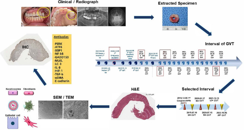
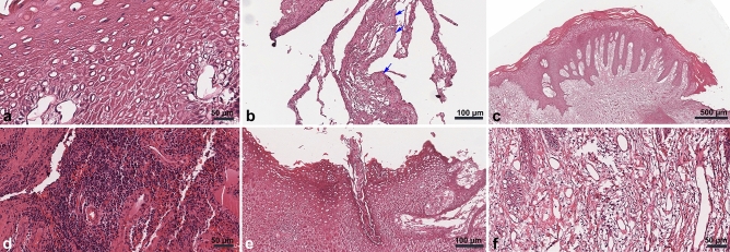
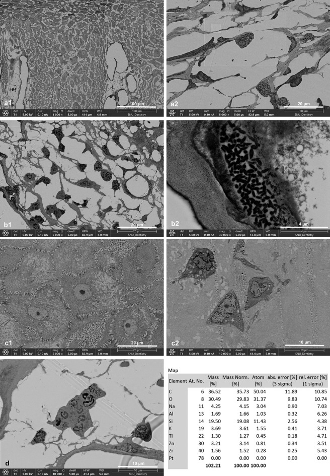
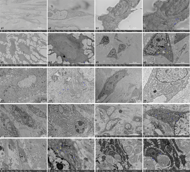
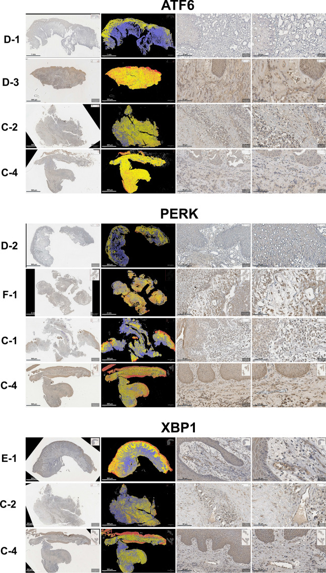
In reconstructive surgery, complications post-fibula free flap (FFF) reconstruction, notably peri-implant hyperplasia, are significant yet understudied. This study analyzed peri-implant hyperplastic tissue surrounding FFF, alongside peri-implantitis and foreign body granulation (FBG) tissues from patients treated at the Department of Oral and Maxillofacial Surgery, Seoul National University Dental Hospital. Using light microscopy, pseudoepitheliomatous hyperplasia, anucleate and pyknotic prickle cells, and excessive collagen deposition were observed in FFF hyperplastic tissue. Ultrastructural analyses revealed abnormal structures, including hemidesmosome dilation, bacterial invasion, and endoplasmic reticulum (ER) swelling. In immunohistochemical analysis, unfolded protein-response markers ATF6, PERK, XBP1, inflammatory marker NFκB, necroptosis marker MLKL, apoptosis marker GADD153, autophagy marker LC3, epithelial-mesenchymal transition, and angiogenesis markers were expressed variably in hyperplastic tissue surrounding FFF implants, peri-implantitis, and FBG tissues. NFκB expression was higher in peri-implantitis and FBG tissues compared to hyperplastic tissue surrounding FFF implants. PERK expression exceeded XBP1 significantly in FFF hyperplastic tissue, while expression levels of PERK, XBP1, and ATF6 were not significantly different in peri-implantitis and FBG tissues. These findings provide valuable insights into the interconnected roles of ER stress, necroptosis, apoptosis, and angiogenesis in the pathogenesis of oral pathologies, offering a foundation for innovative strategies in dental implant rehabilitation management and prevention.
Keywords: Endoplasmic reticulum stress; Fibula free flap; Foreign body granulation; Hyperplastic tissue; Implant; Unfolded protein response.
© 2024. The Author(s).
Conflict of interest statement
The authors declare no competing interests.
Figures
Similar articles
-
Long-term results of osseointegrated implant-based dental rehabilitation in oncology patients reconstructed with a fibula free flap.Clin Implant Dent Relat Res. 2018 Oct;20(5):852-859. doi: 10.1111/cid.12658. Epub 2018 Aug 24. Clin Implant Dent Relat Res. 2018. PMID: 30144257
-
Peri-implant Parameters of Dental Implants Inserted in Prefabricated Microvascular Fibular Flaps: A Retrospective Study.Int J Oral Maxillofac Implants. 2023 Dec 12;38(6):1151-1160. doi: 10.11607/jomi.9952. Int J Oral Maxillofac Implants. 2023. PMID: 38085746
-
Immunohistochemical analysis with apoptosis and autophagy markers in periodontitis and peri-implantitis: Clinical comparative study.J Periodontal Res. 2023 Apr;58(2):456-464. doi: 10.1111/jre.13106. Epub 2023 Feb 8. J Periodontal Res. 2023. PMID: 36755315
-
The role of foreign body response in peri-implantitis: What is the evidence?Periodontol 2000. 2022 Oct;90(1):176-185. doi: 10.1111/prd.12456. Epub 2022 Aug 2. Periodontol 2000. 2022. PMID: 35916872 Free PMC article. Review.
-
Titanium particles in peri-implantitis: distribution, pathogenesis and prospects.Int J Oral Sci. 2023 Nov 23;15(1):49. doi: 10.1038/s41368-023-00256-x. Int J Oral Sci. 2023. PMID: 37996420 Free PMC article. Review.
Cited by
-
Evaluation of risk factors, clinic pathological aspects and implant characteristics in patients of oral peri-implant malignancies.Bioinformation. 2024 Sep 30;20(9):1164-1168. doi: 10.6026/9732063002001164. eCollection 2024. Bioinformation. 2024. PMID: 39917217 Free PMC article.
References
-
- Kearns M, Ermogenous P, Myers S, Ghanem AM. Osteocutaneous flaps for head and neck reconstruction: A focused evaluation of donor site morbidity and patient reported outcome measures in different reconstruction options. Arch. Plast. Surg. 2018;45:495–503. doi: 10.5999/aps.2017.01592. - DOI - PMC - PubMed
Publication types
MeSH terms
Substances
LinkOut - more resources
Full Text Sources
Research Materials
Miscellaneous
