. 2024 May 10;14(1):10696.
doi: 10.1038/s41598-024-61662-7.

Antiviral, anti-inflammatory and antioxidant effects of curcumin and curcuminoids in SH-SY5Y cells infected by SARS-CoV-2

Affiliations

Antiviral, anti-inflammatory and antioxidant effects of curcumin and curcuminoids in SH-SY5Y cells infected by SARS-CoV-2

Tiago Nicoliche et al. Sci Rep. .

Abstract

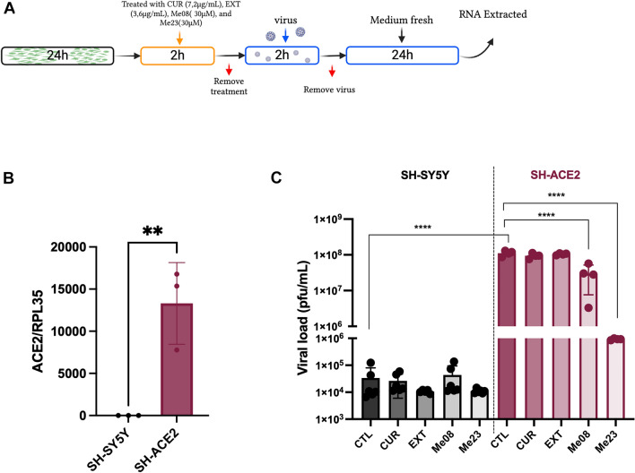
COVID-19, caused by SARS-CoV-2, affects neuronal cells, causing several symptoms such as memory loss, anosmia and brain inflammation. Curcuminoids (Me08 e Me23) and curcumin (CUR) are derived from Curcuma Longa extract (EXT). Many therapeutic actions have been linked to these compounds, including antiviral action. Given the severe implications of COVID-19, especially within the central nervous system, our study aims to shed light on the therapeutic potential of curcuminoids against SARS-CoV-2 infection, particularly in neuronal cells. Here, we investigated the effects of CUR, EXT, Me08 and Me23 in human neuroblastoma SH-SY5Y. We observed that Me23 significantly decreased the expression of plasma membrane-associated transmembrane protease serine 2 (TMPRSS2) and TMPRSS11D, consequently mitigating the elevated ROS levels induced by SARS-CoV-2. Furthermore, Me23 exhibited antioxidative properties by increasing NRF2 gene expression and restoring NQO1 activity following SARS-CoV-2 infection. Both Me08 and Me23 effectively reduced SARS-CoV-2 replication in SH-SY5Y cells overexpressing ACE2 (SH-ACE2). Additionally, all of these compounds demonstrated the ability to decrease proinflammatory cytokines such as IL-6, TNF-α, and IL-17, while Me08 specifically reduced INF-γ levels. Our findings suggest that curcuminoid Me23 could serve as a potential agent for mitigating the impact of COVID-19, particularly within the context of central nervous system involvement.

Keywords: COVID19; Curcumin; Neuroinflammation; Neuronal cell; Oxidative stress.


Conflict of interest statement

The authors declare no competing interests.

Figures

Figure 1
Figure 1
Cytotoxic of CUR, EXT, Me08 and Me23 on SH-SY5Y. Cells were seeded on day 0 and treated after 24 h (Day 1), the MTT was performed 24 h after treatment (Day 2). (A) CUR (90–0.0288 μg/mL), (B) CUR (28–3.6 μg/mL), (C) EXT (90–0.144 μg/mL), (D) Me08 (90–7.5 μM), (E) Me23 (90–7.5 μM). *p < 0.05, ****p < 0.0001. (n = 5–6). Red arrow represents the concentration used in the experiments.
Figure 2
Figure 2
SARS-CoV-2 receptors expression in SH-SY5Y. Cells were treated with CUR, EXT, Me08 or Me23 for 24 h and the gene expression was analyzed by RT-qPCR and protein expression was analyzed by Western-blotting. Gene expression of (A) ACE2 (B) Furin, (C) TMPRSS2, (D) TMPRSS11D. Gene expression was normalized by the endogenous expression of RPL35. (E) Protein expression according to western blot analyses. (F) Histograms reporting the mean ± SD of (G) ACE2, (H) TMPRSS2, (I) TMPRSS11D levels after normalization with the average intensity of the bands from four independent experiments. Protein expression was normalized by the endogenous expression of GAPDH. CTL: cells without treatment, CUR: curcumin, EXT: extract. *p < 0.05. (n = 4–13). The entire blots are presented in Supplementary Figure S1.
Figure 3
Figure 3
ROS quantification after treatment with CUR, EXT, Me08, Me23. Cells were treated with CUR, EXT, Me08 or Me23 for 2 h, after, they were infected with SARS-CoV-2 (MOI 0.2) for 24 h and the ROS was measured by flow cytometry. The percentage of fluorescence was compared to the MOCK. *p < 0.05. (n = 4–6). The schematic diagram illustrating the infection process was based on a previous publication.
Figure 4
Figure 4
NRF2 expression and NQO1 activity after treatment with Me23. Cells were treated with Me23 for 2 h, after, they were infected with SARS-CoV-2 (MOI 0.2) for 24 h and the NRF2 gene expression and NQO1 activity was evaluated. (A) NRF2 gene expression was evaluated by qRT-PCR. (B) NQO1 activity. **p < 0.005. (n = 4–6). MOCK: SH-SY5Y cells not infected with SARS-CoV-2.
Figure 5
Figure 5
ACE2 overexpression increases viral load in SH-SY5Y and potentialize the curcuminoids effect. (A) SH-SY5Y were transduced with a lentivector carrying the ACE2 gene and the cell line SH-ACE2 was created. The overexpression of ACE2 was evaluated by RT-qPCR. (B) SH-SY5Y and SH-ACE2 were treated with CUR, EXT, Me08 or Me23 for 2 h and after they were infected with SARS-CoV-2 (MOI 0.2) for 24 h. (C) The viral load was quantified by RTqPCR. CTL: Cells infected with SARS-CoV-2 and without treatment, CUR: curcumin, EXT: extract. **p < 0.01, ****p < 0.0001. (n = 3–6). The schematic diagram illustrating the infection process was based on a previous publication.
Figure 6
Figure 6
Cytokine’s expression after treatment with CUR, EXT, Me08 and Me23. (A) Cells were treated with CUR, EXT, Me08 or Me23 for 2 h, they were removed, and the cells were infected with SARS-CoV-2 (MOI 0.2) for 2 h, after viral removal, the supernatant was collected after 24 h and the cytokines’ expression was quantified by ELISA. (B) INF-γ, (C) TNF-α, (D) IL-6, (E) IL-17. CTL: Cells infected with SARS-CoV-2 and without treatment, CUR: curcumin, EXT: extract. *p < 0.05. (n = 4). The schematic diagram illustrating the infection process was based on a previous publication.
Figure 7
Figure 7
Schematic representation of the effects of Me23 in SH-SY5Y cells. Me23 (60 μM) reduced TMPRSS2, TMPRSS11D expression, ROS levels, viral load and decreased IL-6, IL-17 and TNF-α.

References

    1. Ding Q, Zhao HJ. Long-term effects of SARS-CoV-2 infection on human brain and memory. Cell Death Discov. 2023;9:1. doi: 10.1038/s41420-023-01512-z. - DOI - PMC - PubMed
    1. Hosp JA, et al. Cognitive impairment and altered cerebral glucose metabolism in the subacute stage of COVID-19. Brain. 2021;144:1263. doi: 10.1093/brain/awab009. - DOI - PMC - PubMed
    1. Halpin SJ, et al. Postdischarge symptoms and rehabilitation needs in survivors of COVID-19 infection: A cross-sectional evaluation. J. Med. Virol. 2021;93:1013. doi: 10.1002/jmv.26368. - DOI - PubMed
    1. Garrigues E, et al. Post-discharge persistent symptoms and health-related quality of life after hospitalization for COVID-19. J. Infect. 2020;81:1. doi: 10.1016/j.jinf.2020.08.029. - DOI - PMC - PubMed
    1. Woo MS, et al. Frequent neurocognitive deficits after recovery from mild COVID-19. Brain Commun. 2020;2:1. doi: 10.1093/braincomms/fcaa205. - DOI - PMC - PubMed

Publication types

MeSH terms