Case Reports
. 2024 May 10;15(1):3969.
doi: 10.1038/s41467-024-48287-0.

Herpes simplex encephalitis due to a mutation in an E3 ubiquitin ligase

Affiliations
Case Reports

Herpes simplex encephalitis due to a mutation in an E3 ubiquitin ligase

Stéphanie Bibert et al. Nat Commun. .

Abstract

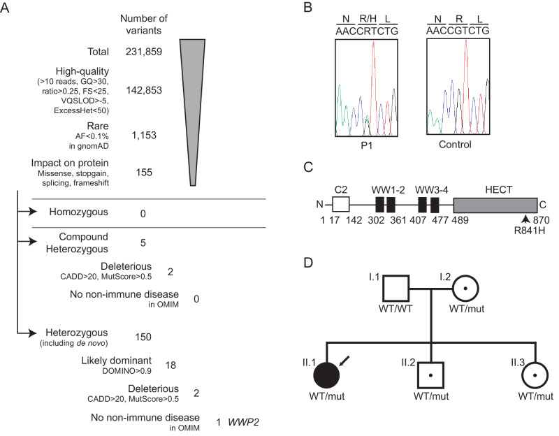
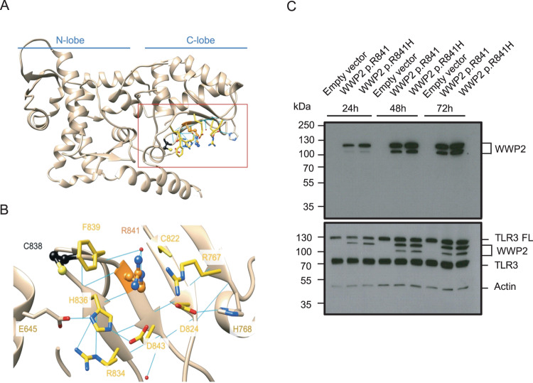
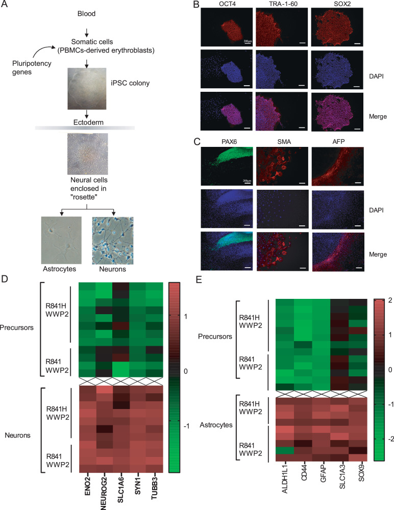
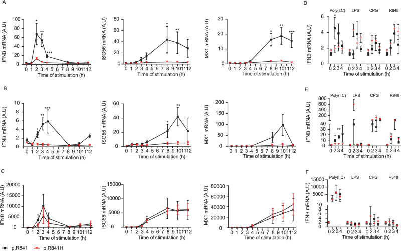
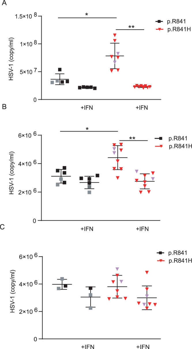
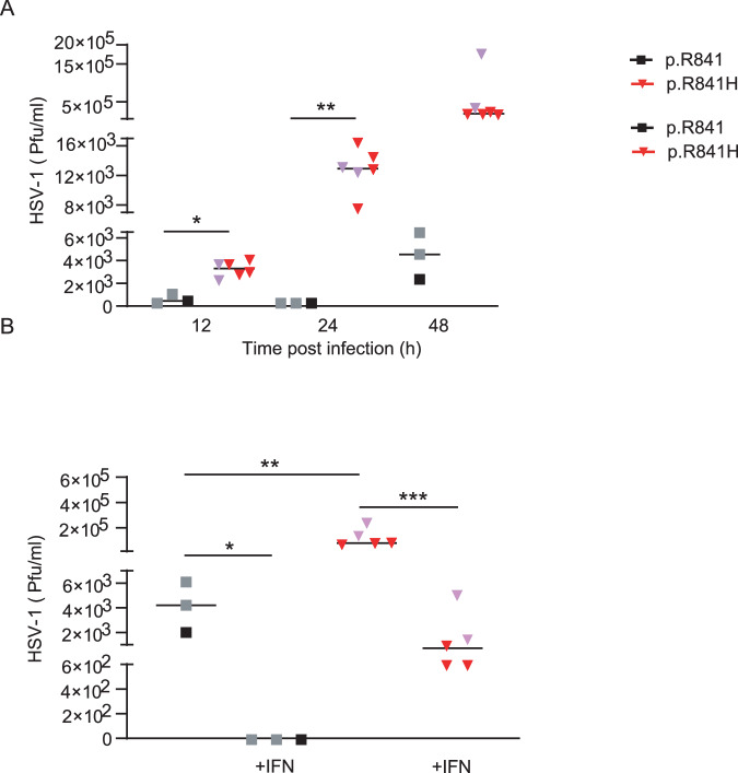
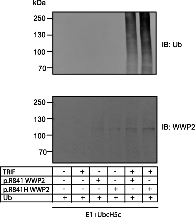
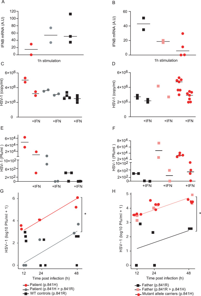
Encephalitis is a rare and potentially fatal manifestation of herpes simplex type 1 infection. Following genome-wide genetic analyses, we identified a previously uncharacterized and very rare heterozygous variant in the E3 ubiquitin ligase WWP2, in a 14-month-old girl with herpes simplex encephalitis. The p.R841H variant (NM_007014.4:c.2522G > A) impaired TLR3 mediated signaling in inducible pluripotent stem cells-derived neural precursor cells and neurons; cells bearing this mutation were also more susceptible to HSV-1 infection compared to control cells. The p.R841H variant increased TRIF ubiquitination in vitro. Antiviral immunity was rescued following the correction of p.R841H by CRISPR-Cas9 technology. Moreover, the introduction of p.R841H in wild type cells reduced such immunity, suggesting that this mutation is linked to the observed phenotypes.


Conflict of interest statement

The authors declare no competing interests.

Figures

Fig. 1
Fig. 1. p.R841H WWP2 variant.
A Filtering process of the variants identified by whole exome sequencing. B Sanger sequencing profiles for the WWP2 p.R841H mutation in genomic DNA from the patient (P1) and her father (as a control). C Schematic representation of WWP2, featuring the different domains as well as the location of the missense variant p.R841H. WWP2 is composed of well-defined domains comprising the C2, WW, and Homologous to the E6-AP Carboxyl Terminus (HECT) domains. D Family pedigree with allele segregation. The patient is indicated by the arrow sign, whereas “mut” refers to the p.R841H allele and “WT” to a wild-type allele. GQ genotype quality, FS Fisher strand, VQSLOD variant quality score log odds ratio, ExcessHet Phred-scaled p-value for exact test of excess heterozygosity, AF allele frequency, gnomAD genome aggregation database, CADD combined annotation dependent depletion.
Fig. 2
Fig. 2. Molecular modeling and expression analysis of p.R841H WWP2 variant.
A Overview of the HECT domain structure and its two main lobes. B Zoom on Arg841 and its neighbors. Arg841 and Cys838 are respectively shown in orange and black ball and stick. The residues involved in the ionic and hydrogen bond network are shown in sticks. Residues with carbons colored in yellow are within 5 A from Arg841 (pdb id:4y07). C The expression of p.R841 and p.R841H WWP2 protein was determined by SDS-PAGE/immunoblotting under reducing conditions in cell lysates of TLR3-expressing 293 T cells after different times of transfection. Results represent 1 experiment among 3. TLR3 FL TLR3 full length.
Fig. 3
Fig. 3. Reprogrammation, derivation, and CNS cells differentiation.
A Schematic diagram of the reprogrammation and differentiation protocols used. B Immunocytochemistry analysis of iPSCs colonies revealed the expession of pluripotent markers OCT4, TRA-1-60, and SOX2. C Immunocytochemistry analysis of the ability of iPSCs-derived embryonic bodies to differentiate into the 3 germ layers. PAX6, SMA, and AFP are markers for ectoderm, mesoderm, and endoderm respectively. Results represent the analysis of 1 iPSCs colony and is representative of results obtained for all iPSCs colonies (n = 2 per donor). D Characterization of neurons. Compared to neuronal precursors, neurons exhibit increased expression of typical neuronal markers assessed by qPCR. E Characterization of astrocytes. Compared to neuronal precursors, astrocytes exhibit increased expression of typical astrocyte markers, assessed by qPCR.
Fig. 4
Fig. 4. WWP2-dependent IFN responses in iPSCs-derived neural stem cells (NSCs).
Induction of IFNβ, ISG56 and Mx1 mRNA after different time of Poly(I:C) stimulation in neural precursor cells (A), neurons (B) and astrocytes (C) from WWP2 p.R841 individuals [black, N = 5 (A), N = 4 (B), N = 5 (C)] and from WWP2 p.R841H individuals [red, N = 8 (A, B), N = 4 (C)]. As a control, the expression of IFNβ was measured in neural precursor cells (D), neurons (E) and astrocytes (F) from WWP2 p.R841 individuals (black) and from WWP2 p.R841H individuals (red), stimulated with LPS [WWP2 p.R841 individuals N = 5 (D, E), N = 4 (F); WWP2 p.R841H individuals N = 8 (D, E), N = 2 (F)], CPG [WWP2 p.R841 individuals N = 5 (D, E), N = 4 (F); WWP2 p.R841H individuals N = 8 (D, E), N = 2 (F)] and R848 [WWP2 p.R841 individuals N = 5 (D, E), N = 4 (F); WWP2 p.R841H individuals N = 8 (D, E), N = 2 (F)] for 2, 3 and 4 h. A.U. stands for arbitrary unit. Results represent the mean ± standard error of 1 representative experiment among 2 (A, B), 3 (C), and 1 (DF). Statistical analyses were performed using an unpaired two-tailed Student t-test. A Left *, **, *** mean for P = 0.0129, P = 0.0011 and P = 0.0001, (A) middle *, ** mean for P = 0.0501 and P = 0.0413, (A) right *, **, *** mean for P = 0.0010, P = 0.0025, P = 0.0021. B Left *, **, *** mean for P = 0.0026, P = 0.0006 and P = 0.0187, (B) middle *, ** mean for P = 0.0299 and P = 0.0022. D * means for P = 0.0475. E *, ** mean for P = 0.0121 and P = 0.0005.
Fig. 5
Fig. 5. WWP2-dependent HSV-1 genomes number in iPSCs-derived neural stem cells (NSCs).
Quantification by polymerase chain reaction of the number of viral genome in neural precursor cells (A), neurons (B) and astrocytes (C) from WWP2 p.R841 individuals [black, N = 5 (A), N = 6 (B), N = 3 (C)] and from WWP2 p.R841H individuals [red, N = 8 (A), N = 9 (B), N = 8 (C)] without or with an IFNα2b pretreatment (+IFN). Results represent the mean ± standard error of 1 experiment among 2 (A) and 1 (B, C). Statistical analyses were performed using an unpaired two-tailed Student t-test. A *, ** mean for P = 0.0028 and P < 0.0001. B *, ** mean for P = 0.0046 and P = 0.0001. The purple triangle and the grey square correspond to patient with HSE and to patient’s father respectively.
Fig. 6
Fig. 6. WWP2-dependent HSV-1 replication in iPSCs-derived neurons.
A Quantification of the number of plaque-forming units (pfu) by plaque-titration at different time after infection of neurons from WWP2 p.R841 individuals (black, N = 3) and from WWP2 p.R841H individuals (red, N = 6). (B) Quantification of the number of plaque-forming units (pfu) by plaque-titration at 48 h after infection of neurons from WWP2 WT individuals (black, N = 3) and from WWP2 p.R841H individuals (red, N = 5) without or with an IFNα2b pretreatment (+IFN). Figure shows median of one experiment. Statistical analyses were performed using an unpaired two-tailed Student t-test. The purple triangle and the grey square correspond to patient with HSE and to patient’s father respectively. A *, ** mean for P = 0.0005 and P = 0.0002. B *, **, *** mean for P = 0.0239, P = 0.0256 and P = 0.0039.
Fig. 7
Fig. 7. Increased TRIF ubiquitination by WWP2 p.R841H compared to WWP2 p.R84.
TRIF, p.R841 WWP2, and p.R841H WWP2 were in vitro translated. An in vitro ubiquitination assay of TRIF was conducted by using an E1 ubiquitin-activating enzyme and UbcH5c together with either p.R841 WWP2 or p.R841H WWP2. Ubiquitin-conjugated TRIF was detected by immunoblot with an anti-ubiquitin antibody. The expression levels of WWP2 were also verified by immunoblots with an anti-WWP2 antibody. Results represent 1 experiment among 2.
Fig. 8
Fig. 8. Rescue of antiviral immunity in iPSCs-derived neurons.
A Induction of IFNβ mRNA after 1 h of Poly(I:C) stimulation in neurons from WWP2 p.R841H (red, N = 2), WWP2 p.H841R (grey, N = 2) individuals and WWP2 p.R841 controls (black, N = 3). B Induction of IFNβ mRNA after 1 h of Poly(I:C) stimulation in neurons from WWP2 p.R841 (black, N = 2), WWP2 p.R841H (grey, N = 2) individuals and WWP2 p.H841 controls (red, N = 4). Results represent the mean ± standard error of 1 experiment among 2. C Quantification of the number of viral genome in neurons from WWP2 p.R841H (red, N = 2), WWP2 p.H841R (grey, N = 2) individuals, and WWP2 p.R841 controls (black, N = 4), without or with an IFNα pretreatment (+IFN). D Quantification of the number of viral genome in neurons from WWP2 p.R841 (black, N = 2), WWP2 p.R841H (grey, N = 1) individuals and WWP2 p.H841 controls (black, N = 7) without or with an IFNα pretreatment (+IFN). E Quantification of the number of plaque-forming units (pfu) by plaque-titration after infection of neurons from WWP2 p.R841H (red, N = 2), WWP2 p.H841R (grey, N = 2) individuals and WWP2 p.R841 controls (black, N = 3) without or with an IFNα2b pretreatment (+IFN). F Quantification of the number of plaque-forming units (pfu) by plaque-titration after infection of neurons from WWP2 p.R841 (black, N = 2), WWP2 p.R841H (grey, N = 2) individuals and WWP2 p.H841 controls (black, N = 4) without or with an IFNα2b pretreatment (+IFN). G Quantification of the number of plaque-forming units (pfu) by plaque-titration at different time after infection of neurons from WWP2 p.R841H (red, N = 2), WWP2 p.H841R (grey, N = 2) individuals and WWP2 p.R841 controls (black, N = 3). A significant effect (*) of the rescue (grey) compared to allele carrier (red) (p = 2.41e-5) was found using a linear regression on log10 + 1 transformed pfu with a time-dependent effect. H Quantification of the number of plaque-forming units (pfu) by plaque-titration at different time after infection of neurons from WWP2 p.R841 (black, N = 2), WWP2 p.R841H (grey, N = 2) individuals and WWP2 p.H841 controls (black, N = 4). A significant effect (*) of the mutant induced (pink) compared to control (black) (p = 2.06e-3) was found using a linear regression on log10 + 1 transformed pfu with a time-dependent effect.

References

    1. Whitley RJ. Herpes simplex encephalitis: adolescents and adults. Antivir. Res. 2006;71:141–148. doi: 10.1016/j.antiviral.2006.04.002. - DOI - PubMed
    1. Whitley RJ. Herpes simplex virus infections of the central nervous system. Continuum. 2015;21:1704–1713. - PubMed
    1. Abel L, et al. Age-dependent Mendelian predisposition to herpes simplex virus type 1 encephalitis in childhood. J. Pediatr. 2010;157:623–629.e1. doi: 10.1016/j.jpeds.2010.04.020. - DOI - PubMed
    1. Koskiniemi M, Piiparinen H, Mannonen L, Rantalaiho T, Vaheri A. Herpes encephalitis is a disease of middle aged and elderly people: polymerase chain reaction for detection of herpes simplex virus in the CSF of 516 patients with encephalitis. The Study Group. J. Neurol. Neurosurg. Psychiatry. 1996;60:174–178. doi: 10.1136/jnnp.60.2.174. - DOI - PMC - PubMed
    1. Kelly D, Kroll JS. Encephalitis–beyond aciclovir. Adv. Exp. Med. Biol. 2004;549:177–183. doi: 10.1007/978-1-4419-8993-2_24. - DOI - PubMed

Publication types