. 2024 May 10;16(1):36.
doi: 10.1038/s41368-024-00304-0.

m1A inhibition fuels oncolytic virus-elicited antitumor immunity via downregulating MYC/PD-L1 signaling

Affiliations

m1A inhibition fuels oncolytic virus-elicited antitumor immunity via downregulating MYC/PD-L1 signaling

Shujin Li et al. Int J Oral Sci. .

Abstract

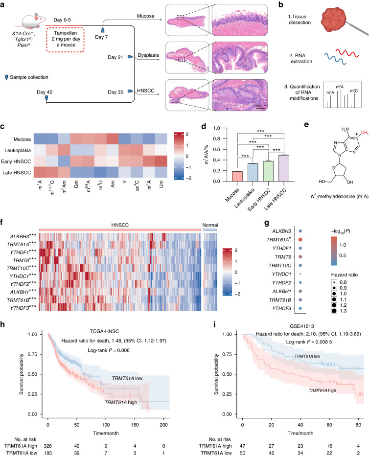
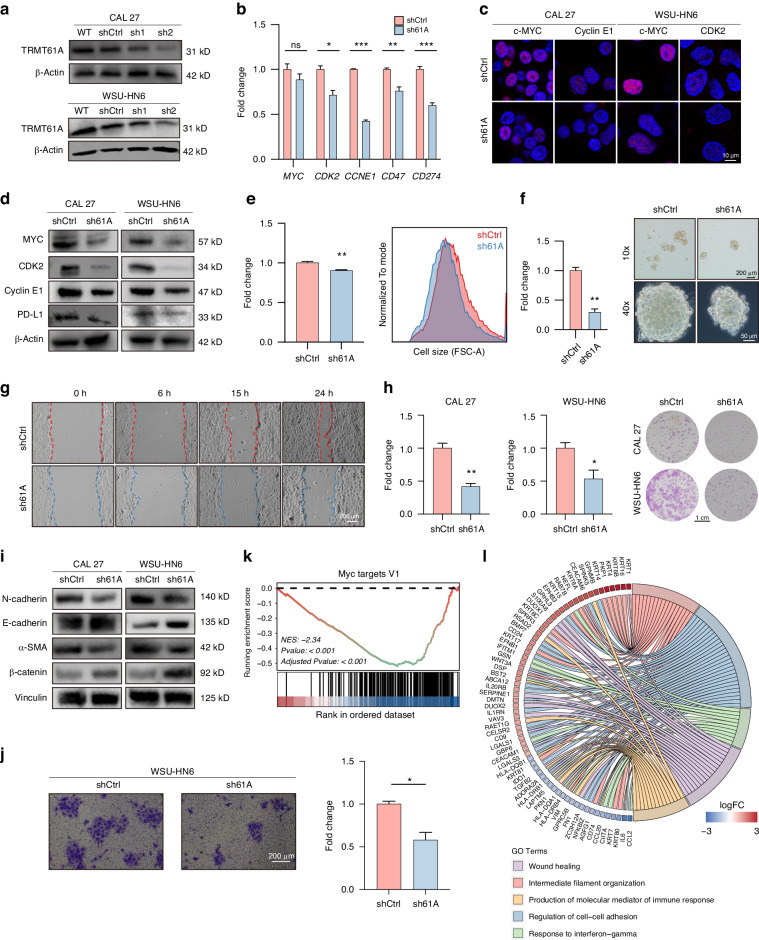
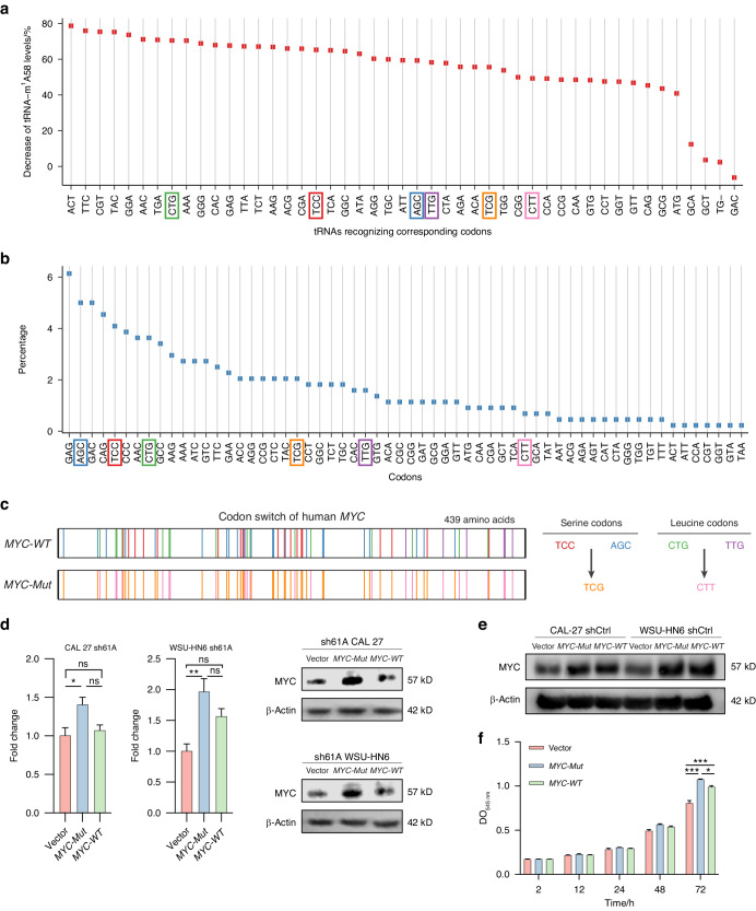
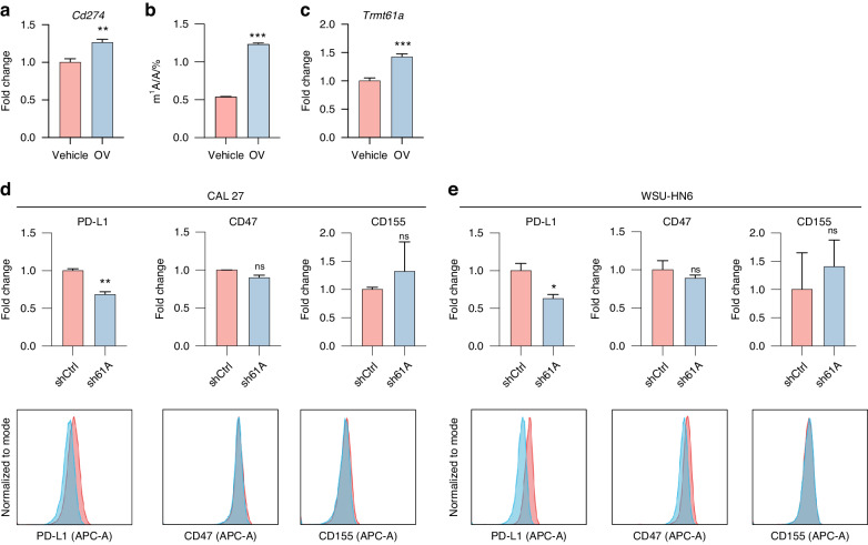
N1-methyladenosine (m1A) RNA methylation is critical for regulating mRNA translation; however, its role in the development, progression, and immunotherapy response of head and neck squamous cell carcinoma (HNSCC) remains largely unknown. Using Tgfbr1 and Pten conditional knockout (2cKO) mice, we found the neoplastic transformation of oral mucosa was accompanied by increased m1A modification levels. Analysis of m1A-associated genes identified TRMT61A as a key m1A writer linked to cancer progression and poor prognosis. Mechanistically, TRMT61A-mediated tRNA-m1A modification promotes MYC protein synthesis, upregulating programmed death-ligand 1 (PD-L1) expression. Moreover, m1A modification levels were also elevated in tumors treated with oncolytic herpes simplex virus (oHSV), contributing to reactive PD-L1 upregulation. Therapeutic m1A inhibition sustained oHSV-induced antitumor immunity and reduced tumor growth, representing a promising strategy to alleviate resistance. These findings indicate that m1A inhibition can prevent immune escape after oHSV therapy by reducing PD-L1 expression, providing a mutually reinforcing combination immunotherapy approach.


Conflict of interest statement

The authors declare no competing interests.

Figures

Fig. 1
Fig. 1
RNA m1A modification is activated during HNSCC initiation and progression. a, b Schematic depicting LC-MS/MS assays. c The levels of various RNA modifications in total RNAs, as detected by LC-MS/MS (n = 3). d The internal m1A/A levels in total RNAs, as detected by LC-MS/MS (n = 3). e The chemical structure of m1A methylation. f Expression of genes related to m1A modification in the TCGA-HNSC dataset. g Cox regression of m1A-associated genes in the TCGA-HNSC dataset. h, i Kaplan‒Meier survival analysis of TRMT61A in the h TCGA-HNSC dataset and i GSE41613 datasets at the best cutoff values. One-way ANOVA followed by Tukey’s multiple comparisons tests (d); Mann–Whitney test (f); Cox regression (gi); log-rank test (h, i). *P < 0.05; **P < 0.01; ***P < 0.001
Fig. 2
Fig. 2
TRMT61A is upregulated in HNSCC tissues and predicts a poor prognosis. a, b Western blot detection (a) and normalized levels (b) of TRMT61A expression in normal oral mucosa, dysplasia, and HNSCC tissues of Tgfbr1/Pten 2cKO mice. c, d Western blot detection (c) and normalized levels (d) of TRMT61A expression in tumor and peritumoral normal tissues of HNSCC patients. e Representative IHC staining results of TRMT61A in normal mucosa, dysplasia, and HNSCC samples with different pathological grades. f Normalized TRMT61A expression in HNSCC (n = 210) compared with dysplasia (n = 69) and oral mucosa (n = 42). g Quantification of the TRMT61A histoscores among pathological grades. h Correlation of TRMT61A with other cancer-associated proteins in the HNSCC tissue microarray database. i Correlation of TRMT61A with PD-L1 in the HNSCC tissue microarray database. j Kaplan‒Meier survival analysis of TRMT61A in the HNSCC tissue microarray database at the best cutoff value. Data were mean with s.e.m. One-way ANOVA followed by Tukey’s multiple comparisons tests (b); Kruskal–Wallis test followed by Dunn’s multiple comparisons tests (f); Kruskal–Wallis test (g); two-tailed paired Student’s t-test (d); Spearman’s correlation (h, i); Cox proportional-hazards model and log-rank test (j). *P < 0.05; **P < 0.01; ***P < 0.001; ns represents no significance
Fig. 3
Fig. 3
TRMT61A promotes cancer invasion and stemness. a Knockdown of TRMT61A by shTRMT61A-2 in WSU-HN6 and CAL 27 cells were confirmed by Western blot. b mRNA levels of shCtrl/sh61A CAL 27 cells were quantified by qPCR (n = 3). c, d Protein levels of shCtrl/sh61A WSU-HN6 and CAL 27 cells were quantified by confocal microscopy (c) and Western blot (d). e Cell sizes of shCtrl/sh61A WSU-HN6 cells quantified by FSC-A (n = 3). Representative flow cytometry histograms are shown to the right. f Sphere formation ability of shCtrl/h61A CAL 27 cells (n = 3). Representative flow cytometry histograms are shown to the right. g Representative images from in vitro wound healing assays of CAL 27 cells. h Colony-forming efficacy of CAL 27 cells (left, n = 3) and WSU-HN6 cells (middle, n = 3). Representative pictures are shown to the right. i Protein levels of epithelial-mesenchymal transition indicators were quantified by Western blot. j Representative images of cell invasion assay (left) and quantitative analysis (right). k, l Analysis of high-throughput sequencing results of sh61A CAL 27 cells against shCtrl CAL 27 cells by a GSEA (k; n = 3) and a GO enrichment analysis (l; n = 3). Data were mean with s.e.m. Two-tailed unpaired Student’s t-test (b, e, f, h, j). *P < 0.05; **P < 0.01; ***P < 0.001; ns represents no significance
Fig. 4
Fig. 4
TRMT61A promotes the translation of human MYC mRNA. a The decrease in the magnitude of the tRNA-m1A58 level in each tRNA after TRMT61A deletion. b The codon frequency of human MYC mRNA. c Schematic diagram of the human MYC codon-switch assay. d Expression of MYC-WT and MYC-Mutant (MYC-Mut) in sh61A CAL 27 and WSU-HN6 cells. Protein levels of MYC were quantified by immunoblotting (n = 4). Representative Western blot results are shown to the right. e Expression of MYC-WT and MYC-Mut in shCtrl CAL 27 and WSU-HN6 cells confirmed by Western blot. f Proliferation of sh61A CAL 27 cells transinfected with vector, MYC-Mut, and MYC-WT detected by SRB assay. Data were mean with s.e.m. Two-tailed unpaired Student’s t-test (d). One-way ANOVA followed by Tukey’s multiple comparisons tests (f). *P < 0.05; **P < 0.01; ***P < 0.001; ns represents no significance
Fig. 5
Fig. 5
TRMT61A promotes PD-L1 expression upon inflammation. a mRNA levels of Cd274 were quantified by qPCR (n = 6 biological replicates per group). b The internal m1A/A levels in total RNAs of tumors injected with vehicle or OV, as detected by LC-MS/MS (n = 3). c mRNA levels of Trmt61a were quantified by qPCR (n = 6 biological replicates per group). d, e Protein levels of PD-L1, CD155, and CD47 were detected by flow cytometry in shCtrl/sh61A CAL 27 (d) and WSU-HN6 (e) cells 24 h post 25 ng/mL IFNγ treatment. Representative flow cytometry results are shown at the bottom. Data were mean with s.e.m.*P < 0.05; **P < 0.01; ***P < 0.001; ns represents no significance by two-tailed unpaired Welch’s t-test
Fig. 6
Fig. 6
TRMT61A confers resistance against oHSV. a CAL 27 expression of genes belonging to MYC target genes or the IFNγ pathway in different groups was identified according to the RNA-seq results. b, d Analysis of high-throughput sequencing results of IFNγ-treated, sh61A CAL 27 cells against IFNγ-treated, CAL 27 shCtrl cells by a GSEA (b, c; n = 3 biological replicates per group), a GO enrichment analysis (d; n = 3 biological replicates per group). e, f Results of the cytotoxicity assay for shCtrl/sh61A CAL 27 (e) or WSU-HN6 (f) cells upon oHSV infection following IFNγ pretreatment at 25 ng/mL for 24 h (n = 3 biological replicates per group). Cell viability was measured three days post infection. Representative log-transformed dose-response curves are shown to the right. Data were mean with s.e.m. Two-tailed unpaired Welch’s t-test (e, f). ***P < 0.001
Fig. 7
Fig. 7
Thiram enhanced oHSV efficacy in two immunocompetent allograft mouse models. a Schematic depicting the study design for 4MOSC1 (up panel) and 4T1 (down panel) tumor inoculation and treatment with oHSV and thiram. b Macroscopic appearance of tumors from 4MOSC1 (left panel) and 4T1 (right panel) at the end of the experiment. c Tumor-growth curve of 4MOSC1 (left panel) and 4T1 tumors (right panel). d Proposed mechanism of combining oHSV and m1A inhibition. e, f Representative flow cytometric analysis images (e) and quantification (f) of CD44 and CD62L expression in the CD8+ T cells from TDLNs of 4MOSC1 tumors. g, h Representative flow cytometric analysis images (g) and quantification (h) of CD44 and CD62L expression in the CD4+ T cells from TDLNs of 4MOSC1 tumors. i, j Representative IHC images of CD3 epsilon (i) and PD-L1 (j) of 4MOSC1 tumors. Two-tailed unpaired Student’s t-test (c, f, h). Data were mean with s.e.m (n = 6 mice per group). *P < 0.05; **P < 0.01; ***P < 0.001; ns represents no significance

References

    1. Sung H, et al. Global cancer statistics 2020: GLOBOCAN estimates of incidence and mortality worldwide for 36 cancers in 185 countries. CA Cancer J. Clin. 2021;71:209–249. doi: 10.3322/caac.21660. - DOI - PubMed
    1. Johnson DE, et al. Head and neck squamous cell carcinoma. Nat. Rev. Dis. Primers. 2020;6:92. doi: 10.1038/s41572-020-00224-3. - DOI - PMC - PubMed
    1. Pulte D, Brenner H. Changes in survival in head and neck cancers in the late 20th and early 21st century: a period analysis. Oncologist. 2010;15:994–1001. doi: 10.1634/theoncologist.2009-0289. - DOI - PMC - PubMed
    1. Gillen SL, Waldron JA, Bushell M. Codon optimality in cancer. Oncogene. 2021;40:6309–6320. doi: 10.1038/s41388-021-02022-x. - DOI - PMC - PubMed
    1. Dominissini D, et al. The dynamic N(1)-methyladenosine methylome in eukaryotic messenger RNA. Nature. 2016;530:441–446. doi: 10.1038/nature16998. - DOI - PMC - PubMed

Publication types

Associated data