. 2024 May 10;24(1):164.
doi: 10.1186/s12935-024-03346-w.

Assessing the role of programmed cell death signatures and related gene TOP2A in progression and prognostic prediction of clear cell renal cell carcinoma

Affiliations

Assessing the role of programmed cell death signatures and related gene TOP2A in progression and prognostic prediction of clear cell renal cell carcinoma

Qingshui Wang et al. Cancer Cell Int. .

Abstract

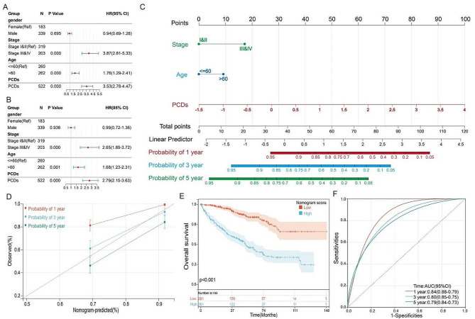
Kidney Clear Cell Carcinoma (KIRC), the predominant form of kidney cancer, exhibits a diverse therapeutic response to Immune Checkpoint Inhibitors (ICIs), highlighting the need for predictive models of ICI efficacy. Our study has constructed a prognostic model based on 13 types of Programmed Cell Death (PCD), which are intertwined with tumor progression and the immune microenvironment. Validated by analyses of comprehensive datasets, this model identifies seven key PCD genes that delineate two subtypes with distinct immune profiles and sensitivities to anti-PD-1 therapy. The high-PCD group demonstrates a more immune-suppressive environment, while the low-PCD group shows better responses to PD-1 treatment. In particular, TOP2A emerged as crucial, with its inhibition markedly reducing KIRC cell growth and mobility. These findings underscore the relevance of PCDs in predicting KIRC outcomes and immunotherapy response, with implications for enhancing clinical decision-making.

Keywords: Immune microenvironment; Immunotherapy; KIRC; PCD; TOP2A.


Conflict of interest statement

The authors declare no competing interests.

The authors declare no conflict of interest for this article.

Figures

Fig. 1
Fig. 1
Variant landscape of PCD genes in KIRC patients. (A) Heatmap and (B) Volcano plot showing differentially expressed PCD genes. Results of (C) Hallmark and (D) KEGG enrichment analyses for the differentially expressed genes. (E) An oncoplot of PCD-related genes in the TCGA cohort
Fig. 2
Fig. 2
Construction of a prognostic model for KIRC patients based on PCD genes. Univariate survival analysis of differentially expressed PCD genes in (A) TCGA, (B) E-MTAB-1980, (C) Braun-2020 cohorts. (D) Venn diagram showing the intersection among the three cohorts. (E & F) LASSO Cox regression to construct a prognostic model for KIRC patients. (G-I) Expression levels of PCDs, survival status, and seven genes in the three KIRC cohorts; (J-L) Impact of PCDs on OS of patients in the three KIRC cohorts
Fig. 3
Fig. 3
Analysis of the clinical correlation of PCDs with KIRC patients.(A)The chromosomal location distribution of 7 genes. the expression correlation analysis of 7 genes in the (B) TCGA, (C) Braun-2020, and (D) E-MTAB-1980 cohorts. (E) Differences in PCDs values across different stages. (F) Differences in PCDs values across different T classifications. (G) Differences in PCDs values across different M classifications. (H) Differences in PCDs values across different N classifications; (I) Differences in PCDs values between recurrent and non-recurrent patients. (J) Differences in PCDs values between living and deceased patients. (K) Gene mutation analysis in the High-PCDs and Low-PCDs subgroups. (L) Correlation analysis of PCDs with the Hallmark signaling pathways. Correlation analysis of PCDs with the G2M checkpoint in the (M) TCGA, (N) Braun-2020, and (O) E-MTAB-1980 cohorts. *, p < 0.05; ***, p < 0.001
Fig. 4
Fig. 4
Relationship between PCDs and the Immune Microenvironment. (A) Results of estimated scores and differential immune cell infiltration between High-PCDs and Low-PCDs subgroups in TCGA, assessed by CIBERSORT and ESTIMATE. In TCGA, the relative cell abundances of macrophages and Tregs between the two groups are calculated using (B) XCELL and (C) CIBERSORT. (D) Differentially expressed genes profile involved in the negative regulation of the Cancer-Immunity Cycle between High-PCDs and Low-PCDs subgroups. (E) Expression of common immune checkpoints between High-PCDs and Low-PCDs subgroups. (F) Expression of immunosuppressive cytokines between High-PCDs and Low-PCDs subgroups. (G) t-SNE plot visualization of all cell subtypes from KIRC patients in the GSE171306 cohort. (H) Bubble plot depicting the expression of model genes across different cell subtypes. ns, p > 0.05; *, p < 0.05; ***, p < 0.001
Fig. 5
Fig. 5
Kaplan-Meier estimator displaying the overall survival curves for High-PCDs and Low-PCDs subgroups, with two non-proportional hazards statistical methods utilized to compare the prognosis of different PCDs. The restricted mean survival (RMS) time difference at six months and one-year post-treatment were compared in (A) Braun-2020-anti-PDL1 cohort and (B) Melanoma-GSE91061-anti-PDL1 cohort. The first three months following immunotherapy were considered to have a delayed clinical effect, hence the long-term survival post three months of treatment was compared using Chi-square (Qua) approach in (C) Braun-2020-anti-PDL1 cohort and (D) Melanoma-GSE91061-anti-PDL1 cohort
Fig. 6
Fig. 6
Establishment and assessment of the nomogram survival model. (A) Univariate analysis for the clinicopathologic characteristics and PCDs in TCGA cohort. (B) Multivariate analysis for the clinicopathologic characteristics and PCDs in TCGA cohort. (C) A nomogram was established to predict the prognosis of Kidney Renal Clear Cell Carcinoma (KIRC) patients. (D) Calibration plots showing the probability of 1-, 3-, and 5-year overall survival in TCGA cohort. (E) Kaplan-Meier analyses for the two KIRC groups based on the nomogram score. (G) Receiver operator characteristic (ROC) analysis of the nomogram in TCGA cohort
Fig. 7
Fig. 7
TOP2A is overexpressed in KIRC and associated with poor prognosis. (A) Area under the curve (AUC) analysis of seven genes distinguishing KIRC tissue from adjacent non-cancerous tissue; Expression of TOP2A in KIRC tissue and adjacent non-cancerous tissue in (B) GSE14994, (C) GSE36895, (D) GSE40435, (E) GSE46699, (F) GSE53757, (G) GSE66272, (H) TCGA cohort. (I) Expression analysis of the TOP2A gene at different stages in the TCGA cohort. (J) Expression analysis of the TOP2A gene in different grades in the TCGA cohort. (K) Expression analysis of the TOP2A gene in different grades in the E-MTAB-1980 cohort. OS analysis of TOP2A in (L) TCGA, (M) Braun-2020, and (N) E-MTAB-1980 cohorts. (O) Disease-free survival (DFS) analysis of TOP2A in TCGA cohort. ***, p < 0.001
Fig. 8
Fig. 8
TOP2A promotes proliferation and metastasis of KIRC cells. RT-PCR detection of TOP2A expression in (A) 786-O and (B) ACHN cells after knockdown of TOP2A. CCK8 assay to measure changes in proliferation ability after knockdown of TOP2A in (C) 786-O and (D) ACHN cells. Effect of TOP2A knockdown on (E) migration and (G) invasion ability of 786-O cells. Effect of TOP2A knockdown on (F) migration and (H) invasion ability of ACHN cells. Impact of TO P2A knockdown on (I) proliferation and (J) metastasis of ACHN cells in zebrafish. ns, p > 0.05; *, p < 0.05; **, p < 0.01; ***, p < 0.001

References

    1. Hsieh JJ, Purdue MP, Signoretti S, Swanton C, Albiges L, Schmidinger M, Heng DY, Larkin J, Ficarra V. Renal cell carcinoma. Nat Rev Dis Primers. 2017;3:17009. doi: 10.1038/nrdp.2017.9. - DOI - PMC - PubMed
    1. Yin X, Wang Z, Wang J, Xu Y, Kong W, Zhang J. Development of a novel gene signature to predict prognosis and response to PD-1 blockade in clear cell renal cell carcinoma. Oncoimmunology. 2021;10:1933332. doi: 10.1080/2162402X.2021.1933332. - DOI - PMC - PubMed
    1. Ljungberg B, Bensalah K, Canfield S, Dabestani S, Hofmann F, Hora M, Kuczyk MA, Lam T, Marconi L, Merseburger AS, Mulders P, Powles T, Staehler M, et al. EAU guidelines on renal cell carcinoma: 2014 update. Eur Urol. 2015;67:913–24. doi: 10.1016/j.eururo.2015.01.005. - DOI - PubMed
    1. Motzer RJ, Penkov K, Haanen J, Rini B, Albiges L, Campbell MT, Venugopal B, Kollmannsberger C, Negrier S, Uemura M, Lee JL, Vasiliev A, Miller WH, Jr, et al. Avelumab plus Axitinib versus Sunitinib for Advanced Renal-Cell Carcinoma. N Engl J Med. 2019;380:1103–15. doi: 10.1056/NEJMoa1816047. - DOI - PMC - PubMed
    1. Rini BI, Plimack ER, Stus V, Gafanov R, Hawkins R, Nosov D, Pouliot F, Alekseev B, Soulières D, Melichar B, Vynnychenko I, Kryzhanivska A, Bondarenko I, et al. Pembrolizumab plus Axitinib versus Sunitinib for Advanced Renal-Cell Carcinoma. N Engl J Med. 2019;380:1116–27. doi: 10.1056/NEJMoa1816714. - DOI - PubMed

LinkOut - more resources