

Sagittal Full-Spine vs. Sectional Cervical Lateral Radiographs: Are the Measurements of Cervical Alignment Interchangeable?
- PMID: 38731030
- PMCID: PMC11084776
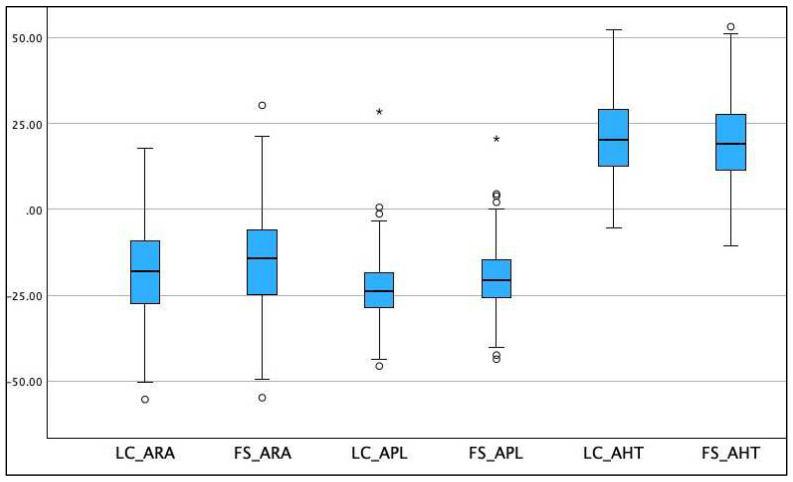
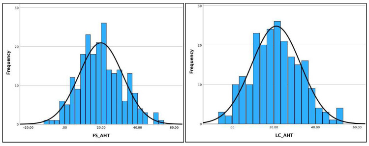
- DOI: 10.3390/jcm13092502
Sagittal Full-Spine vs. Sectional Cervical Lateral Radiographs: Are the Measurements of Cervical Alignment Interchangeable?
Abstract
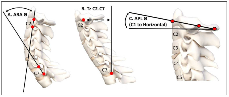
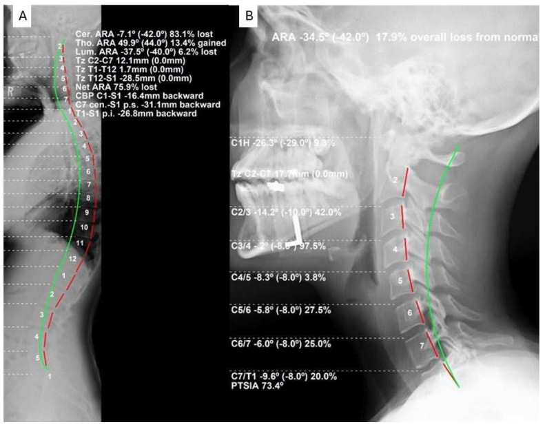
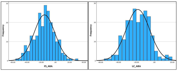
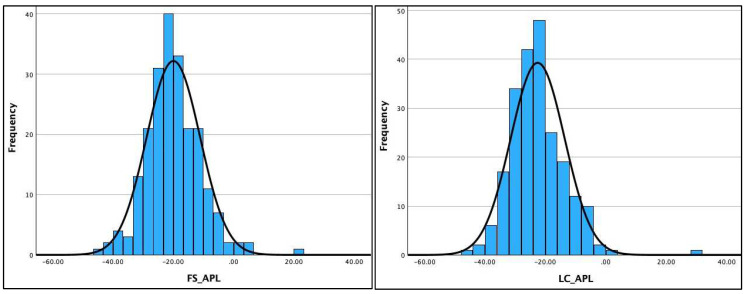
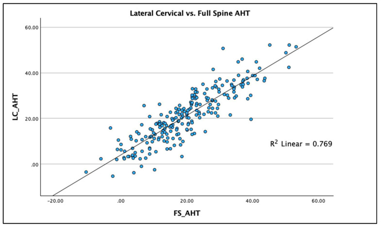
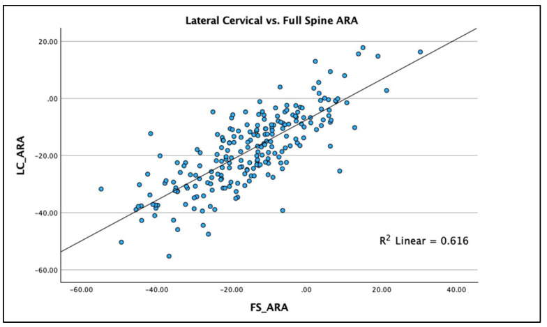
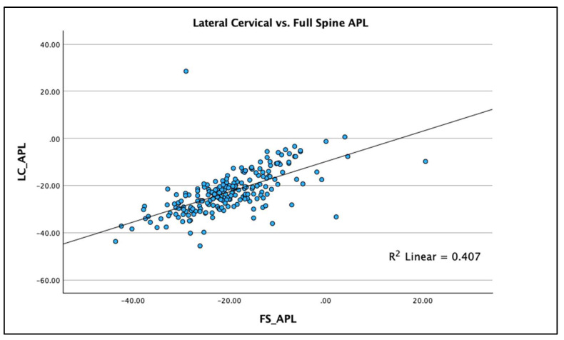
(1) Background: This study assessed the relationship between cervical spine parameters taken on standing full-spine lateral radiographic images compared to sectional lateral cervical radiographs. (2) Methods: Full-spine (FS) and sectional lateral cervical (LC) radiographs from four spine treatment facilities across the USA retrospectively provided data collected on 220 persons to assess the comparison of three sagittal cervical radiographic measurements between the two views. The measures included cervical lordosis using the absolute rotation angle from C2-C7, sagittal cervical translation of C2-C7, and atlas plane angle to horizontal. Linear correlation and R2 models were used for statistical comparison of the measures for the two views. (3) Results: The mean values of the three measurements were statistically different from each other: C2-C7 translation (FS = 19.84 ± 11.98 vs. LC = 21.18 ± 11.8), C2-C7 lordosis (FS = -15.3 ± 14.63 vs. LC = -18.32 ± 13.16), and atlas plane (FS = -19.99 ± 8.88 vs. LC = -22.56 ± 8.93), where all values were p < 0.001. Weak-to-moderate-to-strong correlations existed between the full-spine and sectional lateral cervical radiographic variables. The R2 values varied based on the measurement were R2 = 0.768 (p < 0.001) for sagittal cervical translation of C2-C7 (strong), R2 = 0.613 (p < 0.001) for the absolute rotation angle C2-C7 (moderate), and R2 = 0.406 (p < 0.001) for the atlas plane line (weak). Though a linear correlation was identified, there were consistent intra-person differences between the measurements on the full spine versus sectional lateral cervical radiographic views, where the full-spine view consistently underestimated the magnitude of the variables. (4) Conclusion: Key sagittal cervical radiographic measurements on the full spine versus sectional lateral cervical radiographic views show striking intra-person differences. The findings of this study confirm that full spine versus sectional lateral cervical radiographic views provide different biomechanical magnitudes of cervical sagittal alignment, and caution should be exercised by health care providers as these are not interchangeable. We recommend the LC view for measurement of cervical sagittal alignment variables.
Keywords: X-ray; cervical lateral radiograph; cervical lordosis; cervical spine; neck pain; radiography; sagittal balance; whole-spine lateral radiograph.
Conflict of interest statement
Authors J.E.M., J.O.J. and I.M.M. declare no competing interests. J.W.H. is a compensated researcher for CBP Non-Profit, Inc. P.A.O. is a compensated consultant for Chiropractic BioPhysics, NonProfit, Inc. D.E.H. is the CEO of Chiropractic BioPhysics® (CBP®) and provides post-graduate education to health care providers and physicians. Spine rehabilitation devices are distributed through his company. D.E.H. is the president of CBP Non-Profit, Inc., a not-for-profit spine research foundation.
Figures
Similar articles
-
Factors determining cervical spine sagittal balance in asymptomatic adults: correlation with spinopelvic balance and thoracic inlet alignment.Spine J. 2015 Apr 1;15(4):705-12. doi: 10.1016/j.spinee.2013.06.059. Epub 2013 Sep 8. Spine J. 2015. PMID: 24021619
-
C7 sagittal vertical axis is the determinant of the C5-C7 angle in cervical sagittal alignment.Spine J. 2017 May;17(5):622-626. doi: 10.1016/j.spinee.2016.11.007. Epub 2016 Nov 15. Spine J. 2017. PMID: 27871819
-
Correlation of cervical sagittal alignment parameters on full-length spine radiographs compared with dedicated cervical radiographs.Scoliosis Spinal Disord. 2016 Apr 7;11:12. doi: 10.1186/s13013-016-0072-0. eCollection 2016. Scoliosis Spinal Disord. 2016. PMID: 27299161 Free PMC article.
-
Radiographic Comparison between Cervical Spine Lateral and Whole-Spine Lateral Standing Radiographs.Global Spine J. 2016 Mar;6(2):118-23. doi: 10.1055/s-0035-1556584. Epub 2015 Jun 24. Global Spine J. 2016. PMID: 26933612 Free PMC article.
-
The 3 Sagittal Morphotypes That Define the Normal Cervical Spine: A Systematic Review of the Literature and an Analysis of Asymptomatic Volunteers.J Bone Joint Surg Am. 2020 Oct 7;102(19):e109. doi: 10.2106/JBJS.19.01384. J Bone Joint Surg Am. 2020. PMID: 33027127
References
Grants and funding
LinkOut - more resources
Full Text Sources
Miscellaneous
