

Parasitic Granulomatous Dermatitis Caused by Pelodera spp. in Buffalo on Marajó Island, Pará
- PMID: 38731331
- PMCID: PMC11083330
- DOI: 10.3390/ani14091328
Parasitic Granulomatous Dermatitis Caused by Pelodera spp. in Buffalo on Marajó Island, Pará
Abstract
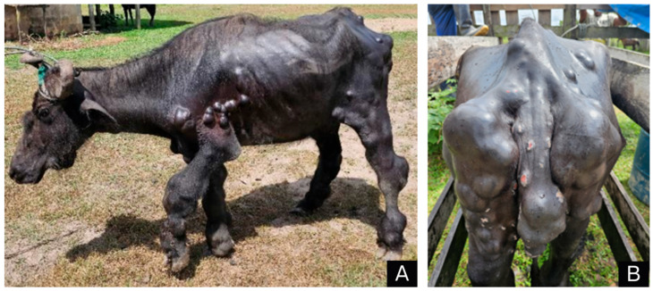
This is the first report of parasitic granulomatous dermatitis caused by Pelodera spp. in a buffalo. The affected buffalo was about seven years old, was a female of the Murrah breed and belonged to a property located on Marajó Island in the State of Pará. During the clinical examination, the animal was in a standing position and presented several multifocal nodular and placoid masses throughout the body, mostly on the forelimbs, hindlimbs, abdomen, mammary glands, perineum, vulva and tail. These masses were also observed on the nasal mucosa, head, neck, back and chest. On macroscopic examination, the skin had several multifocal-to-coalescent sessile nodular and placoid lesions. Histopathology of the skin showed a marked reduction in the number of hair follicles. In the superficial dermis, there was significant multifocal-to-coalescent inflammatory infiltration, consisting of macrophages, epithelioid macrophages, lymphocytes, plasma cells and multinucleated giant cells. In the remaining hair follicles, there were numerous cross and longitudinal sections of small rhabditoid nematodes characterized by a thin cuticle, platymyarian musculature, an intestinal tract, a rhabditiform esophagus and lateral alae (morphologically compatible with Pelodera spp.). The diagnosis of parasitic dermatitis was confirmed by histopathological skin lesions associated with the presence of intralesional rhabditiform larvae morphologically compatible with Pelodera spp.
Keywords: Amazon biome; Bubalus bubalis; parasitic disease; rhabditoid nematodes.
Conflict of interest statement
The authors declare no conflicts of interest.
Figures


Similar articles
-
Pelodera (syn. Rhabditis) strongyloides as a cause of dermatitis--a report of 11 dogs from Finland.Acta Vet Scand. 2006 Sep 5;48(1):18. doi: 10.1186/1751-0147-48-18. Acta Vet Scand. 2006. PMID: 16987397 Free PMC article.
-
Pelodera strongyloides infection in Pacific harbor seals (Phoca vitulina richardii) from California.J Zoo Wildl Med. 2013 Sep;44(3):799-802. doi: 10.1638/2013-0027.1. J Zoo Wildl Med. 2013. PMID: 24063118
-
Pelodera strongyloides in the critically endangered Apennine brown bear (Ursus arctos marsicanus).Res Vet Sci. 2022 Jul;145:50-53. doi: 10.1016/j.rvsc.2022.02.016. Epub 2022 Feb 9. Res Vet Sci. 2022. PMID: 35168109
-
Vesicular Contact Reaction May Progress into Erythema Multiforme.Acta Dermatovenerol Croat. 2016 Dec;24(4):307-309. Acta Dermatovenerol Croat. 2016. PMID: 28128086 Review.
-
Role of In Vivo Reflectance Confocal Microscopy in the Analysis of Melanocytic Lesions.Acta Dermatovenerol Croat. 2018 Apr;26(1):64-67. Acta Dermatovenerol Croat. 2018. PMID: 29782304 Review.
References
-
- I.B.B.E.—Instituto Brasileiro de Geografia e Estatística; de Bubalinos, R. (Búfalos) no Brasil. [(accessed on 1 October 2023)]; Available online: https://www.ibge.gov.br/explica/producao-agropecuaria/bubalinos/br.
-
- Damasceno F.A., Viana J.M., Tinôco I.D.F.F., Gomes R.C.C., Schiassi L. Adaptação de bubalinos ao ambiente tropical. Rev. Eletr. Nutr. 2010;7:1370–1381.
-
- Da Silva W.C., Araújo L.J.S., Silva L.K.X., Reis A.D.S.B. Lesões de pele diagnosticadas em búfalos (Bubalis bubalis) na região do baixo Amazonas. Rev. Bras. Ciênc. Vet. 2021;28:146–150. doi: 10.4322/rbcv.2021.027. - DOI
-
- Barbosa J.D. ((Instituto de Medicina Veterinária, Universidade Federal do Pará, Castanhal, Brazil)). Doenças de pele em búfalos no bioma Amazônico. Personal communication. 2023.
Publication types
LinkOut - more resources
Full Text Sources
Miscellaneous
