

Cutaneous Mucormycosis in Buffalos in the Brazilian Amazon Biome
- PMID: 38731337
- PMCID: PMC11083279
- DOI: 10.3390/ani14091327
Cutaneous Mucormycosis in Buffalos in the Brazilian Amazon Biome
Abstract
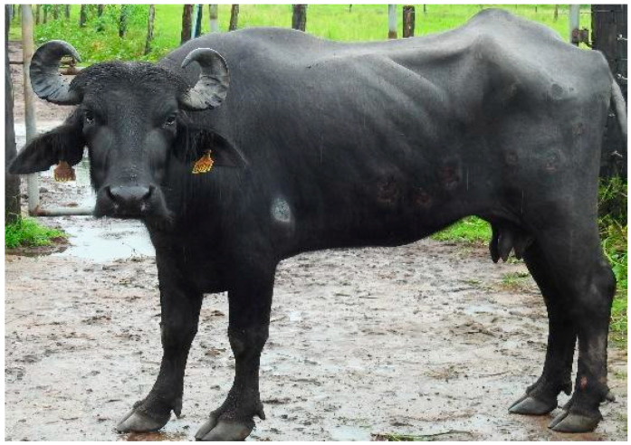
This is the first description of cutaneous mucormycosis in buffalo in the Brazilian Amazon biome. All buffalo showed apathy, inappetence, weight loss, reluctance to move, and prolonged sternal decubitus. Of the four affected animals, two died 15 and 30 days after the appearance of clinical signs. In the initial phase, the skin lesions were rounded areas with dry central regions, sensitive to palpation, with protruding edges and diameters ranging from 8 cm to 15 cm. These areas of necrosis were isolated or coalescing and present mainly on the limbs and sides. In an advanced stage of the disease, there was detachment of the skin from the necrotic areas with extensive wound formation, which sometimes exposed the subcutaneous tissue. The histopathology of the skin showed a multifocal inflammatory infiltrate composed of intact and degenerated eosinophils surrounded by epithelioid macrophages. At the center of these areas was a focally extensive area of epidermal ulceration characterized by intact and degenerated neutrophils, the necrosis of epithelial cells, and the accumulation of fibrin and erythrocytes. The mycological culture was positive for Rhizopus sp. The diagnosis of cutaneous dermatitis caused by Rhizopus sp. was based on clinical signs, macroscopic and histopathological findings, and the identification of the fungus by mycological and molecular techniques.
Keywords: Brazil; Bubalus bubalis; Rhizopus sp.; fungal infection; skin diseases.
Conflict of interest statement
The authors declare no conflicts of interest.
Figures


References
-
- Santos C.L.R.D., Santos J.B.D., Cunha M.C.D., Nunes S.R.F., Bezerra D.C., Torres J.R.D.S., Chaves N.P. Nível tecnológico e organizacional da cadeia produtiva da bubalinocultura de corte no estado do Maranhão. Arq. Inst. Biol. 2016;83:1–8. doi: 10.1590/1808-1657000022014. - DOI
-
- I.B.B.E.–Instituto Brasileiro de Geografia e Estatística Rebanho de Bubalinos (Búfalos) No Brasil. [(accessed on 1 October 2023)]; Available online: https://www.ibge.gov.br/explica/producao-agropecuaria/bubalinos/br.
LinkOut - more resources
Full Text Sources
