. 2024 Apr 26;29(9):1999.
doi: 10.3390/molecules29091999.

Gallic Acid Can Promote Low-Density Lipoprotein Uptake in HepG2 Cells via Increasing Low-Density Lipoprotein Receptor Accumulation

Affiliations

Gallic Acid Can Promote Low-Density Lipoprotein Uptake in HepG2 Cells via Increasing Low-Density Lipoprotein Receptor Accumulation

Dongying Zhang et al. Molecules. .

Abstract

Gallic acid (GA) is a type of polyphenolic compound that can be found in a range of fruits, vegetables, and tea. Although it has been confirmed it improves non-alcoholic fatty liver disease (NAFLD), it is still unknown whether GA can improve the occurrence of NAFLD by increasing the low-density lipoprotein receptor (LDLR) accumulation and alleviating cholesterol metabolism disorders. Therefore, the present study explored the effect of GA on LDLR and its mechanism of action. The findings indicated that the increase in LDLR accumulation in HepG2 cells induced by GA was associated with the stimulation of the epidermal growth factor receptor-extracellular regulated protein kinase (EGFR-ERK1/2) signaling pathway. When the pathway was inhibited by EGFR mab cetuximab, it was observed that the activation of the EGFR-ERK1/2 signaling pathway induced by GA was also blocked. At the same time, the accumulation of LDLR protein and the uptake of LDL were also suppressed. Additionally, GA can also promote the accumulation of forkhead box O3 (FOXO3) and suppress the accumulation of hepatocyte nuclear factor-1α (HNF1α), leading to the inhibition of proprotein convertase subtilisin/kexin 9 (PCSK9) mRNA expression and protein accumulation. This ultimately results in increased LDLR protein accumulation and enhanced uptake of LDL in cells. In summary, the present study revealed the potential mechanism of GA's role in ameliorating NAFLD, with a view of providing a theoretical basis for the dietary supplementation of GA.

Keywords: LDLR; NAFLD; PCSK9; cholesterol metabolism; gallic acid.


Conflict of interest statement

The authors declare that they have no known competing financial interests or personal relationships that could have appeared to influence the work reported in this paper.

Figures

Figure 1
Figure 1
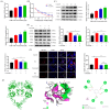
The administration of GA resulted in an upregulation of LDLR accumulation and enhanced LDL uptake in HepG2 cells: (A) GA molecular structure; (B,D) HepG2 cells were subjected to GA treatment at various concentrations and durations, followed by quantification of LDLR protein levels via Western Blot analysis; (C) HepG2 cells were exposed to various concentrations of GA (5, 10, 20, 40, and 80 μM) for a duration of 24 h, and the assessment of cell viability was conducted using the MTT assay; and (E) HepG2 cells were exposed to different concentrations of GA (10, 20, and 40 μM) for a duration of 20 h. Subsequently, the cells were treated with Dil-LDL (20 μg/mL) for a period of 4 h. The uptake activity of LDL was then visualized using fluorescence microscopy. Values are presented as means ± SEM (n = 3). *: p < 0.05, **: p < 0.01, ***: p < 0.001 compared with control group.
Figure 2
Figure 2
The EGFR-ERK1/2 signaling pathway was activated by GA, leading to an increase in LDLR accumulation and improved uptake of LDL in HepG2 cells: (A) The HepG2 cell line was exposed to gallic acid at concentrations of 10, 20, and 40 μM for a duration of 24 h. The LDLR mRNA levels were measured by RT-PCR. (B) The HepG2 cells were exposed to actinomycin D (Act D: 5 µg/mL) for 30 min, and then treated with or without 20 μM GA at intervals of 0.5 h, 1 h, 2 h, 3 h, and 4 h for RNA extraction. The impact of GA on LDLR mRNA half-life was evaluated using RT-qPCR. (C) The HepG2 cell line was treated with varying concentrations of GA (10, 20, and 40 μM) for a duration of 24 h. Subsequently, the effect of GA on the levels of p-EGFR, EGFR, p-ERK1/2, and ERK1/2 proteins was evaluated through Western Blot analysis. (D) The EGFR signaling pathway in HepG2 cells was blocked by treating them with cetuximab for 1 h, followed by a 24 h treatment with GA (20 μM). Western Blot analysis was performed to evaluate the effects of 20 μM GA treatment on LDLR, p-EGFR, EGFR, p-ERK1/2, and ERK1/2 protein levels in HepG2 cells. (E) The HepG2 cells were first exposed to cetuximab for 1 h to block the EGFR signaling pathway. This was followed by treatment with GA (20 μM) for 20 h and then Dil-LDL (20 μg/mL) for 4 h. The effect of GA on the LDL uptake activity of the inhibited EGFR signaling pathway was then observed using inverted fluorescence microscopy. (F) 3D schematic diagram (EGFR: green; GA: red), hydrogen bond receptor surface schematic diagram, and 2D schematic diagram of docking between gallic acid and EGFR extracellular domain. Values are presented as means ± SEM (n = 3). *: p < 0.05, **: p < 0.01, ***: p < 0.001 compared with control group.
Figure 3
Figure 3
Repression of PCSK9 accumulation and its binding with LDLR in HepG2 cells is inhibited by GA: (A) The catalytic structural domain of PCSK9 (green) and the EGF-A region of LDLR (yellow) both bind to form the PCSK9: EGF-A complex. (B) The HepG2 cell line was treated with GA at 10, 20, and 40 μM concentrations for a period of 24 h. After the treatment, the levels of PCSK9 mRNA and protein were analyzed using Western Blot and RT-PCR. (C) 3D schematic diagram (PCSK9: green; GA: red), hydrogen bond receptor surface schematic diagram, and 2D schematic diagram of GA docking with PCSK9. Values are presented as means ± SEM (n = 3). *: p < 0.05, **: p < 0.01, ***: p < 0.001 compared with control group.
Figure 4
Figure 4
GA decreased PCSK9 accumulation in HepG2 cells by increasing FOXO3 accumulation and inhibiting HNF1α and SREBP2 accumulation: (A) HepG2 cells were exposed to GA (10, 20, and 40 μM) for a duration of 24 h, as well as FOXO3, HNF1α, and SREBP2 mRNA and protein levels were measured by Western Blot and RT-PCR and (B,C) GA (10, 20, 40 μM) was used to treat HepG2 cells for 24 h, and FOXO3, HNF1α, and SREBP2 cytosis and nuclear protein levels were measured through Western Blot. Values are presented as means ± SEM (n = 3). *: p < 0.05, **: p < 0.01, ***: p < 0.001 compared with control group.
Figure 5
Figure 5
Schematic representation of the pathway mechanism by which GA can promote LDL uptake by HepG2 cells by increasing LDLR accumulation.

Similar articles

Cited by

References

    1. He Y., Rodrigues R.M., Wang X., Seo W., Ma J., Hwang S., Fu Y., Trojnar E., Matyas C., Zhao S., et al. Neutrophil-to-hepatocyte communication via LDLR-dependent miR-223-enriched extracellular vesicle transfer ameliorates nonalcoholic steatohepatitis. J. Clin. Investig. 2021;131:e141513. doi: 10.1172/JCI141513. - DOI - PMC - PubMed
    1. Li H., Yu X.H., Ou X., Ouyang X.P., Tang C.K. Hepatic cholesterol transport and its role in non-alcoholic fatty liver disease and atherosclerosis. Prog. Lipid Res. 2021;83:101109. doi: 10.1016/j.plipres.2021.101109. - DOI - PubMed
    1. Pouwels S., Sakran N., Graham Y., Leal A., Pintar T., Yang W., Kassir R., Singhal R., Mahawar K., Ramnarain D. Non-alcoholic fatty liver disease (NAFLD): A review of pathophysiology, clinical management and effects of weight loss. BMC Endocr. Disord. 2022;22:63. doi: 10.1186/s12902-022-00980-1. - DOI - PMC - PubMed
    1. Li Y.H., Hou J. Research progress in the regulation of cholesterol metabolism and its involvement in the occurrence and development of liver cancer. Chin. J. Cancer Biother. 2021;28:1215–1218.
    1. Zhang Q., Xu C., Tang Z.K. Regulation Mechanism of Intracellular Cholesterol Level. Prog. Biochem. BioPhys. 2022;49:292–302.

MeSH terms