. 2024 Apr 30;29(9):2080.
doi: 10.3390/molecules29092080.

Application of Cinnamomum burmannii Essential Oil in Promoting Wound Healing

Affiliations

Application of Cinnamomum burmannii Essential Oil in Promoting Wound Healing

Xiangsheng Zhang et al. Molecules. .

Abstract

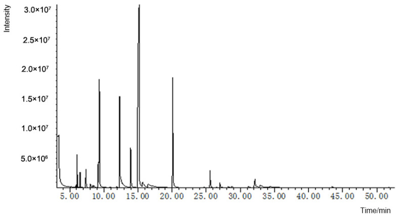
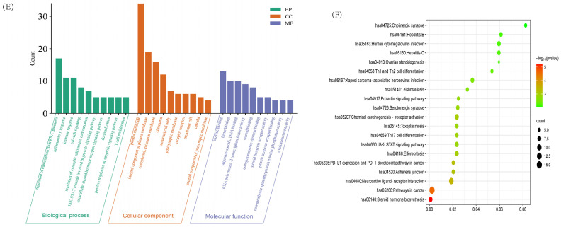
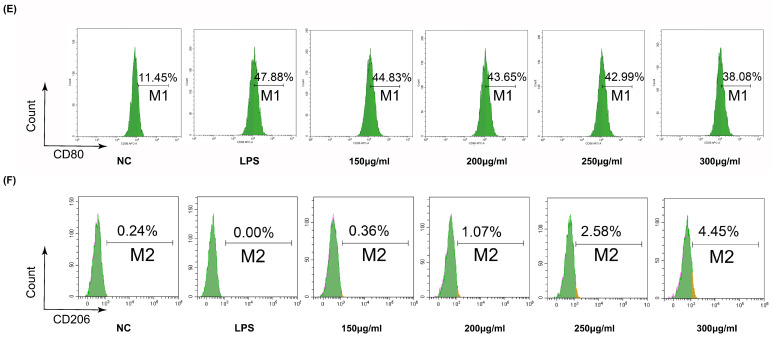
Skin wounds, leading to infections and death, have a huge negative impact on healthcare systems around the world. Antibacterial therapy and the suppression of excessive inflammation help wounds heal. To date, the application of wound dressings, biologics and biomaterials (hydrogels, epidermal growth factor, stem cells, etc.) is limited due to their difficult and expensive preparation process. Cinnamomum burmannii (Nees & T. Nees) Blume is an herb in traditional medicine, and its essential oil is rich in D-borneol, with antibacterial and anti-inflammatory effects. However, it is not clear whether Cinnamomum burmannii essential oil has the function of promoting wound healing. This study analyzed 32 main components and their relative contents of essential oil using GC-MS. Then, network pharmacology was used to predict the possible targets of this essential oil in wound healing. We first proved this essential oil's effects in vitro and in vivo. Cinnamomum burmannii essential oil could not only promote the proliferation and migration of skin stromal cells, but also promote M2-type polarization of macrophages while inhibiting the expression of pro-inflammatory cytokines. This study explored the possible mechanism by which Cinnamomum burmannii essential oil promotes wound healing, providing a cheap and effective strategy for promoting wound healing.

Keywords: Cinnamomum burmannii; essential oil; macrophages; wound healing.


Conflict of interest statement

The authors declare no conflict of interest.

Figures

Figure 1
Figure 1
GC-MS analysis of Cinnamomum burmannii essential oil total ion flow diagram.
Figure 2
Figure 2
Network pharmacology predicts the possible factors of BEO promoting wound healing. (A) Compound–disease targets were intersected using a Venn diagram. (B) The herb–component–target network for Cinnamomum burmannii essential oil in wound-healing treatment. Yellow: herb; Pink: component; Purple: target. (C) The top 20 core genes of the PPI network. (D) Protein–protein interaction (PPI) network of intersecting targets. Green: protein target; Yellow: core protein target; Pink: Visual analysis of core protein target interactions. (E) GO analysis. (F) KEGG analysis. BP: biological process, CC: cellular component, MF: molecular function.
Figure 2
Figure 2
Network pharmacology predicts the possible factors of BEO promoting wound healing. (A) Compound–disease targets were intersected using a Venn diagram. (B) The herb–component–target network for Cinnamomum burmannii essential oil in wound-healing treatment. Yellow: herb; Pink: component; Purple: target. (C) The top 20 core genes of the PPI network. (D) Protein–protein interaction (PPI) network of intersecting targets. Green: protein target; Yellow: core protein target; Pink: Visual analysis of core protein target interactions. (E) GO analysis. (F) KEGG analysis. BP: biological process, CC: cellular component, MF: molecular function.
Figure 3
Figure 3
Effects of BEO on fibroblasts and epithelial cells in vitro. (A,B) The proliferation effect of BEO on HaCaT and L929 cells was determined by CCK8. Wound scratch assay of L929 cells and HaCaT cells, which were incubated with PBS (Ctrl), 150 μg/mL, 200 μg/mL, 250 μg/mL and 300 μg/mL BEO for 48 h. Migration rate (C,D) and representative scratch images (E,F) of each group are shown. Scale bar: 100 μm. n = 3. *, p < 0.05; **, p < 0.01; ****, p < 0.0001; ns, not significant.
Figure 3
Figure 3
Effects of BEO on fibroblasts and epithelial cells in vitro. (A,B) The proliferation effect of BEO on HaCaT and L929 cells was determined by CCK8. Wound scratch assay of L929 cells and HaCaT cells, which were incubated with PBS (Ctrl), 150 μg/mL, 200 μg/mL, 250 μg/mL and 300 μg/mL BEO for 48 h. Migration rate (C,D) and representative scratch images (E,F) of each group are shown. Scale bar: 100 μm. n = 3. *, p < 0.05; **, p < 0.01; ****, p < 0.0001; ns, not significant.
Figure 4
Figure 4
BEO attenuated the inflammatory response in RAW 264.7 cells stimulated by LPS. (A) qPCR of IL-1β, IL-6 and TNF-α levels in RAW 264.7 cells that were stimulated by LPS at 24 h from different groups. (B) The level of IL-6 and TNF-α secreted by RAW 264.7 cells treated with BEO was determined by ELISA. (C) Effects of 150 μg/mL, 200 μg/mL, 250 μg/mL and 300 μg/mL BEO on the expression of NF-κB/p-IκBα protein. (D) The cell typing of RAW 264.7 was detected by flow cytometry. Polarization rate (D) and sectional images (E,F) of each group are shown. n = 3. *, p < 0.05; **, p < 0.01; ***, p < 0.001; **** p < 0.0001; ns, not significant.
Figure 4
Figure 4
BEO attenuated the inflammatory response in RAW 264.7 cells stimulated by LPS. (A) qPCR of IL-1β, IL-6 and TNF-α levels in RAW 264.7 cells that were stimulated by LPS at 24 h from different groups. (B) The level of IL-6 and TNF-α secreted by RAW 264.7 cells treated with BEO was determined by ELISA. (C) Effects of 150 μg/mL, 200 μg/mL, 250 μg/mL and 300 μg/mL BEO on the expression of NF-κB/p-IκBα protein. (D) The cell typing of RAW 264.7 was detected by flow cytometry. Polarization rate (D) and sectional images (E,F) of each group are shown. n = 3. *, p < 0.05; **, p < 0.01; ***, p < 0.001; **** p < 0.0001; ns, not significant.
Figure 5
Figure 5
BEO accelerates skin wound healing in mice. (A) Representative images of skin wounds of mice treated by PBS, EGF, 10% BEO, 20% BEO and 20% BEO after 8 days. (B,C) PBS, EGF, 10% BEO, 20% BEO, and 20% BEO groups. n = 3. (D) Representative images of the healed skin area on day 8 with hematoxylin–eosin (HE), Masson trichromatic staining and immunohistochemistry of α-SMA and CD3 antibodies; scale bar: 200 μm. *, p < 0.05; **, p < 0.01; ns, not significant.
Figure 5
Figure 5
BEO accelerates skin wound healing in mice. (A) Representative images of skin wounds of mice treated by PBS, EGF, 10% BEO, 20% BEO and 20% BEO after 8 days. (B,C) PBS, EGF, 10% BEO, 20% BEO, and 20% BEO groups. n = 3. (D) Representative images of the healed skin area on day 8 with hematoxylin–eosin (HE), Masson trichromatic staining and immunohistochemistry of α-SMA and CD3 antibodies; scale bar: 200 μm. *, p < 0.05; **, p < 0.01; ns, not significant.
Figure 6
Figure 6
BEO reduces inflammatory factor expression, as well as promoting CD206 mRNA expression. (A,B) The types of spleen T cells were detected by flow cytometry. (C) q-PCR levels of CD-80 and CD-206 in each group on day 8 of the wound surface. (D) q-PCR levels of IL-1β, IL-6, and TNF-α in each group on day 8 of the wound surface. *, p < 0.05; **, p < 0.01; ***, p < 0.001; ****, p < 0.0001; ns, not significant.
Figure 6
Figure 6
BEO reduces inflammatory factor expression, as well as promoting CD206 mRNA expression. (A,B) The types of spleen T cells were detected by flow cytometry. (C) q-PCR levels of CD-80 and CD-206 in each group on day 8 of the wound surface. (D) q-PCR levels of IL-1β, IL-6, and TNF-α in each group on day 8 of the wound surface. *, p < 0.05; **, p < 0.01; ***, p < 0.001; ****, p < 0.0001; ns, not significant.
Figure 7
Figure 7
Graphical abstract: mechanism by which Cinnamomum burmannii essential oils promote wound healing.

Similar articles

Cited by

References

    1. Deng P., Yao L., Chen J., Tang Z., Zhou J. Chitosan-based hydrogels with injectable, self-healing and antibacterial properties for wound healing. Carbohydr. Polym. 2022;276:118718. doi: 10.1016/j.carbpol.2021.118718. - DOI - PubMed
    1. Kour H., Raina R., Verma P.K., Khan A.M., Bhat M.A., Nashiruddullah N. Evaluation of the wound healing activity of ethanolic extract of Bergenia ciliata (Haw.) Sternb. rhizome with excision wound model in Wistar rats. J. Ethnopharmacol. 2021;281:114527. doi: 10.1016/j.jep.2021.114527. - DOI - PubMed
    1. Sen C.K., Gordillo G.M., Roy S., Kirsner R., Lambert L., Hunt T.K., Gottrup F., Gurtner G.C., Longaker M.T. Human skin wounds: A major and snowballing threat to public health and the economy. Wound Repair Regen. 2009;17:763. doi: 10.1111/j.1524-475X.2009.00543.x. - DOI - PMC - PubMed
    1. Garraud O., Hozzein W.N., Badr G. Wound healing: Time to look for intelligent, ‘natural’ immunological approaches? BMC Immunol. 2017;18((Suppl. S1)):23. doi: 10.1186/s12865-017-0207-y. - DOI - PMC - PubMed
    1. Stan D., Tanase C., Avram M., Apetrei R., Mincu N.B., Mateescu A.L., Stan D. Wound healing applications of creams and “smart” hydrogels. Exp. Dermatol. 2021;30:1218. doi: 10.1111/exd.14396. - DOI - PMC - PubMed

LinkOut - more resources