. 2024 May 4;29(9):2134.
doi: 10.3390/molecules29092134.

Stress Granule Core Protein-Derived Peptides Inhibit Assembly of Stress Granules and Improve Sorafenib Sensitivity in Cancer Cells

Affiliations

Stress Granule Core Protein-Derived Peptides Inhibit Assembly of Stress Granules and Improve Sorafenib Sensitivity in Cancer Cells

Juan Li et al. Molecules. .

Abstract

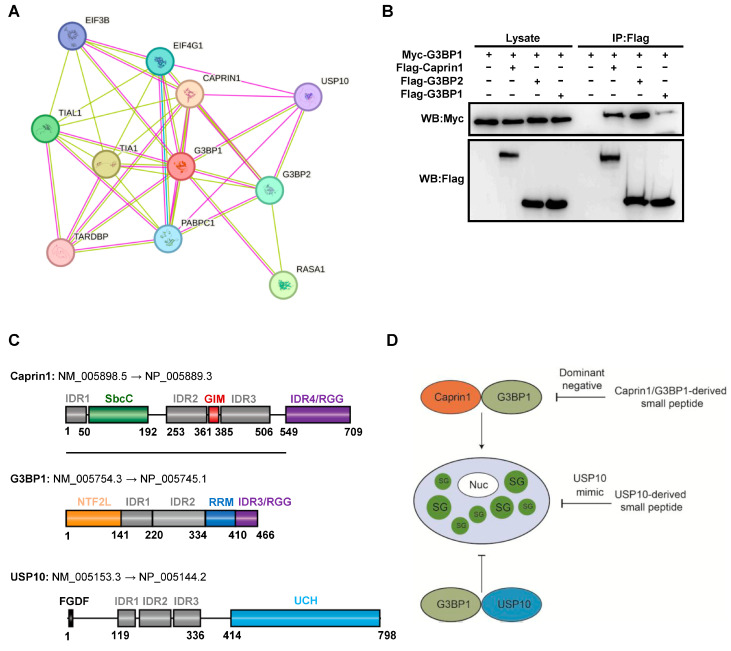
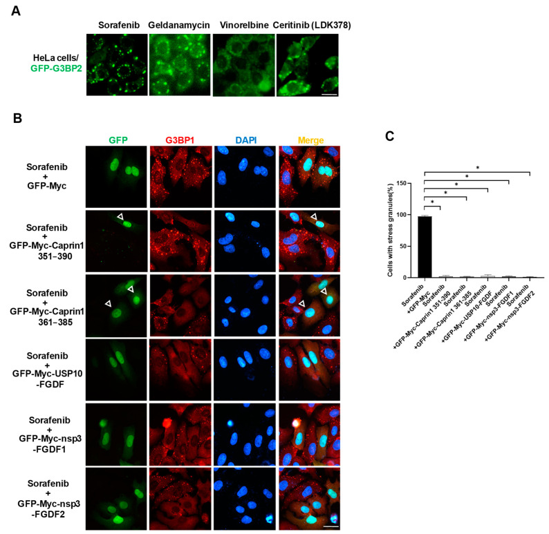
Upon a variety of environmental stresses, eukaryotic cells usually recruit translational stalled mRNAs and RNA-binding proteins to form cytoplasmic condensates known as stress granules (SGs), which minimize stress-induced damage and promote stress adaptation and cell survival. SGs are hijacked by cancer cells to promote cell survival and are consequently involved in the development of anticancer drug resistance. However, the design and application of chemical compounds targeting SGs to improve anticancer drug efficacy have rarely been studied. Here, we developed two types of SG inhibitory peptides (SIPs) derived from SG core proteins Caprin1 and USP10 and fused with cell-penetrating peptides to generate TAT-SIP-C1/2 and SIP-U1-Antp, respectively. We obtained 11 SG-inducing anticancer compounds from cell-based screens and explored the potential application of SIPs in overcoming resistance to the SG-inducing anticancer drug sorafenib. We found that SIPs increased the sensitivity of HeLa cells to sorafenib via the disruption of SGs. Therefore, anticancer drugs which are competent to induce SGs could be combined with SIPs to sensitize cancer cells, which might provide a novel therapeutic strategy to alleviate anticancer drug resistance.

Keywords: Caprin1; G3BP1; USP10; anticancer drug resistance; sorafenib; stress granules.


Conflict of interest statement

The authors declare that the research was conducted in the absence of any commercial or financial relationships that could be construed as potential conflicts of interest.

Figures

Figure 1
Figure 1
Design strategy for SG core protein-derived SG inhibitory peptides. (A) G3BP1 protein–protein interaction network. (B) Co-IP showing the G3BP1 and Caprin1 interaction and G3BP1/2 dimerization. HEK293T cells were transfected with indicated expression plasmids and cell lysates were subjected to Co-IP using the anti-Flag antibody, followed by Western blot analysis. (C) Schematic representation of functional domains in human Caprin1, G3BP1, and USP10 indicated in different colored boxes. IDR, intrinsically disordered region; GIM, G3BP1-interacting motif; RRM, RNA recognition motif; UCH, Ubiquitin C-terminal hydrolase. (D) Schematic representation of design rationale for SIPs derived from SG core proteins Caprin1, G3BP1, and USP10.
Figure 2
Figure 2
Identification of Caprin1-derived SG inhibitory peptides. (A) Co-IP showing the interaction between G3BP1 and serial fragments of Caprin1. HEK293T cells were transfected with indicated expression plasmids and cell lysates were subjected to Co-IP using the anti-Myc antibody, followed by Western blot analysis. (B,C) GST pull-down showing the interaction between G3BP1 and serial fragments or internal deletions of Caprin1. HEK293T cells were transfected with indicated expression plasmids and cell lysates were subjected to GST pull-down using GST-G3BP1 beads, followed by Western blot analysis. (D) GST pull-down showing that Caprin1 fragment 361–385 competed with full-length Caprin1 to bind G3BP1 in a dose-dependent manner. HEK293T cells were transfected with indicated expression plasmids and cell lysates were subjected to GST pull-down using GST or GST-G3BP1 beads, followed by Western blot analysis. (E) Statistical analysis of the binding efficiency of full-length Caprin1 to G3BP1 shown in panel (D). * p ≤ 0.05.
Figure 3
Figure 3
Overexpression of Caprin1-derived fragments inhibits SG assembly. (A) Amino acid sequences of Caprin1 351–390 (SIP-C1) and 361–385 (SIP-C2). (B) Immunofluorescence showing that overexpression of Caprin1 fragments 351–390 and 361–385 inhibited AS-induced SGs. HeLa cells were transfected with indicated expression plasmids, treated with 0.5 mM AS for 45 min and subjected to immunofluorescence staining using the anti-G3BP1 antibody. Solid triangles indicate cells with SGs while empty triangles indicate cells without SGs. Scale bars: 20 µm. (C) Statistical analysis of SG induction efficiency shown in panel (B), which is reflected by the percentage of SG-positive cells among cells successfully transfected with expression plasmids. * p ≤ 0.05.
Figure 4
Figure 4
Identification of G3BP1-derived SG inhibitory peptides. (A,B) Co-IP showing the interaction between Caprin1 and serial fragments of G3BP1. HEK293T cells were transfected with indicated expression plasmids and cell lysates were subjected to Co-IP using the anti-Myc antibody, followed by Western blot analysis. (C) Immunofluorescence showing that only overexpression of G3BP1 fragment 1–141 (NTF2L) effectively inhibited AS-induced SGs. HeLa cells were transfected with indicated expression plasmids, treated with 0.5 mM AS for 45 min and subjected to immunofluorescence staining using the anti-Caprin1 antibody. Solid triangles indicate cells with SGs while empty triangles indicate cells without SGs. Scale bars: 20  µm. (D) Statistical analysis of SG induction efficiency shown in panel (C), which is reflected by the percentage of SG-positive cells among cells successfully transfected with expression plasmids. * p ≤ 0.05.
Figure 5
Figure 5
Identification of FGDF motif-containing SG inhibitory peptides derived from USP10 and viral protein nsp3. (A) Amino acid sequences of USP10-derived SIP (USP10-FGDF) and nsp3-derived SIPs (nsp3-FGDF1 and nsp3-FGDF2) containing FGDF motif. (B) Immunofluorescence showing that overexpression of FGDF motif-containing fragments USP10-FGDF, nsp3-FGDF1, and nsp3-FGDF2 effectively inhibited AS-induced SGs. HeLa cells were transfected with indicated expression plasmids, treated with 0.5 mM AS for 45 min and subjected to immunofluorescence staining using the anti-G3BP1 antibody. Empty triangles indicate cells without SGs. Scale bars: 20 µm. (C) Statistical analysis of SG induction efficiency shown in panel (B), which is reflected by the percentage of SG-positive cells among cells successfully transfected with expression plasmids. * p ≤ 0.05.
Figure 6
Figure 6
Overexpression of SIP fragments blocks the formation of SGs induced by sorafenib. (A) Screen for SG-inducing anticancer compounds. HeLa cells stably expressing GFP-G3BP2 were treated with 10 μM compounds (geldanamycin, vinorelbine, and ceritinib) or 50 μM sorafenib for 2 h and captured for fluorescent images. (B) Immunofluorescence showing that overexpression of SIP fragments effectively inhibited sorafenib-induced SGs. HeLa cells were transfected with indicated expression plasmids, treated with 50 μM sorafenib for 2 h and subjected to immunofluorescence staining using the anti-G3BP1 antibody. Empty triangles indicate cells without SGs. Scale bars: 20 µm. (C) Statistical analysis of SG induction efficiency shown in panel (B), which is reflected by the percentage of SG-positive cells among cells successfully transfected with expression plasmids. * p ≤ 0.05.
Figure 7
Figure 7
Synthesized peptides TAT-SIP-C1/2 and SIP-U1-Antp inhibit sorafenib-induced SGs and promote the sensitivity to sorafenib in HeLa cells. (A) Immunofluorescence showing that TAT-SIP-C1/2 and SIP-U1-Antp significantly reduced sorafenib-induced SGs. HeLa cells were pretreated with peptides for 2 h followed by incubation with 50 μM sorafenib for an additional 2 h, and then subjected to immunofluorescence staining using the anti-G3BP1 antibody. Nuclei of HeLa cells were co-stained with DAPI. Scale bars: 20 µm. (B) Statistical analysis of SG induction efficiency shown in panel (A), which is reflected by the percentage of SG-positive cells among all cells. (C) Annexin V/PI flow cytometry assay showing that TAT-SIP-C1/2 and SIP-U1-Antp significantly increased sorafenib-induced cell death. HeLa cells were pretreated with peptides for 2 h followed by incubation with 50 μM sorafenib for an additional 2 h, and then subjected to Annexin V/PI flow cytometry assay. (D) Statistical analysis of cell death shown in panel (C), which is reflected by the percentage of dead cells (a combined population of Annexin V+PI+, Annexin V+PI, and Annexin VPI+) among all cells. * p ≤ 0.05.
Figure 8
Figure 8
Working model of SIPs that increase the efficacy of sorafenib in cancer cells. Sorafenib imposes stress on cancer cells, which respond to induce SGs that promote stress adaptation and cell survival. SIP-C1/2 and SIP-U1 derived from SG core proteins Caprin1 and USP10 exert a dominant effect and USP10-mimic effect on the SG-promoting function of G3BP1, leading to inhibition on SG assembly and cell survival. Combined treatment of SG-inducing anticancer drugs with SIPs might alleviate SG-associated drug resistance.

Similar articles

Cited by

References

    1. Anderson P., Kedersha N., Ivanov P. Stress granules, P-bodies and cancer. Biochim. Biophys. Acta. 2015;1849:861–870. doi: 10.1016/j.bbagrm.2014.11.009. - DOI - PMC - PubMed
    1. El-Naggar A.M., Sorensen P.H. Translational control of aberrant stress responses as a hallmark of cancer. J. Pathol. 2018;244:650–666. doi: 10.1002/path.5030. - DOI - PubMed
    1. Wang F., Li J., Fan S., Jin Z., Huang C. Targeting stress granules: A novel therapeutic strategy for human diseases. Pharmacol. Res. 2020;161:105143. doi: 10.1016/j.phrs.2020.105143. - DOI - PMC - PubMed
    1. Decker C.J., Parker R. P-bodies and stress granules: Possible roles in the control of translation and mRNA degradation. Cold Spring Harb. Perspect. Biol. 2012;4:a012286. doi: 10.1101/cshperspect.a012286. - DOI - PMC - PubMed
    1. Wheeler J.R., Matheny T., Jain S., Abrisch R., Parker R. Distinct stages in stress granule assembly and disassembly. eLife. 2016;5:e18413. doi: 10.7554/eLife.18413. - DOI - PMC - PubMed

MeSH terms

LinkOut - more resources