

Poria cocos Attenuated DSS-Induced Ulcerative Colitis via NF-κB Signaling Pathway and Regulating Gut Microbiota
- PMID: 38731645
- PMCID: PMC11085930
- DOI: 10.3390/molecules29092154
Poria cocos Attenuated DSS-Induced Ulcerative Colitis via NF-κB Signaling Pathway and Regulating Gut Microbiota
Abstract
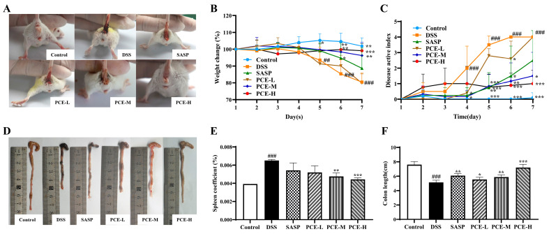
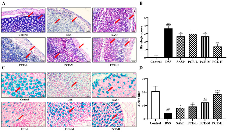
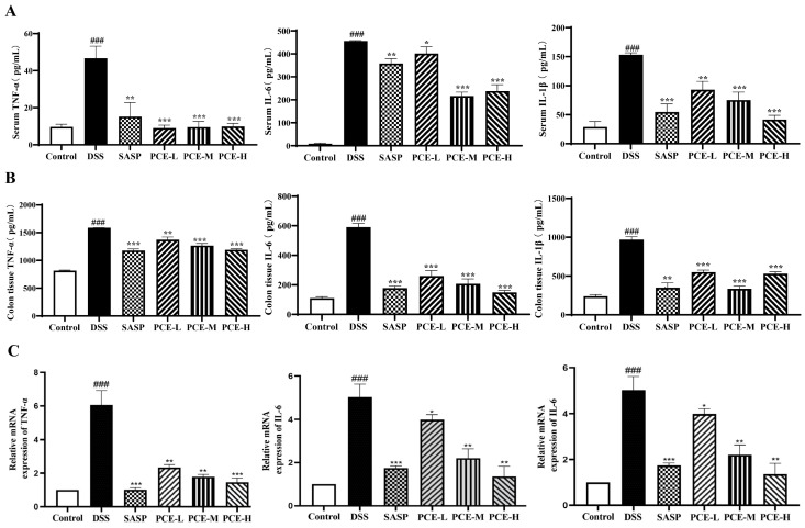
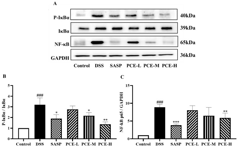
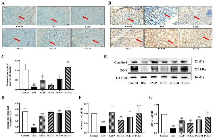
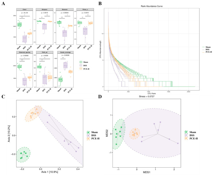
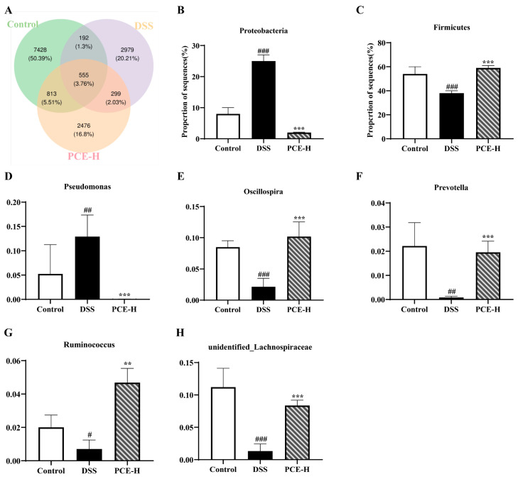
Ulcerative colitis (UC), as a chronic inflammatory disease, presents a global public health threat. However, the mechanism of Poria cocos (PC) in treating UC remains unclear. Here, LC-MS/MS was carried out to identify the components of PC. The protective effect of PC against UC was evaluated by disease activity index (DAI), colon length and histological analysis in dextran sulfate sodium (DSS)-induced UC mice. ELISA, qPCR, and Western blot tests were conducted to assess the inflammatory state. Western blotting and immunohistochemistry techniques were employed to evaluate the expression of tight junction proteins. The sequencing of 16S rRNA was utilized for the analysis of gut microbiota regulation. The results showed that a total of fifty-two nutrients and active components were identified in PC. After treatment, PC significantly alleviated UC-associated symptoms including body weight loss, shortened colon, an increase in DAI score, histopathologic lesions. PC also reduced the levels of inflammatory cytokines TNF-α, IL-6, and IL-1β, as evidenced by the suppressed NF-κB pathway, restored the tight junction proteins ZO-1 and Claudin-1 in the colon, and promoted the diversity and abundance of beneficial gut microbiota. Collectively, these findings suggest that PC ameliorates colitis symptoms through the reduction in NF-κB signaling activation to mitigate inflammatory damage, thus repairing the intestinal barrier, and regulating the gut microbiota.
Keywords: Poria cocos; gut microbiota; intestinal barrier function; ulcerative colitis.
Conflict of interest statement
The authors declare no conflicts of interest.
Figures
Similar articles
-
Trilobatin ameliorates dextran sulfate sodium-induced ulcerative colitis in mice via the NF-κB pathway and alterations in gut microbiota.PLoS One. 2024 Jun 24;19(6):e0305926. doi: 10.1371/journal.pone.0305926. eCollection 2024. PLoS One. 2024. PMID: 38913606 Free PMC article.
-
Poria cocos polysaccharide alleviates dextran sulphate sodium-induced ulcerative colitis in mice by modulating intestinal inflammatory responses and microbial dysbiosis.Int J Biol Macromol. 2024 Dec;283(Pt 2):137450. doi: 10.1016/j.ijbiomac.2024.137450. Epub 2024 Nov 8. Int J Biol Macromol. 2024. PMID: 39522895
-
Ethanol extract of Centella asiatica alleviated dextran sulfate sodium-induced colitis: Restoration on mucosa barrier and gut microbiota homeostasis.J Ethnopharmacol. 2021 Mar 1;267:113445. doi: 10.1016/j.jep.2020.113445. Epub 2020 Oct 3. J Ethnopharmacol. 2021. PMID: 33022343
-
Exopolysaccharide of Levilactobacillus brevis M-10 Improved Physiological and Biochemical Indicators and Gut Microbiota in DSS-Induced Colitis Mice.Curr Microbiol. 2025 Mar 24;82(5):204. doi: 10.1007/s00284-025-04190-5. Curr Microbiol. 2025. PMID: 40126646 Review.
-
Research Progress in Ulcerative Colitis: The Role of Traditional Chinese Medicine on Gut Microbiota and Signaling Pathways.Am J Chin Med. 2024;52(8):2277-2336. doi: 10.1142/S0192415X24500885. Epub 2024 Dec 31. Am J Chin Med. 2024. PMID: 39756829 Review.
Cited by
-
The Role of Wolfiporia cocos (F. A. Wolf) Ryvarden and Gilb. Polysaccharides in Regulating the Gut Microbiota and Its Health Benefits.Molecules. 2025 Mar 7;30(6):1193. doi: 10.3390/molecules30061193. Molecules. 2025. PMID: 40141970 Free PMC article. Review.
-
Huanglian Ejiao Decoction Alleviates Ulcerative Colitis in Mice Through Regulating the Gut Microbiota and Inhibiting the Ratio of Th1 and Th2 Cells.Drug Des Devel Ther. 2025 Jan 17;19:303-324. doi: 10.2147/DDDT.S468608. eCollection 2025. Drug Des Devel Ther. 2025. PMID: 39845151 Free PMC article.
-
Immune dysregulation in ulcerative colitis: pathogenic mechanisms and therapeutic strategies of traditional Chinese medicine.Front Cell Dev Biol. 2025 Jun 5;13:1610435. doi: 10.3389/fcell.2025.1610435. eCollection 2025. Front Cell Dev Biol. 2025. PMID: 40538978 Free PMC article. Review.
References
MeSH terms
Substances
Grants and funding
- 2023AH051751/Natural Science Foundation of the Higher Education Institutions of Anhui Province
- 2023CXMMTCM007/Research Funds of Center for Xin'an Medicine and Modernization of Traditional Chinese Medi-cine of IHM
- GXXT-2019-043/The University Synergy Innovation Program of Anhui Province
- 2022jc33/Key Science and Technology Program of Wuhu City
- 2023yf090/Key Science and Technology Program of Wuhu City
LinkOut - more resources
Full Text Sources
Medical
