. 2024 Apr 25;25(9):4694.
doi: 10.3390/ijms25094694.

Single-Cell Transcriptional Profile Construction of Rat Pituitary Glands before and after Sexual Maturation and Identification of Novel Marker Spp1 in Gonadotropes

Affiliations

Single-Cell Transcriptional Profile Construction of Rat Pituitary Glands before and after Sexual Maturation and Identification of Novel Marker Spp1 in Gonadotropes

Qing-Hua Huang et al. Int J Mol Sci. .

Abstract

The mammalian pituitary gland drives highly conserved physiological processes such as somatic cell growth, pubertal transformation, fertility, and metabolism by secreting a variety of hormones. Recently, single-cell transcriptomics techniques have been used in pituitary gland research. However, more studies have focused on adult pituitary gland tissues from different species or different sexes, and no research has yet resolved cellular differences in pituitary gland tissue before and after sexual maturation. Here, we identified a total of 15 cell clusters and constructed single-cell transcriptional profiles of rats before and after sexual maturation. Furthermore, focusing on the gonadotrope cluster, 106 genes were found to be differentially expressed before and after sexual maturation. It was verified that Spp1, which is specifically expressed in gonadotrope cells, could serve as a novel marker for this cell cluster and has a promotional effect on the synthesis and secretion of follicle-stimulating hormone. The results provide a new resource for further resolving the regulatory mechanism of pituitary gland development and pituitary hormone synthesis and secretion.

Keywords: SPP1; puberty; rat pituitary; sexual maturity; single-cell RNA sequencing.


Conflict of interest statement

The authors declare no conflicts of interest.

Figures

Figure 1
Figure 1
Comparison of pituitary hormone secretion levels in 4- and 12-week-old rats. (A) ELISA to detect the secretion of FSH in 4- and 12-week-old rats (n = 8). (B) ELISA to detect the secretion of LH in 4- and 12-week-old rats (n = 8). (C) ELISA to detect the secretion of GH in 4- and 12-week-old rats (n = 8). (D) ELISA to detect the secretion of ACTH in 4- and 12-week-old rats (n = 8). (E) ELISA to detect the secretion of TSH in 4- and 12-week-old rats (n = 8). (F) ELISA to detect the secretion of PRL in 4- and 12-week-old rats (n = 8). *, p < 0.05; **, p < 0.01; ***, p < 0.001.
Figure 2
Figure 2
The major cell clusters identified in male rats’ pituitary. (A,B) UMAP visualization of unintergrated clusters. (C) Manual assignment of cells to expected cell types based on gene expression of known markers. (D) Heatmap of cluster marker genes. Heatmap shows the top 5 average expressions of cluster marker genes. (E) The Dot-plot of the percentage (pct. exp.) and average expression (avg. exp.) of marker genes. Cort, corticotropes; End, endothelial; Epi, epithelial; Gon, gonadotrope; Imm, immune cells; Lac, lactotropes; Mel, melanotropes; Mes, mesenchymal; Mki67+, Mki67+ proliferating cells; Per, pericytes; PL, posterior lobe; Som, somatotropes; SomLac, somatolactotropes; Fsc, folliculostellate cells.
Figure 3
Figure 3
UMAP maps of marker genes in distinct pituitary hormone cell clusters. Classical pituitary endocrine cell markers are all shown by UMAP maps. Color bar indicates natural log transformed normalized expression.
Figure 4
Figure 4
Cell type composition between 4- and 12-week-old male rat pituitary cells. (A) UMAP visualization of 4- and 12-week-old male rat pituitary cells. Dots: single cells. (B) Bar graph of cell type composition between 4- and 12-week-old male rats.
Figure 5
Figure 5
Enrichment analysis of differentially expressed genes in gonadotrope cells before and after sexual maturation. (A) Volcanic map of differentially expressed genes in the gonadotrope cell. (B) Major enrichment and meaningful GO terms of differentially expressed genes in the gonadotrope cell.
Figure 6
Figure 6
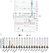
Validation of differentially expressed genes. (A) Differential expression of selected genes in sequencing results. The orange column represents pituitary tissue samples of 4-week-old rats, and the green column represents pituitary tissue samples of 12-week-old rats. (B) Validation of differentially expressed genes by RT-qPCR. The black column represents pituitary tissue samples of 4-week-old rats, and the yellow column represents pituitary tissue samples of 12-week-old rats. ns, no statistical difference; *, p < 0.05; **, p < 0.01; ***, p < 0.001.
Figure 7
Figure 7
Spp1 promotes FSH synthesis and secretion. (A) Pituitary single-cell transcriptomic analysis of Spp1 mRNA enrichment in different cell populations. (B) Immunofluorescence analysis OPN (green), FSHB (red), and LHB (purple) in pituitary tissue of 4-week-old and 12-week-old rats. The white arrows point to representative co-localization. (C) RT-qPCR analysis Spp1 mRNA expression in LβT2 cells transfected with siNC and three Spp1 siRNAs (n = 3). (D) RT-qPCR analysis Fshb mRNA expression transfected with siNC and Spp1 siRNA in LβT2 cells (n = 3). (E) ELISA analysis secretion level of FSH transfected with siNC and Spp1 siRNA in LβT2 cells (n = 9). (F) RT-qPCR analysis Spp1 mRNA expression in LβT2 cells transfected with vector and different quality of Spp1 plasmid (n = 3). (G) RT-qPCR analysis Fshb mRNA expression transfected with vector and Spp1 plasmid (1000 ng) in LβT2 cells (n = 3). (H) ELISA analysis secretion level of FSH transfected with vector and Spp1 plasmid (1000 ng) in LβT2 cells (n = 9). **, p < 0.01; ***, p < 0.001. Cort, corticotropes; End, endothelial; Epi, epithelial; Gon, gonadotrope; Imm, immune cells; Lac, lactotropes; Mel, melanotropes; Mes, mesenchymal; Mki67+, Mki67+ proliferating cells; Per, pericytes; PL, posterior lobe; Som, somatotropes; SomLac, somatolactotropes; Fsc, folliculostellate cells; Others, other cells.

Similar articles

References

    1. Alatzoglou K.S., Gregory L.C., Dattani M.T. Development of the Pituitary Gland. Compr. Physiol. 2020;10:389–413. doi: 10.1002/cphy.c150043. - DOI - PubMed
    1. Sheng H.Z., Moriyama K., Yamashita T., Li H., Potter S.S., Mahon K.A., Westphal H. Multistep control of pituitary organogenesis. Science. 1997;278:1809–1812. doi: 10.1126/science.278.5344.1809. - DOI - PubMed
    1. Pottin K., Hyacinthe C., Retaux S. Conservation, development, and function of a cement gland-like structure in the fish Astyanax mexicanus. Proc. Natl. Acad. Sci. USA. 2010;107:17256–17261. doi: 10.1073/pnas.1005035107. - DOI - PMC - PubMed
    1. Kato Y., Yoshida S., Kato T. Missing pieces of the pituitary puzzle: Participation of extra-adenohypophyseal placode-lineage cells in the adult pituitary gland. Cell Tissue Res. 2023;394:487–496. doi: 10.1007/s00441-023-03829-8. - DOI - PubMed
    1. Harris J., Gouhier A., Drouin J. Mechanisms in endocrinology: Pioneer transcription factors in pituitary development and tumorigenesis. Eur. J. Endocrinol. 2021;184:R1–R15. doi: 10.1530/EJE-20-0866. - DOI - PubMed