. 2024 Apr 26;25(9):4734.
doi: 10.3390/ijms25094734.

Cinnamaldehyde Protects against P. gingivalis Induced Intestinal Epithelial Barrier Dysfunction in IEC-6 Cells via the PI3K/Akt-Mediated NO/Nrf2 Signaling Pathway

Affiliations

Cinnamaldehyde Protects against P. gingivalis Induced Intestinal Epithelial Barrier Dysfunction in IEC-6 Cells via the PI3K/Akt-Mediated NO/Nrf2 Signaling Pathway

Chethan Sampath et al. Int J Mol Sci. .

Abstract

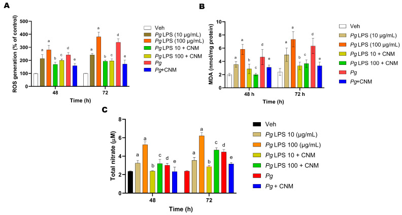
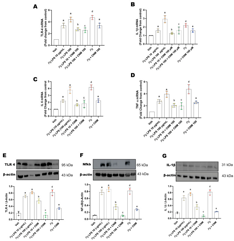
Porphyromonas gingivalis (Pg), a Gram-negative oral pathogen, promotes and accelerates periodontitis-associated gut disorders. Intestinal epithelial barrier dysfunction is crucial in the pathogenesis of intestinal and systemic diseases. In this study, we sought to elucidate the protective role of cinnamaldehyde (CNM, an activator of Nrf2) against P. gingivalis (W83) and Pg-derived lipopolysaccharide (Pg-LPS) induced intestinal epithelial barrier dysfunction via antioxidative mechanisms in IEC-6 cells. IEC-6 (ATCC, CRL-1592) cells were pretreated with or without CNM (100 µM), in the presence or absence of P. gingivalis (strain W83, 109 MOI) or Pg-LPS (1, 10, and 100 µg/mL), respectively, between 0-72 h time points by adopting a co-culture method. Intestinal barrier function, cytokine secretion, and intestinal oxidative stress protein markers were analyzed. P. gingivalis or Pg-LPS significantly (p < 0.05) increased reactive oxygen species (ROS) and malondialdehyde (MDA) levels expressing oxidative stress damage. Pg-LPS, as well as Pg alone, induces inflammatory cytokines via TLR-4 signaling. Furthermore, infection reduced Nrf2 and NAD(P)H quinone dehydrogenase 1 (NQO1). Interestingly, inducible nitric oxide synthase (iNOS) protein expression significantly (p < 0.05) increased with Pg-LPS or Pg infection, with elevated levels of nitric oxide (NO). CNM treatment suppressed both Pg- and Pg-LPS-induced intestinal oxidative stress damage by reducing ROS, MDA, and NO production. Furthermore, CNM treatment significantly upregulated the expression of tight junction proteins via increasing the phosphorylation levels of PI3K/Akt/Nrf2 suppressing inflammatory cytokines. CNM protected against Pg infection-induced intestinal epithelial barrier dysfunction by activating the PI3K/Akt-mediated Nrf2 signaling pathway in IEC-6 cells.

Keywords: Nrf2; P. gingivalis; cinnamaldehyde; inflammation; intestinal epithelial barrier; nitric oxide; oxidative stress.


Conflict of interest statement

The authors declare no conflicts of interest.

Figures

Figure 1
Figure 1
Screening of cytotoxic effects of Pg-LPS, Pg, and cinnamaldehyde (CNM) on IEC-6 cells. (A) IEC-6 cells were treated with 0, 1, 10, 100 µg/mL of Pg-LPS, Pg alone and (B) 25, 50, 100 µM of CNM for 6, 12, 24, 48, and 72 h. The cell growth inhibitory rate was measured using an MTT assay. The values are expressed as a percentage of the control (uninfected with Pg-LPS or Pg) for each time point, respectively (0 to 72 h). (CE) protective effects of CNM (100 µM) against Pg-LPS (10 and 100 µg/mL) and Pg-induced damage on IEC-6 cell viability. The results from three independent experiments are represented in the form of means ± SD (n = 3, three independent experiments and, in each experiment, we had triplicates values for all the results). * p < 0.05 compared with control (non-infected) cells. # p < 0.05 compared with control (infected) cells. PgP. Gingivalis; LPS-lipopolysaccharide.
Figure 1
Figure 1
Screening of cytotoxic effects of Pg-LPS, Pg, and cinnamaldehyde (CNM) on IEC-6 cells. (A) IEC-6 cells were treated with 0, 1, 10, 100 µg/mL of Pg-LPS, Pg alone and (B) 25, 50, 100 µM of CNM for 6, 12, 24, 48, and 72 h. The cell growth inhibitory rate was measured using an MTT assay. The values are expressed as a percentage of the control (uninfected with Pg-LPS or Pg) for each time point, respectively (0 to 72 h). (CE) protective effects of CNM (100 µM) against Pg-LPS (10 and 100 µg/mL) and Pg-induced damage on IEC-6 cell viability. The results from three independent experiments are represented in the form of means ± SD (n = 3, three independent experiments and, in each experiment, we had triplicates values for all the results). * p < 0.05 compared with control (non-infected) cells. # p < 0.05 compared with control (infected) cells. PgP. Gingivalis; LPS-lipopolysaccharide.
Figure 2
Figure 2
Cinnamaldehyde (CNM) inhibits Pg-LPS and Pg induced intracellular ROS, MDA, and NO changes in IEC-6 cells. (A) IEC-6 cells were pretreated with CNM (l00 µM) for 4 h prior to exposure to Pg-LPS or Pg at 48 and 72 h. The cells were incubated with DCFH2-DA for 15 min. ROS generation was detected by fluorescence values of ROS were measured by using a fluorescence microplate reader. The fluorescence intensity read at 48 and 72 h for the vehicle was 46.27 ± 12.34 and 57.24 ± 28.61, respectively. (B) The cells were pretreated with CNM (100 µM) for 4 h and then exposed to Pg-LPS or Pg at 48 and 72 h. The MDA levels in the supernatant were determined by using commercial kits. (C) The NO concentration in the supernatant was determined by the Griess reaction. Data are presented as the means ± SEM of three independent experiments. a, d: p < 0.05 compared to the control group; b: p < 0.05 compared to Pg-LPS 10 µg/mL, c: p < 0.05 compared to Pg-LPS 100 µg/mL, e: p < 0.01 compared to the Pg group.
Figure 3
Figure 3
Cinnamaldehyde (CNM) treatment suppresses Keap-1, activates Nrf2, NQO1, and PI3K/Akt phosphorylation protein expression in Pg-LPS and Pg induced IEC-6 cells. The IEC-6 cell was pre-incubated with CNM (100 µM) for 4 h and exposed to Pg-LPS or Pg for 72 h. (A) Keap-1, (B) Nrf2, (C) NQO1, (D) p-PI3K, and (E) p-AKT protein expressions in IEC-6 cells at 72 h. β-Actin was used as a loading control. Bar graphs showed a ratio of target gene or protein with β-actin. Data were analyzed using one-way ANOVA by using GraphPad prism software. Values are mean ± SD (n = 3). a, d: p < 0.05 compared to the control group; b: p < 0.05 compared to Pg-LPS 10 µg/mL, c: p < 0.05 compared to Pg-LPS 100 µg/mL, e: p < 0.01 compared to the Pg group.
Figure 4
Figure 4
Cinnamaldehyde (CNM) suppresses iNOS and activates GCH-1 protein expression in Pg-LPS and Pg induced IEC-6 cells. IEC-6 cell was pre-incubated with CNM (100 µM) for 4 h and exposed to Pg-LPS or Pg for 72 h. (A) iNOS and (B) GCH-1 protein expression in IEC-6 cells. Stripped blots were re-probed with β-actin. Bar graphs showed a ratio of target protein with β-actin. Data were analyzed using one-way ANOVA by using GraphPad Prism software. Bars represent mean values, with error bars representing SD. Data are for three independent experiments (n = 3). a, d: p < 0.05 compared to the control group; b: p < 0.05 compared to Pg-LPS 10 µg/mL, c: p < 0.05 compared to Pg-LPS 100 µg/mL, e: p < 0.01 compared to the Pg group.
Figure 5
Figure 5
Cinnamaldehyde (CNM) reduces cytokine expression in Pg-LPS and Pg induced IEC-6 cells. The IEC-6 cell was pre-incubated with CNM (100 µM) for 4 h and exposed either with Pg-LPS or Pg for 72 h. The cell lysates were subjected to RT-qPCR, as well as western blot analysis. (A) TLR-4, (B) IL-1β, (C) IL 6, and (D) TNF α transcript levels. Representative immunoblot and densitometric analysis data for (E) TLR-4, (F) Nfκb, and (G) IL-1β. β-Actin was used as a loading control. Stripped blots were re-probed with β-actin. Bar graphs showed a ratio of target protein with β-actin. Data were analyzed using one-way ANOVA by using GraphPad Prism software. a, d: p < 0.05 compared to the control group; b: p < 0.05 compared to Pg-LPS 10 µg/mL, c: p < 0.05 compared to Pg-LPS 100 µg/mL, e: p < 0.01 compared to the Pg group.
Figure 6
Figure 6
Effects of cinnamaldehyde (CNM) on ZO-1 and OC-1 protein expression and intestinal TJ permeability changes in Pg-LPS and Pg induced IEC-6 cells. IEC-6 cell was pre-incubated with CNM (100 µM) for 4 h and exposed to either Pg-LPS or Pg for 72 h. The cell lysates were subjected to RT-qPCR, as well as western blot analysis with an (A) anti-ZO-1 and (B) anti-OC-1, antibody. (C) The permeability of FD4 across the cell monolayer was measured after exposure to Pg-LPS and Pg, which were pre-treated with cinnamaldehyde (CNM). a, d: p < 0.05 compared to the control group; b: p < 0.05 compared to Pg-LPS 10 µg/mL, c: p < 0.05 compared to Pg-LPS 100 µg/mL, e: p < 0.01 compared to the Pg group.
Figure 7
Figure 7
Cinnamaldehyde (CNM) prevents Pg-LPS and Pg induced apoptosis in IEC-6 cells. IEC-6 cell was pre-incubated with CNM (100 µM) for 4 h and exposed to either Pg-LPS or Pg for 72 h. Representative immunoblots and densitometric analysis data for (A) Bax, and (B) Bcl2 in IEC-6 cells. Blots showing same β-actin were stripped and re-probed. Data were normalized with band intensities for β-actin. Bar graphs depict ratios of target proteins to β-actin. Data are for four independent experiments. a, d: p < 0.05 compared to the control group; b: p < 0.05 compared to Pg-LPS 10 µg/mL, c: p < 0.05 compared to Pg-LPS 100 µg/mL, e: p < 0.01 compared to the Pg group.

References

    1. Srivastava R., Srivastava M., Verma P., Gautam A. Metabolic syndrome and periodontal disease: An overview for physicians. J. Fam. Med. Prim. Care. 2019;8:3492. doi: 10.4103/jfmpc.jfmpc_866_19. - DOI - PMC - PubMed
    1. Lira-Junior R., Boström E.A. Oral-gut connection: One step closer to an integrated view of the gastrointestinal tract? Mucosal Immunol. 2018;11:316–318. doi: 10.1038/mi.2017.116. - DOI - PubMed
    1. Damgaard C., Danielsen A.K., Enevold C., Massarenti L., Nielsen C.H., Holmstrup P., Belstrøm D. Porphyromonas gingivalis in saliva associates with chronic and aggressive periodontitis. J. Oral Microbiol. 2019;11:1653123. doi: 10.1080/20002297.2019.1653123. - DOI - PMC - PubMed
    1. He J., Huang W., Pan Z., Cui H., Qi G., Zhou X., Chen H. Quantitative analysis of microbiota in saliva, supragingival, and subgingival plaque of Chinese adults with chronic periodontitis. Clin. Oral Investig. 2012;16:1579–1588. doi: 10.1007/s00784-011-0654-4. - DOI - PubMed
    1. Liu Y., Huang W., Wang J., Ma J., Zhang M., Lu X., Liu J., Kou Y. Multifaceted Impacts of Periodontal Pathogens in Disorders of the Intestinal Barrier. Front. Immunol. 2021;12:693479. doi: 10.3389/fimmu.2021.693479. - DOI - PMC - PubMed

MeSH terms

LinkOut - more resources