

Paricalcitol Has a Potent Anti-Inflammatory Effect in Rat Endothelial Denudation-Induced Intimal Hyperplasia
- PMID: 38732029
- PMCID: PMC11084681
- DOI: 10.3390/ijms25094814
Paricalcitol Has a Potent Anti-Inflammatory Effect in Rat Endothelial Denudation-Induced Intimal Hyperplasia
Abstract
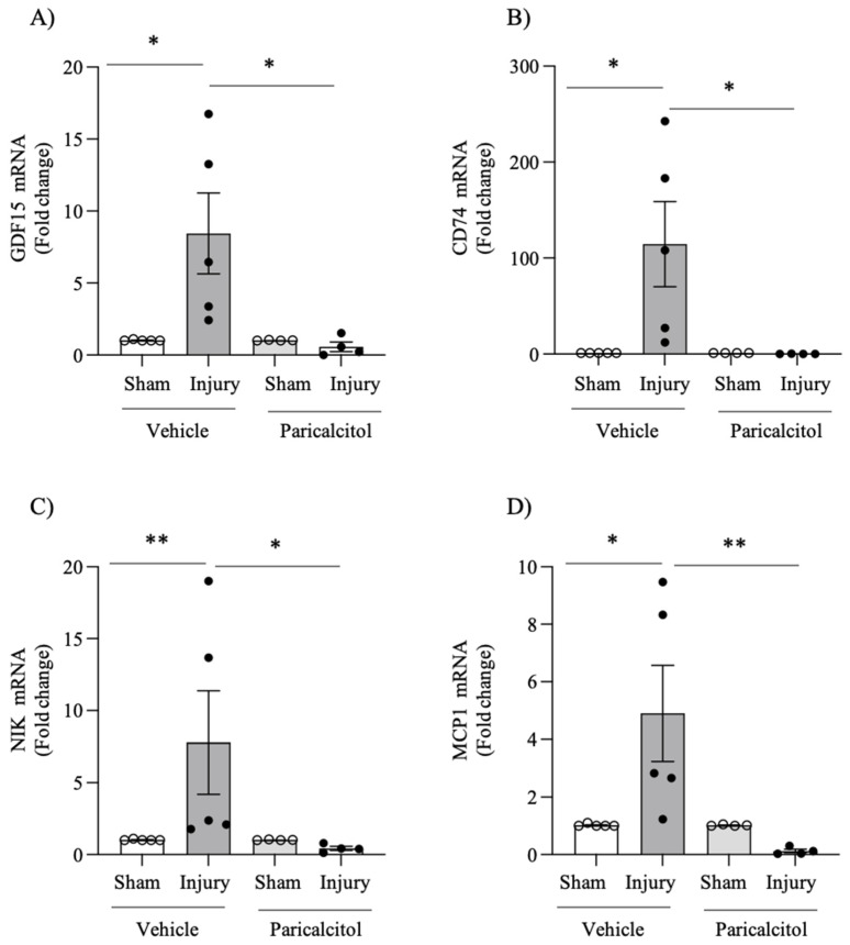
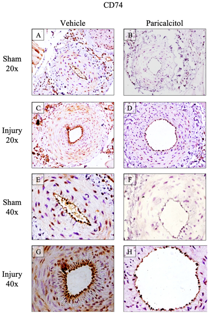
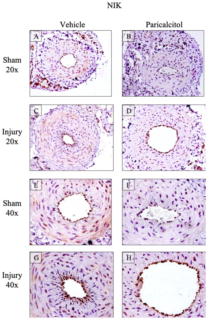
Neointimal hyperplasia is the main cause of vascular graft failure in the medium term. Vitamin D receptor activation modulates the biology of vascular smooth muscle cells and has been reported to protect from neointimal hyperplasia following endothelial injury. However, the molecular mechanisms are poorly understood. We have now explored the impact of the selective vitamin D receptor activator, paricalcitol, on neointimal hyperplasia, following guidewire-induced endothelial cell injury in rats, and we have assessed the impact of paricalcitol or vehicle on the expression of key cell stress factors. Guidewire-induced endothelial cell injury caused neointimal hyperplasia and luminal stenosis and upregulated the expression of the growth factor growth/differentiation factor-15 (GDF-15), the cytokine receptor CD74, NFκB-inducing kinase (NIK, an upstream regulator of the proinflammatory transcription factor NFκB) and the chemokine monocyte chemoattractant protein-1 (MCP-1/CCL2). Immunohistochemistry confirmed the increased expression of the cellular proteins CD74 and NIK. Paricalcitol (administered in doses of 750 ng/kg of body weight, every other day) had a non-significant impact on neointimal hyperplasia and luminal stenosis. However, it significantly decreased GDF-15, CD74, NIK and MCP-1/CCL2 mRNA expression, which in paricalcitol-injured arteries remained within the levels found in control vehicle sham arteries. In conclusion, paricalcitol had a dramatic effect, suppressing the stress response to guidewire-induced endothelial cell injury, despite a limited impact on neointimal hyperplasia and luminal stenosis. This observation identifies novel molecular targets of paricalcitol in the vascular system, whose differential expression cannot be justified as a consequence of improved tissue injury.
Keywords: inflammation; intimal hyperplasia; paricalcitol; peripheral vascular disease; vitamin D receptor.
Conflict of interest statement
A.O. has received grants from Sanofi and consultancy or speaker fees or travel support from Adviccene, Alexion, Astellas, Astrazeneca, Amicus, Amgen, Boehringer Ingelheim, Fresenius Medical Care, GSK, Bayer, Sanofi-Genzyme, Menarini, Mundipharma, Kyowa Kirin, Lilly, Freeline, Idorsia, Chiesi, Otsuka, Novo-Nordisk, Sysmex and Vifor Fresenius Medical Care Renal Pharma and Spafarma and is Director of the Catedra UAM-Astrazeneca of chronic kidney disease and electrolytes. He has stock in Telara Farma.
Figures


Similar articles
-
NIK Is a Mediator of Inflammation and Intimal Hyperplasia in Endothelial Denudation-Induced Vascular Injury.Int J Mol Sci. 2024 Oct 25;25(21):11473. doi: 10.3390/ijms252111473. Int J Mol Sci. 2024. PMID: 39519026 Free PMC article.
-
Cilostazol suppression of arterial intimal hyperplasia is associated with decreased expression of sialyl Lewis X homing receptors on mononuclear cells and E-selectin in endothelial cells.J Vasc Surg. 2012 Feb;55(2):506-16. doi: 10.1016/j.jvs.2011.07.087. J Vasc Surg. 2012. PMID: 22264805
-
Monocyte chemoattractant protein 1-mediated migration of mesenchymal stem cells is a source of intimal hyperplasia.Arterioscler Thromb Vasc Biol. 2013 Jun;33(6):1271-9. doi: 10.1161/ATVBAHA.112.300773. Epub 2013 Apr 18. Arterioscler Thromb Vasc Biol. 2013. PMID: 23599443
-
Resident Arterial Cells and Circulating Bone Marrow-Derived Cells both Contribute to Intimal Hyperplasia in a Rat Allograft Carotid Transplantation Model.Cell Physiol Biochem. 2017;42(4):1303-1312. doi: 10.1159/000478959. Epub 2017 Jul 11. Cell Physiol Biochem. 2017. Retraction in: Cell Physiol Biochem. 2018 Mar 25;45:2577. doi: 10.1159/000488305. PMID: 28715799 Retracted.
-
Postoperative Intimal Hyperplasia: Review from My Research.Int J Angiol. 2024 Feb 5;33(3):135-138. doi: 10.1055/s-0044-1779300. eCollection 2024 Sep. Int J Angiol. 2024. PMID: 39131804 Free PMC article. Review.
Cited by
-
NIK Is a Mediator of Inflammation and Intimal Hyperplasia in Endothelial Denudation-Induced Vascular Injury.Int J Mol Sci. 2024 Oct 25;25(21):11473. doi: 10.3390/ijms252111473. Int J Mol Sci. 2024. PMID: 39519026 Free PMC article.
-
Demonstration of the Protective Effect of Vinpocetine in Diabetic Cardiomyopathy.J Clin Med. 2024 Aug 8;13(16):4637. doi: 10.3390/jcm13164637. J Clin Med. 2024. PMID: 39200779 Free PMC article.
-
Immunotherapy for hypertensive end-organ damage: a new therapeutic strategy.Essays Biochem. 2025 Mar 25;0(0):EBC20243000. doi: 10.1042/EBC20243000. Essays Biochem. 2025. PMID: 40134277 Free PMC article. Review.
References
-
- Dosluoglu H.H., Lall P., Harris L.M., Dryjski M.L. Long-Term Limb Salvage and Survival after Endovascular and Open Revascularization for Critical Limb Ischemia after Adoption of Endovascular-First Approach by Vascular Surgeons. J. Vasc. Surg. 2012;56:361–371. doi: 10.1016/J.JVS.2012.01.054. - DOI - PubMed
MeSH terms
Substances
Grants and funding
LinkOut - more resources
Full Text Sources
Research Materials
Miscellaneous
