. 2024 Apr 29;25(9):4862.
doi: 10.3390/ijms25094862.

Dihydrotestosterone Augments the Angiogenic and Migratory Potential of Human Endothelial Progenitor Cells by an Androgen Receptor-Dependent Mechanism

Affiliations

Dihydrotestosterone Augments the Angiogenic and Migratory Potential of Human Endothelial Progenitor Cells by an Androgen Receptor-Dependent Mechanism

Mirel Adrian Popa et al. Int J Mol Sci. .

Abstract

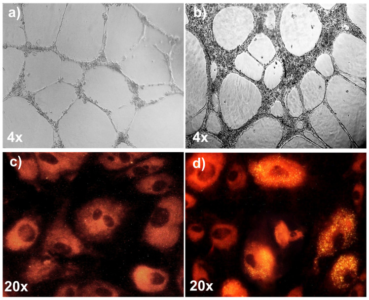
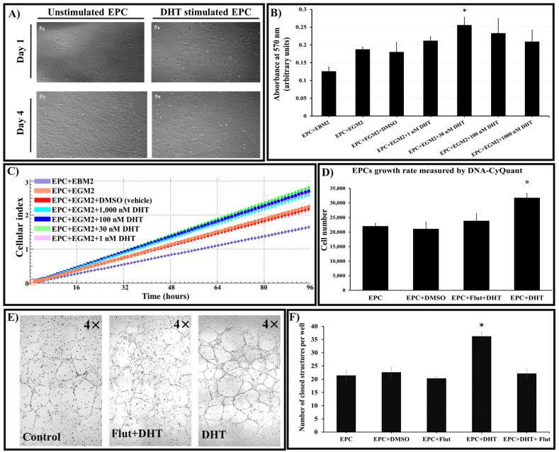
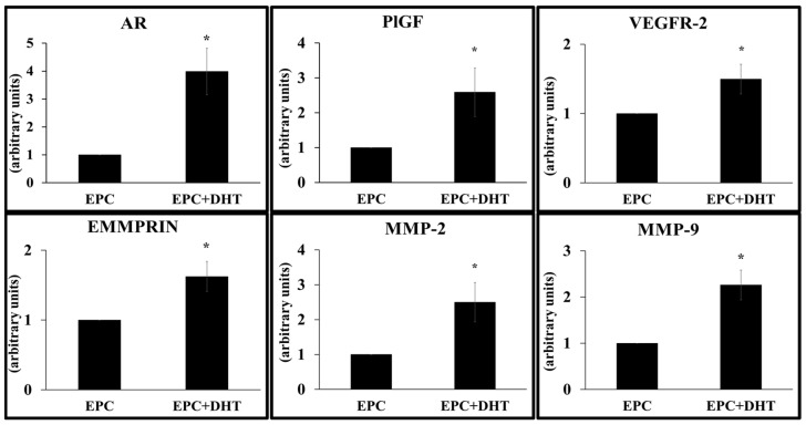
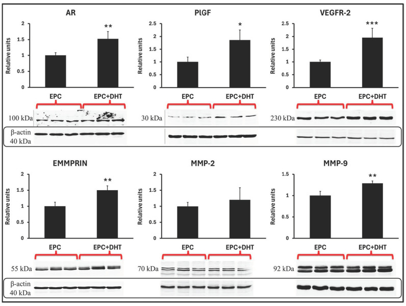
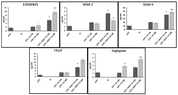
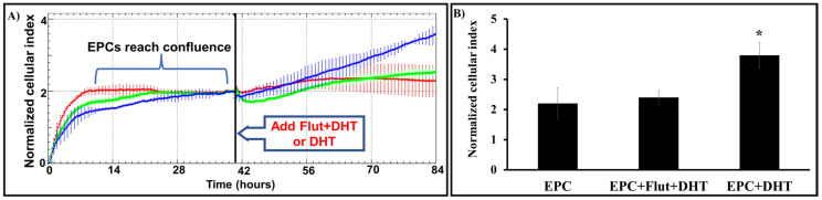
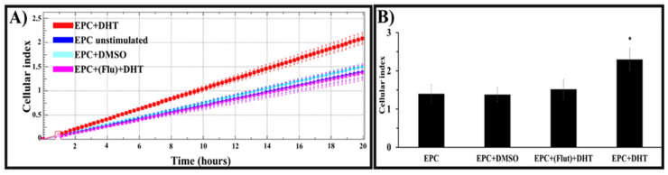
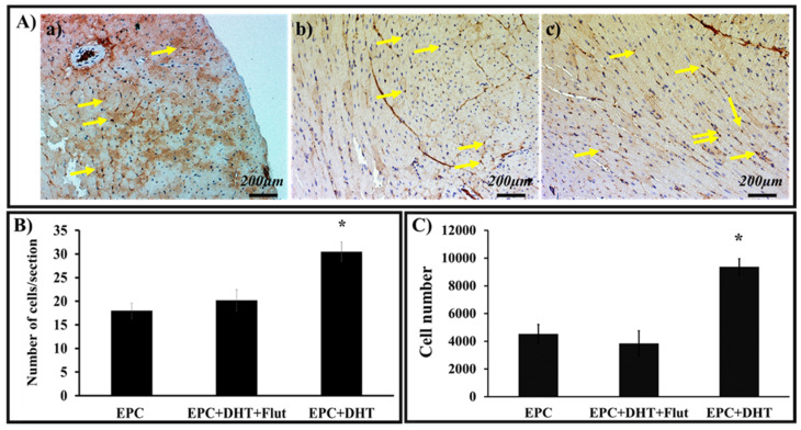
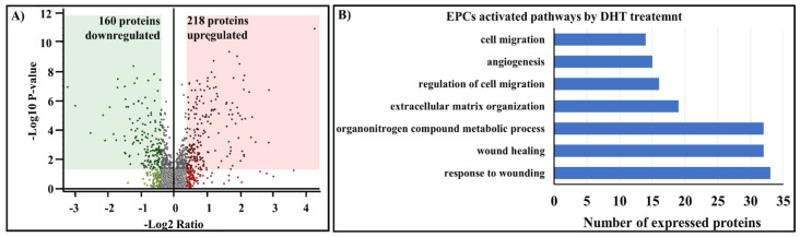
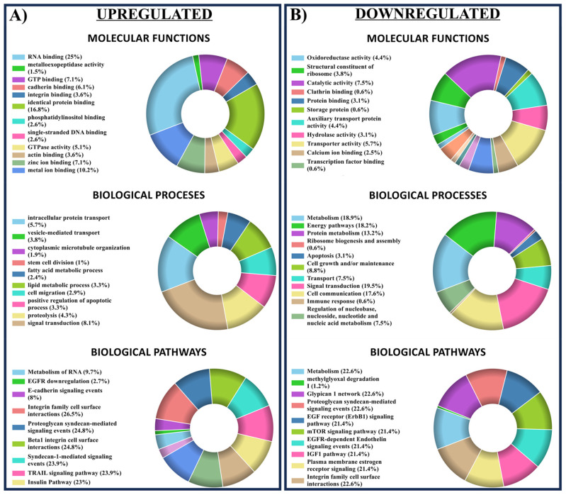
Endothelial progenitor cells (EPCs) play a critical role in cardiovascular regeneration. Enhancement of their native properties would be highly beneficial to ensuring the proper functioning of the cardiovascular system. As androgens have a positive effect on the cardiovascular system, we hypothesized that dihydrotestosterone (DHT) could also influence EPC-mediated repair processes. To evaluate this hypothesis, we investigated the effects of DHT on cultured human EPCs' proliferation, viability, morphology, migration, angiogenesis, gene and protein expression, and ability to integrate into cardiac tissue. The results showed that DHT at different concentrations had no cytotoxic effect on EPCs, significantly enhanced the cell proliferation and viability and induces fast, androgen-receptor-dependent formation of capillary-like structures. DHT treatment of EPCs regulated gene expression of androgen receptors and the genes and proteins involved in cell migration and angiogenesis. Importantly, DHT stimulation promoted EPC migration and the cells' ability to adhere and integrate into murine cardiac slices, suggesting it has a role in promoting tissue regeneration. Mass spectrometry analysis further highlighted the impact of DHT on EPCs' functioning. In conclusion, DHT increases the proliferation, migration, and androgen-receptor-dependent angiogenesis of EPCs; enhances the cells' secretion of key factors involved in angiogenesis; and significantly potentiates cellular integration into heart tissue. The data offer support for potential therapeutic applications of DHT in cardiovascular regeneration and repair processes.

Keywords: angiogenesis; dihydrotestosterone; endothelial progenitor cells; migration.


Conflict of interest statement

The authors declare no conflicts of interest.

Figures

Figure 1
Figure 1
Functional characterization of EPCs isolated from umbilical-cord blood. (a) Isolated EPCs were cultured on Matrigel® substrate. At 4 h (a) and at 24 h (b) after seeding, the cells formed robust capillary-like tubes. At 24 h after seeding, compared to controls (c) isolated EPCs avidly took up acetylated LDL (yellow dots), as assessed by fluorescence microscopy (d).
Figure 2
Figure 2
Exposure of EPCs to DHT does not alter cell morphology and significantly increases cell proliferation and function by an androgen-receptor-dependent mechanism. (A) Morphological evaluation of EPCs at days 1 and 4 after seeding showed that 30 nM DHT treatment does not alter the characteristic spindle-shaped morphology indicative of their endothelial identity. (B) EPCs were cultured in EGM2 medium supplemented with varying concentrations of DHT (1, 30, 100, and 1000 nM) or vehicle. MTT assay shows that after 96 h of stimulation, the presence of 30 nM DHT significantly increases cell proliferation compared to control (EPC+EGM2). (C) Analysis on an xCELLigence real-time cell analyzer shows that at all concentrations used, DHT induces in EPCs (cultured as above) a similar mitogenic effect and an increase in cell viability (compared to controls). (D) Assessment of EPC proliferation by quantification of DNA content using the CyQuant assay. The DHT treatment (30 nM) results in a ~30% increase in EPCs’ DNA content compared to controls (EPC and EPC+DMSO). Given prior treatment of cells with 10 µM flutamide (Flut), an androgen-receptor antagonist, DHT significantly reduces EPC proliferation, indicating a role for androgen receptors in proliferation. (E) Compared to controls, DHT-exposed EPCs exhibit increased formation of tube-like structures in Matrigel-coated wells. Note the inhibitory effect of flutamide (Flut+DHT). (F) Morphometric analyses showing that DHT treatment significantly enhances (50%) the formation of closed capillary-like structures compared to controls (EPC, EPC+DMSO); flutamide significantly reduces this property, indicating a role for AR in EPCs’ formation of tube-like structures. n = 3. * p value < 0.05. EGM2 = endothelial growth media 2; EBM2 = endothelial basal medium 2; DMSO = dimethyl sulfoxide.
Figure 3
Figure 3
Quantification of the gene expression in DHT-stimulated EPCs by qRT-PCR. EPCs exposure to 30 nM DHT (EPC+DHT) increases gene expression of androgen receptors (AR), EMMPRIN, MMP-2, MMP-9 as well as VEGFR-2 and PlGF. Data are expressed as means (±) SEM of each gene relative to GAPDH and normalized to an arbitrary value of 1. n = 3. * p value < 0.05.
Figure 4
Figure 4
Western blot analysis of proteins expressed by EPCs stimulated with DHT (EPC+DHT) shows significant increases in the levels of the proteins AR, PlGF, EMMPRIN, VEGFR-2, and MMP-9. Compared to controls (EPCs), DHT induces a significant increase in the expression of all proteins except for the MMP-2 protein. All results were normalized to levels of β-actin expression; * p value < 0.05, ** p value < 0.001, *** p value < 0.0001, n = 3.
Figure 5
Figure 5
DHT-exposed EPCs were seeded in the upper chamber of a Transwell plate, and the heart tissue slices (Si) were placed in the lower chamber. After 24 and 48 h, the medium was collected and centrifuged and the proteins in the supernatant were quantified using Human Angiogenesis Kit Panel A—Luminex assay. Note that the secretion of the proteins EMMPRIN, MMP-9, angiogenin, and VEGF is significantly increased in the media collected from DHT-stimulated EPCs (EPC+DHT+Si 24 h/48 h) upon indirect contact with heart fragments compared to controls (EPC+Si 24 h/48 h). * p < 0.05; ** p < 0.001. n = 3.
Figure 6
Figure 6
DHT increases the migration capacity of EPCs by a mechanism dependent on androgen receptors. The cells plated on our E-Plate patented device reached confluence, and after ~40 h, DHT or flutamide+DHT were added to the wells. Real-time measurements of the cell migration were conducted via the xCELLigence system. The representative histogram (A) and the cumulative normalized cellular index (B) show that compared to that of non-stimulated cells (red), the speed of migration of DHT-stimulated EPCs (blue) was significantly higher. Exposure of cells to flutamide (green) prior to exposure to DHT (EPC+Flut+DHT) reduces the EPCs’ migration by ~50%. * Value p < 0.05, n = 3.
Figure 7
Figure 7
Effect of DHT on EPCs’ migration towards ventricular slices, as quantified by real-time measurements of cellular impedance using xCELLigence’s system (CIM-Plate). As shown by the representative histogram (A) and the cumulative cellular index (B), after 20 h of measurements, the cell index, indicating the number of migrating cells, is significantly higher for DHT-treated EPCs (red) compared to controls (untreated cells (blue) and vehicle (DMSO)-treated cells (cyan)). Exposure of cells to flutamide (pink) significantly reduces the effect of the hormone. Experiments were performed in quadruplicate, n = 3; * p value < 0.05.
Figure 8
Figure 8
Adhesion and integration of DHT-treated EPCs in ventricular tissue, as detected by immunohistochemistry and quantified by ImageJ and qRT-PCR. EPCs exposed or not exposed to 30 nM DHT in the presence or absence of flutamide (Flut) were co-cultured with heart fragments, washed, and embedded in paraffin. Heart sections were incubated successively with anti-human nuclear primary antibody and HRP-labelled secondary antibody. After the peroxidation reaction, the human EPCs nuclei were stained brown (yellow arrows), while the mouse cell nuclei were blue. (A) Compared to controls (a), the number of brown-stained human nuclei was higher in heart fragments co-cultured with stimulated EPCs (b); Flut considerably reduces the integration of EPCs (c). (B) Digital counting of stained heart sections confirmed the observations that the number of human cell nuclei, indicating the integration of DHT-exposed EPCs (EPC+DHT), is significantly higher compared to controls (EPC) and that the Flut-induced AR blockade reduces the number of adherent cells (EPC+DHT+Flut). For each section, four different fields were analyzed (n = 3). (C) Quantification of the number of adhered/integrated EPCs co-cultured with heart sections, as assessed by human DNA analysis (qRT-PCR). n = 3. * p value < 0.05 compared to untreated cells.
Figure 9
Figure 9
Nano-liquid chromatography–mass spectrometric analysis of DHT-treated EPCs and non-treated cells. (A) Qualitative analysis by Proteome Discoverer identified 2521 proteins, of which 160 proteins are downregulated and 218 proteins are upregulated. The volcano plot highlights proteins that have undergone significant changes in abundance between the two groups, as determined by spectral abundance of the EPCs proteins. The green and red rectangles in the scatterplot represent the threshold values for the log2 normalized ratio (−0.33) and −log10 p value (0.05), respectively, indicating biological alteration corroborated with statistical significance. (B) Gene ontology analysis of differentially expressed proteins from EPCs. DHT-exposed EPCs exhibit deregulation of proteins involved in tissue regeneration mechanisms. The x-axis represents the number of differentially expressed proteins, and the y-axis lists biological processes extracted from the FunRich database (FunRich version 3.1.3) associated with the proteins showing statistically significant evidence of regulation.
Figure 10
Figure 10
Bioinformatic analyses (using FunRich 3.1.3—Uniprot database) cluster the DHT-stimulated EPC proteins that are upregulated (A) and downregulated (B) into categories related to their molecular functions, biological processes, and signaling pathways. The proteins participating in different cellular processes, identified by bioinformatic tools, correspond to a false-discovery-rate-corrected p-value of <0.05. The EPCs proteins with different associations are represented as percentages of the total cellular protein components.

Similar articles

References

    1. Asahara T., Murohara T., Sullivan A., Silver M., Van Der Zee R., Li T., Witzenbichler B., Schatteman G., Isner J.M. Isolation of putative progenitor endothelial cells for angiogenesis. [(accessed on 1 September 2018)];Science. 1997 275:964–967. doi: 10.1126/science.275.5302.964. Available online: http://www.ncbi.nlm.nih.gov/pubmed/9020076. - DOI - PubMed
    1. Di Stefano R., Felice F., Feriani R., Balbarini A. Endothelial progenitor cells, cardiovascular risk factors and lifestyle modifications. [(accessed on 23 April 2024)];Intern. Emerg. Med. 2013 8:47–49. doi: 10.1007/s11739-013-0915-0. Available online: https://link.springer.com/content/pdf/10.1007%2Fs11739-013-0915-0.pdf. - DOI - PubMed
    1. Vasa M., Fichtlscherer S., Aicher A., Adler K., Urbich C., Martin H., Zeiher A.M., Dimmeler S. Number and migratory activity of circulating endothelial progenitor cells inversely correlate with risk factors for coronary artery disease. [(accessed on 2 September 2018)];Circ. Res. 2001 89:e1–e7. doi: 10.1161/hh1301.093953. Available online: http://www.ncbi.nlm.nih.gov/pubmed/11440984. - DOI - PubMed
    1. Fadini G.P., Losordo D., Dimmeler S. Critical reevaluation of endothelial progenitor cell phenotypes for therapeutic and diagnostic use. [(accessed on 24 September 2018)];Circ. Res. 2012 110:624–637. doi: 10.1161/CIRCRESAHA.111.243386. Available online: http://www.ncbi.nlm.nih.gov/pubmed/22343557. - DOI - PMC - PubMed
    1. Rosano G.M.C., Spoletini I., Vitale C. Cardiovascular disease in women, is it different to men? The role of sex hormones. [(accessed on 23 April 2024)];Climacteric. 2017 20:125–128. doi: 10.1080/13697137.2017.1291780. Available online: https://www.tandfonline.com/doi/full/10.1080/13697137.2017.1291780. - DOI - DOI - PubMed