Review
. 2024 May 2;25(9):4973.
doi: 10.3390/ijms25094973.

Exploring Salivary Epithelial Dysfunction in Sjögren's Disease

Affiliations
Review

Exploring Salivary Epithelial Dysfunction in Sjögren's Disease

Braxton Noll et al. Int J Mol Sci. .

Abstract

Sjögren's Disease (SjD) is an autoimmune disease of the exocrine tissues. Etiological events result in the loss of epithelial homeostasis alongside extracellular matrix (ECM) destruction within the salivary and lacrimal glands, followed by immune cell infiltration. In this review, we have assessed the current understanding of epithelial-mesenchymal transition (EMT)-associated changes within the salivary epithelium potentially involved in salivary dysfunction and SjD pathogenesis. We performed a PubMed literature review pertaining to the determination of pathogenic events that lead to EMT-related epithelial dysfunction and signaling in SjD. Molecular patterns of epithelial dysfunction in SjD salivary glands share commonalities with EMT mediating wound healing. Pathological changes altering salivary gland integrity and function may precede direct immune involvement while perpetuating MMP9-mediated ECM destruction, inflammatory mediator expression, and eventual immune cell infiltration. Dysregulation of EMT-associated factors is present in the salivary epithelium of SjD and may be significant in initiating and perpetuating the disease. In this review, we further highlight the gap regarding mechanisms that drive epithelial dysfunction in salivary glands in the early or subclinical pre-lymphocytic infiltration stages of SjD.

Keywords: Sjögren’s disease; epithelial–mesenchymal transition; extracellular matrix; salivary gland hypofunction.


Conflict of interest statement

The authors have no conflicts of interest.

Figures

Figure 1
Figure 1
Associations between diagnostic markers anti-SSA and/or focus score [FS] and their relationship to epithelial disease progression and activity in SjD. Legend: Relationships regarding the presence of anti-SSA and/or focus score [FS] with symptoms and manifestations of SjD are shown. Features positively associated with both anti-SSA and FS diagnostic markers are listed under the shared header. Positively associated symptoms/manifestations represented under plus signs (+) are listed under their respective diagnostic feature (anti-SSA or FS). Symptoms/manifestations not clearly associated with FS are listed under the blue (−/≠) symbol. Overall, diagnostic markers (anti-SSA and FS) overlap or represent patients sharing a large degree of clinical manifestations. However, neither criterium provides an adequate measure or association with glandular function or atrophy. RF is rheumatoid factor; ANA is antinuclear autoantibody. References: Abd-Allah NM et al., 2019; Carrubi F et al., 2015; Daniels TE et al., 2011; Fayyaz et al., 2016; Kakugawa T et al., 2018; Leehan KM et al., 2018; Radfar L et al. 2002; Risselada AP et al. 2013, 2014; Theander E et al. 2015; Wei P et al. 2015; Wise CM and Woodruff RD, 1993.
Figure 2
Figure 2
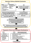
Proposed model for salivary epithelium-driven pathogenesis of SjD in subclinical salivary epithelial dysfunction and clinical histological manifestation phases.
Figure 3
Figure 3
Potential disruptions in acinar cell function and homeostasis by MMP9 in SjD. Legend. Epithelial function and integrity are reliant upon both internal and external factors for homeostasis. External factors may include the integrity of cell–cell connections (tight junctions) and cell connections with the extracellular matrix [ECM]. Changes or loss of these external factors can disrupt epithelial homeostasis and initiate pathologic changes, which have been observed in the glandular epithelium of SjD patients. Disruption of both tight junctions and cell–ECM connections has been attributed to the overexpression of matrix metalloproteinase 9 [MMP9], which may be overexpressed or more active due to reduced levels of the TIMP metallopeptidase inhibitor 1 [TIMP1] in the salivary epithelium of both sicca and SjD patients. MMP9 can degrade multiple components within cell–cell junctions and the surrounding ECM. (1) Degradation of tight junctions by MMP9 can alter the directional secretory abilities of epithelial cells. (2) Loss of tight junctions causes cellular components located within apical/basolateral sections to diffuse among the plasma membrane, inhibiting directional (polarized) secretion. Tight junctions also facilitate paracrine-mediated signaling, which is subsequently compromised within the epithelium. (3) Breakdown of the ECM and tight junctions increases the permeability of the epithelium, in turn facilitating lymphocytic infiltration mostly by CD8+ T-cells into acini. ECM degradation products reciprocally act as chemo-attractants for lymphocytes, further driving tissue infiltration. Latent growth factors such as vascular endothelial growth factor [VEGF], fibroblast growth factor 2 [FGF2], and transforming growth factor beta 1 [TGF-β] sequestered within the ECM are released and proteolytically activated by MMP9. (4) After subsequent damage to tissues, re-epithelization of the damaged region by resident stem/progenitor cells relies on external cues from the surrounding ECM; however, MMP9 overexpression and excessive breakdown of ECM structures could impair healing and repair of the glandular epithelium in SjD patients.

References

    1. Noll B. Ph.D. Thesis. The University of North Carolina at Charlotte; Charlotte, NC, USA: 2022. Dissecting the Salivary Gland: Epithelial-Centered Dysfunction in Primary Sjögren’s Syndrome.
    1. Parisis D., Chivasso C., Perret J., Soyfoo M.S., Delporte C. Current State of Knowledge on Primary Sjögren’s Syndrome, an Autoimmune Exocrinopathy. J. Clin. Med. 2020;9:2299. doi: 10.3390/jcm9072299. - DOI - PMC - PubMed
    1. Seror R., Nocturne G., Mariette X. Current and future therapies for primary Sjögren syndrome. Nat. Rev. Rheumatol. 2021;17:475–486. doi: 10.1038/s41584-021-00634-x. - DOI - PubMed
    1. Molina C., Alliende C., Aguilera S., Kwon Y.J., Leyton L., Martínez B., Leyton C., Pérez P., González M.J. Basal lamina disorganisation of the acini and ducts of labial salivary glands from patients with Sjogren’s syndrome: Association with mononuclear cell infiltration. Ann. Rheum. Dis. 2006;65:178–183. doi: 10.1136/ard.2004.033837. - DOI - PMC - PubMed
    1. Holdgate N., St Clair E.W. Recent advances in primary Sjogren’s syndrome. F1000Research. 2016;5:F1000 Faculty Rev-1412. doi: 10.12688/f1000research.8352.1. - DOI - PMC - PubMed