Review
. 2024 Apr 30;14(9):937.
doi: 10.3390/diagnostics14090937.

Concepts of Cardiac Dyssynchrony and Dynamic Approach

Affiliations
Review

Concepts of Cardiac Dyssynchrony and Dynamic Approach

Bianca Iulia Catrina et al. Diagnostics (Basel). .

Abstract

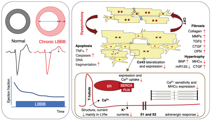
Cardiac conduction involves electrical activity from one myocyte to another, creating coordinated contractions in each. Disruptions in the conducting system, such as left bundle branch block (LBBB), can result in premature activation of specific regions of the heart, leading to heart failure and increased morbidity and mortality. Structural alterations in T-tubules and the sarcoplasmic reticulum can lead to dyssynchrony, a condition that can be treated by cardiac resynchronization therapy (CRT), which stands as a cornerstone in this pathology. The heterogeneity in patient responses underscored the necessity of improving the diagnostic approach. Vectocardiography, ultra-high-frequency ECG, 3D echocardiography, and electrocardiographic imaging seem to offer advanced precision in identifying optimal candidates for CRT in addition to the classic diagnostic methods. The advent of His bundle pacing and left bundle branch pacing further refined the approach in the treatment of dyssynchrony, offering more physiological pacing modalities that promise enhanced outcomes by maintaining or restoring the natural sequence of ventricular activation. HOT-CRT emerges as a pivotal innovation combining the benefits of CRT with the precision of His bundle or left bundle branch area pacing to optimize cardiac function in a subset of patients where traditional CRT might fall short.

Keywords: HOT-CRT; His; dyssynchrony; left bundle branch block; resynchronization.


Conflict of interest statement

The authors declare no conflicts of interest.

Figures

Figure 6
Figure 6
Cardiac resynchronization indications from ESC 2021 pacing guidelines (Photo extracted from Kenneth A. Ellenbogen et al. The evolving state of cardiac resynchronization therapy and conduction system pacing: 25 years of research at EP Europace journal, EP Europace, Volume 25, Issue 8, August 2023, by permission of Oxford University Press) [63].
Figure 1
Figure 1
Processes contributing to structural, electrical, and contractile remodeling in the dyssynchronous heart, as seen on functional measurements (left) and on a cellular and molecular level (right) (Photo extracted from Kenneth A. Ellenbogen et al. The evolving state of cardiac resynchronization therapy and conduction system pacing: 25 years of research at EP Europace journal, EP Europace, Volume 25, Issue 8, August 2023, by permission of Oxford University Press).
Figure 2
Figure 2
Optimization of AV delay after CRT by pulsed Doppler mitral inflow pattern. (Photo extracted from Galderisi, M. et al. Doppler echocardiography and myocardial dyssynchrony: a practical update of old and new ultrasound technologies. Cardiovasc Ultrasound 5, 28 (2007)) [25].
Figure 3
Figure 3
Estimation of interventricular mechanical delay using the conventional Doppler technique. The duration from the occurrence of the ECG Q wave to the onset of the right ventricular outflow tract is 122 ms, whereas the time from the Q wave to the onset of the left ventricular outflow tract is 211 ms (left panel). An interventricular mechanical delay (IVMD) of 89 milliseconds is obtained, which signifies the presence of substantial interventricular dyssynchrony (Photo extracted from Galderisi, M. et al. Doppler echocardiography and myocardial dyssynchrony: a practical update of old and new ultrasound technologies. Cardiovasc Ultrasound 5, 28 (2007) [25].
Figure 4
Figure 4
M-mode evaluation at midventricular level demonstrating a septal to posterior wall delay of 324 msec (from personal archive).
Figure 5
Figure 5
Time-to-peak longitudinal strain of the 16 segments. Images (AJ) represent different image acquisition incidents (Photo extracted from Song Y. et al., Left Ventricular Longitudinal Dyssynchrony by CMR Feature Tracking Is Related to Adverse Prognosis in Advanced Arrhythmogenic Cardiomyopathy. Front Cardiovasc Med. 2021 Oct 11;8:712832) [30].
Figure 7
Figure 7
Longitudinal Dissociation Within the His Bundle (Photo extracted from Vijayaraman P. et al., ACC’s Electrophysiology Council. His Bundle Pacing. J Am Coll Cardiol. 2018 Aug 21;72(8):927–947) [67].
Figure 8
Figure 8
Recording of the LBB potential. (A) The LBB potential can be recorded when the pacing lead helix is approaching the LBB. (B) The LBB potential becomes larger when the lead is closer to or at the LBB. (C,D) The LBB potential cannot be recorded in patients with LBBB, unless LBB is corrected by HBP (9). AVN, atrioventricular node; HB, His bundle; LBB, left bundle branch; LBBB, left bundle branch block; RBB, right bundle branch; IVS, interventricular septum; PoHis, His potential; PoLBB, left bundle branch potential. (Photo extracted from Liu P., Wang Q., Sun H., Qin X., Zheng Q. Left Bundle Branch Pacing: Current Knowledge and Future Prospects. Front Cardiovasc Med. 2021 Mar 23;8:630399) [69].

References

    1. Veeraraghavan R., Gourdie R.G., Poelzing S. Mechanisms of cardiac conduction: A history of revisions. Am. J. Physiol. Heart Circ. Physiol. 2014;306:H619–H627. doi: 10.1152/ajpheart.00760.2013. - DOI - PMC - PubMed
    1. Ghio S., Constantin C., Klersy C., Serio A., Fontana A., Campana C., Tavazzi L. Interventricular and intraventricular dyssynchrony are common in heart failure patients, regardless of QRS duration. Eur. Heart J. 2004;25:571–578. doi: 10.1016/j.ehj.2003.09.030. - DOI - PubMed
    1. Kirk J.A., Kass D.A. Cellular and Molecular Aspects of Dyssynchrony and Resynchronization. Card. Electrophysiol. Clin. 2015;7:585–597. doi: 10.1016/j.ccep.2015.08.011. - DOI - PMC - PubMed
    1. Prinzen F.W., Hunter W.C., Wyman B.T., McVeigh E.R. Mapping of regional myocardial strain and work during ventricular pacing: Experimental study using magnetic resonance imaging tagging. J. Am. Coll. Cardiol. 1999;33:1735–1742. doi: 10.1016/S0735-1097(99)00068-6. - DOI - PMC - PubMed
    1. Park R.C., Little W.C., O’Rourke R.A. Effect of alteration of left ventricular activation sequence on the left ventricular end-systolic pressure-volume relation in closed-chest dogs. Circ. Res. 1985;57:706–717. doi: 10.1161/01.RES.57.5.706. - DOI - PubMed