. 2024 May 2;14(9):957.
doi: 10.3390/diagnostics14090957.

Abnormal Static Sagittal Cervical Curvatures following Motor Vehicle Collisions: A Retrospective Case Series of 41 Patients before and after a Crash Exposure

Affiliations

Abnormal Static Sagittal Cervical Curvatures following Motor Vehicle Collisions: A Retrospective Case Series of 41 Patients before and after a Crash Exposure

Jason W Haas et al. Diagnostics (Basel). .

Abstract

Previous investigations have found a correlation between abnormal curvatures and a variety of patient complaints such as cervical pain and disability. However, no study has shown that loss of the cervical curve is a direct result of exposure to a motor vehicle collision (MVC). This investigation presents a retrospective consecutive case series of patients with both a pre-injury cervical lateral radiograph (CLR) and a post-injury CLR after exposure to an MVC. Computer analysis of digitized vertebral body corners on CLRs was performed to investigate the possible alterations in the geometric alignment of the sagittal cervical curve.

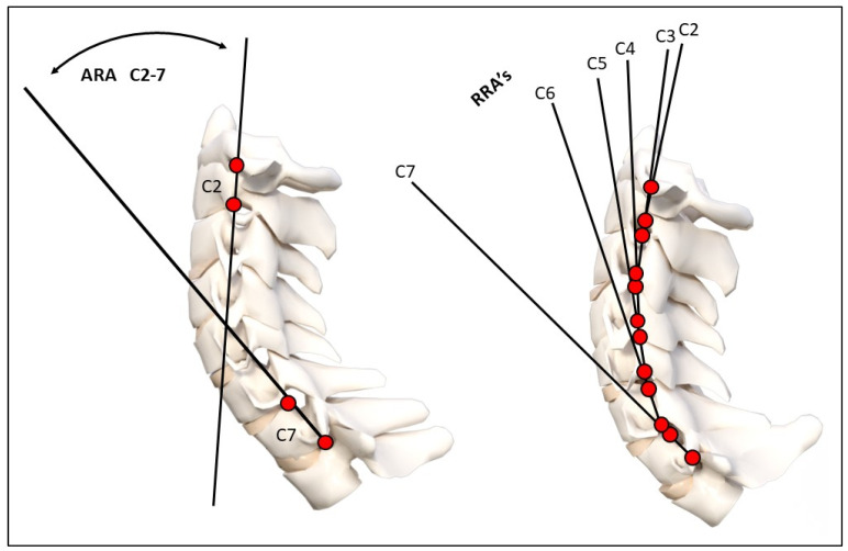
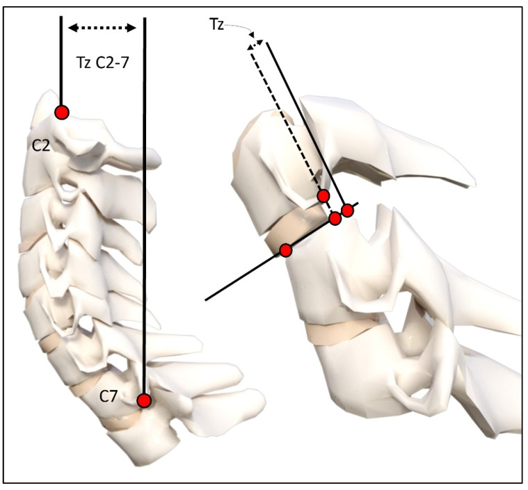
Methods: Three spine clinic records were reviewed over a 2-year period, looking for patients where both an initial lateral cervical X-ray and an examination were performed prior to the patient being exposed to a MVC; afterwards, an additional exam and radiographic analysis were obtained. A total of 41 patients met the inclusion criteria. Examination records of pain intensity on numerical pain rating scores (NPRS) and neck disability index (NDI), if available, were analyzed. The CLRs were digitized and modeled in the sagittal plane using curve fitting and the least squares error approach. Radiographic variables included total cervical curve (ARA C2-C7), Chamberlain's line to horizontal (skull flexion), horizontal translation of C2 relative to C7, segmental translations (retrolisthesis and anterolisthesis), and circular modelling radii.

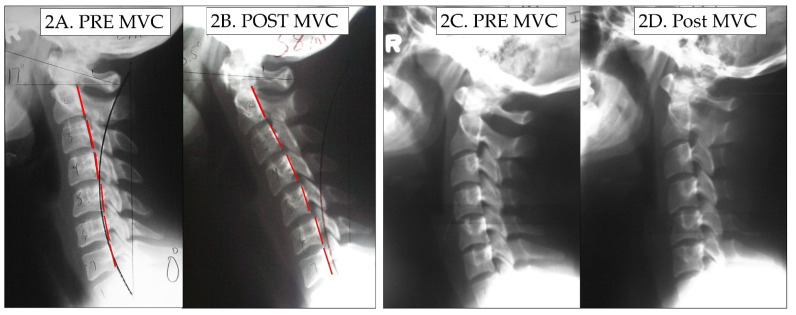
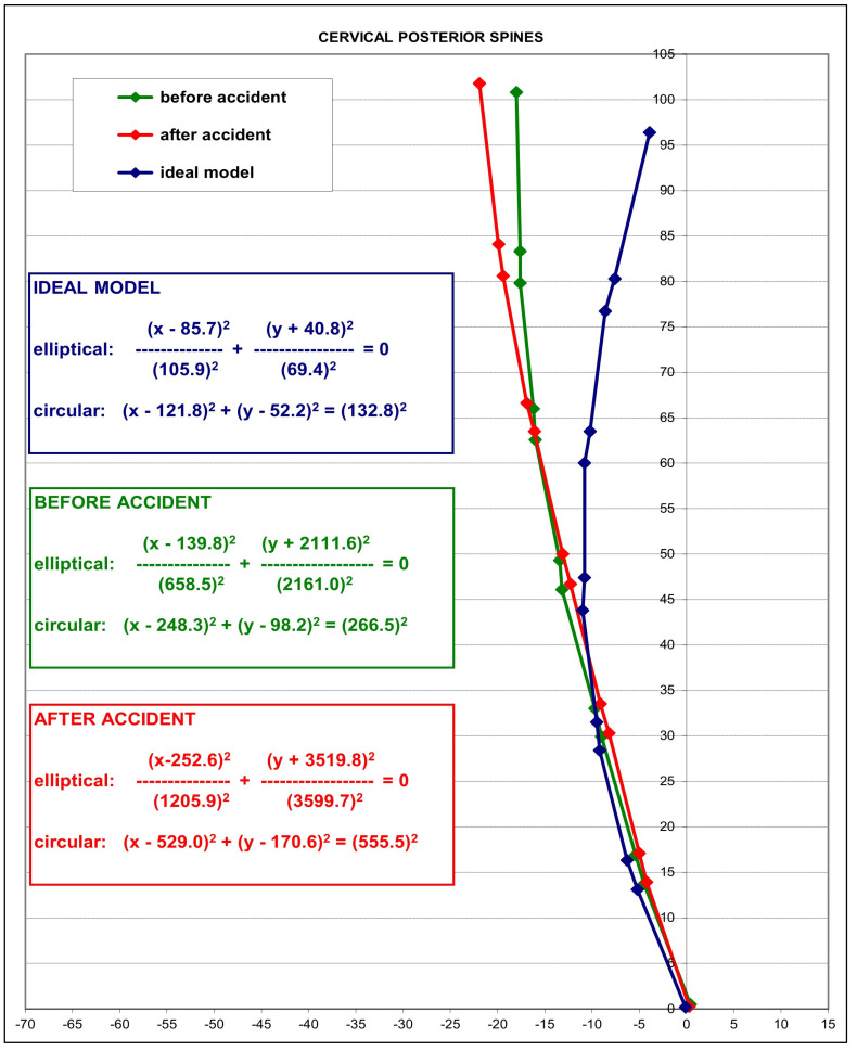
Results: There were 15 males and 26 females with an age range of 8-65 years. Most participants were drivers (28) involved in rear-end impacts (30). The pre-injury NPRS was 2.7 while the post injury was 5.0; p < 0.001. The NDI was available on 24/41 (58.5%) patients and increased after the MVC from 15.7% to 32.8%, p < 0.001. An altered cervical curvature was identified following exposure to MVC, characterized by an increase in the mean radius of curvature (265.5 vs. 555.5, p < 0.001) and an approximate 8° reduction of lordosis from C2-C7; p < 0.001. The mid-cervical spine (C3-C5) showed the greatest curve reduction with an averaged localized mild kyphosis at these levels. Four participants (10%) developed segmental translations that were just below the threshold of instability, segmental translations < 3.5 mm.

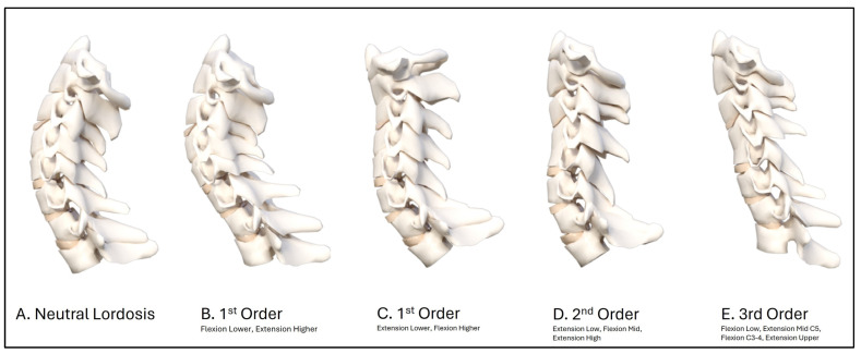
Conclusions: The post-exposure MVC cervical curvature was characterized by an increase in radius of curvature, an approximate 8° reduction in C2-C7 lordosis, a mild kyphosis of the mid-cervical spine, and a slight increase in anterior translation of C2-C7 sagittal balance. The modelling result indicates that the post-MVC cervical sagittal alignment approximates a second-order buckling alignment, indicating a significant alteration in curve geometry. Future biomechanics experiments and clinical investigations are needed to confirm these findings.

Keywords: buckling; cervical lateral radiograph; cervical lordosis; motor vehicle collision; neck pain; trauma.


Conflict of interest statement

Authors E.A.K. and I.M.M. declare no competing interests. J.W.H. is a compensated researcher for CBP Non-Profit, Inc. P.A.O. is a compensated consultant for Chiropractic BioPhysics, NonProfit, Inc. J.R.F. is the CEO of PostureCo, Inc., and receives compensation for the sale of PostureRay® software; this software was not used in this investigation. D.E.H. is the CEO of Chiropractic BioPhysics® (CBP®) and provides post-graduate education to healthcare providers and physicians. Spine rehabilitation devices are distributed through his company; none of these devices were used in this manuscript. D.E.H. is the president of CBP Non-Profit, Inc., a not-for-profit spine research foundation.

Figures

Figure 1
Figure 1
Radiographic mensuration techniques evaluated. Top image: Harrison posterior tangent method analysis of the cervical lordosis ARA C2–C7 and RRAs (note: RRA’s were represented using curve fitting and circular modelling). Bottom image: horizontal translation of C2–C7 and intersegmental translation between individual vertebrae. ARA: absolute rotation angle, RRA: relative rotation angle. These measurements have excellent examiner reliability and small examiner error magnitudes [40,41].
Figure 1
Figure 1
Radiographic mensuration techniques evaluated. Top image: Harrison posterior tangent method analysis of the cervical lordosis ARA C2–C7 and RRAs (note: RRA’s were represented using curve fitting and circular modelling). Bottom image: horizontal translation of C2–C7 and intersegmental translation between individual vertebrae. ARA: absolute rotation angle, RRA: relative rotation angle. These measurements have excellent examiner reliability and small examiner error magnitudes [40,41].
Figure 2
Figure 2
Before and after MVC alteration in the cervical lordosis. In (2A), a female patient’s initial cervical curve, and in (2B), the post-MVC cervical curve is shown to be significantly altered. In (2C), a second female patient’s initial cervical lordosis, and in (2D), the same patient’s post-MVC lateral cervical radiograph is shown with a significant reduction in lordosis, especially throughout the mid-cervical segments. The red dashed line represents the path of the patient’s posterior vertebral body margins in the sagittal plane from C2–C7.
Figure 3
Figure 3
Modeling results. In blue, an average idealized model of cervical lordosis is shown [38]. In green, the pre-MVC patient average model is shown. In red, the post-MVC patient model is shown. Modelling methods are described in detail in the report previously presented [38].
Figure 4
Figure 4
A selection of possible buckled modes for the sagittal cervical during and after exposure to an impact or inertial loading event such as a motor vehicle crash collision. Increasing complexity is shown by increasing the number of directional or slope changes. (A) The normal neutral cervical lordosis is shown. (B) A 1st order buckled mode is shown with flexion in the lower cervical and extension in the upper cervical spine. (C) An opposite first-order buckled mode in comparison to B where there is extension in the lower cervical and flexion in the upper cervical spine. (D) A second-order buckled mode is shown with an extension in the lower cervical, a flexion in the mid cervical, and an extension in the upper cervical region. (E) A third-order buckled mode is shown with a flexion in the lower, an extension at C5, a flexion at C3–C4, and an extension at C0–C2.
Figure 5
Figure 5
Segmental translation modeling results. In 4/41 patients (10%), segmental translations were identified on the post-MVC radiographs that were different than the ones identified on the initial pre-MVC radiograph. Each of these patient’s model is shown pre- and post-MVC as a change in color and coded by their number out of 41. Acc.: = MVC. # represents the patient number in the sample of 41 patients. # represents the patient number in the sample of 41 patients.

Similar articles

Cited by

References

    1. Quinlan K.P., Annest J.L., Myers B., Ryan G., Hill H. Neck strains and sprains among motor vehicle occupants-United States, 2000. Accid. Anal. Prev. 2004;36:21–27. doi: 10.1016/s0001-4575(02)00110-0. - DOI - PubMed
    1. Curatolo M., Bogduk N., Ivancic P.C., McLean S.A., Siegmund G.P., Winkelstein B.A. The role of tissue damage in whiplash-associated disorders: Discussion paper 1. Spine. 2011;36((Suppl. S25)):S309–S315. doi: 10.1097/BRS.0b013e318238842a. - DOI - PMC - PubMed
    1. Hendriks E.J., Scholten-Peeters G.G., Van der Windt D.A., Neeleman-van der Steen C.W., Oostendorp R.A., Verhagen A.P. Prognostic factors for a poor recovery in acute whiplash patients. Pain. 2005;114:408–416. doi: 10.1016/j.pain.2005.01.006. - DOI - PubMed
    1. Sterling M., Jull G., Kenardy J. Physical and psychological factors maintain long-term predictive capacity post-whiplash injury. Pain. 2006;122:102–108. doi: 10.1016/j.pain.2006.01.014. - DOI - PubMed
    1. Rebbeck T., Sindhusake D., Cameron I.D., Rubin G., Feyer A.M., Walsh J., Gold M., Schofield W.N. A prospective cohort study of health outcomes following whiplash associated disorders in an Australian population. Inj. Prev. 2006;12:93–98. doi: 10.1136/ip.2005.010421. - DOI - PMC - PubMed

LinkOut - more resources