. 2024 Jul 8;227(3):iyae077.
doi: 10.1093/genetics/iyae077.

TWISP: a transgenic worm for interrogating signal propagation in Caenorhabditis elegans

Affiliations

TWISP: a transgenic worm for interrogating signal propagation in Caenorhabditis elegans

Anuj Kumar Sharma et al. Genetics. .

Abstract

Genetically encoded optical indicators and actuators of neural activity allow for all-optical investigations of signaling in the nervous system. But commonly used indicators, actuators, and expression strategies are poorly suited for systematic measurements of signal propagation at brain scale and cellular resolution. Large-scale measurements of the brain require indicators and actuators with compatible excitation spectra to avoid optical crosstalk. They must be highly expressed in every neuron but at the same time avoid lethality and permit the animal to reach adulthood. Their expression must also be compatible with additional fluorescent labels to locate and identify neurons, such as those in the NeuroPAL cell identification system. We present TWISP, a transgenic worm for interrogating signal propagation, that addresses these needs and enables optical measurements of evoked calcium activity at brain scale and cellular resolution in the nervous system of the nematode Caenorhabditis elegans. In every neuron we express a nonconventional optical actuator, the gustatory receptor homolog GUR-3 + PRDX-2, under the control of a drug-inducible system QF + hGR, and a calcium indicator GCAMP6s, in a background with additional fluorophores from the NeuroPAL cell ID system. We show that this combination, but not others tested, avoids optical crosstalk, creates strong expression in the adult, and generates stable transgenic lines for systematic measurements of signal propagation in the worm brain.

Keywords: Caenorhabditis elegans; GUR-3; PRDX-2; calcium imaging; dexamethasone; drug-inducible gene expression; functional connectivity; neurons; optogenetics.


Conflict of interest statement

Conflicts of interest The author(s) declare no conflicts of interest.

Figures
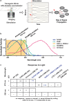
Fig. 1.
Fig. 1.
Strategy for all-neuron random-access stimulation and calcium imaging without optical cross talk. a) Schematic of a transgenic C. elegans for measuring signal propagation via optogenetic stimulation and calcium imaging. b) Previously reported action spectra for several neural actuators, compared with the absorbance spectra of GCaMP6s. Adapted from: Chen et al. (2013), Husson et al. (2013), Klapoetke et al. (2014), Bhatla and Horvitz (2015), and Dana et al. (2016). Shaded area indicates the action spectral range of GUR-3 + PRDX-2. 505 nm light is used to excite GCaMP6s close to its absorbance peak of 498 nm. c) Optogenetic proteins were expressed in every neuron under a rab-3 promoter. Behavior response to 1.5 mW/mm2 illumination of either 505 nm or 475 nm light is shown. For animals expressing rhodopsin-based optogenetic proteins, behavior is measured with and without the necessary co-factor all-trans retinal (ATR). TsChR and GUR-3 + PRDX-2 (highlighted in blue text) show good compatibility with GCaMP6s imaging wavelengths.
Fig. 2.
Fig. 2.
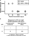
High concentration injections of actuator-containing plasmids are not viable for transgenesis, but higher expression is desirable. a) Injection concentration for plasmids containing either GUR-3 + PRDX-2 or TsChR and the viability of transgene expressing progeny is shown. Each symbol indicates one trial. b) Light-evoked behavior response of worms expressing TsChR (50 ng/µl injection) in all neurons upon 1.5 mW/mm2 475 nm blue-light illumination, with and without the co-factor ATR (all-trans retinal). Most worms had very low expression, as estimated from reporter expression. Rare worms with high expression showed stronger responses.
Fig. 3.
Fig. 3.
Drug-inducible expression enables robust light response while avoiding lethality a) Injections of plasmids containing actuators under the control of the QF + hGR drug-inducible expression system are viable at higher injection concentrations (black filled shapes) than injections of plasmids for direct expression of the actuators (gray open shapes, same as Fig. 2). b) Exposure to the drug dexamethasone (Dex) evokes actuator expression and confers robust light response to 475 nm illumination.
Fig. 4.
Fig. 4.
Drug induced actuator expression modulates health and growth. Animals are observed as they are propagated on plates across multiple generations either on or off the drug dexamethasone. Observations of the animal's health and developmental stage are made at time points indicated by the location of the circle on the timeline. For each observation, the developmental stage of the most advance animal found on the plate is reported (listed inside the circle). L4s were always selected for transfer to new plates. Health and generation times improve for the progeny of animals that had previously been exposed to the drug but subsequently cultivated on regular NGM plates.
Fig. 5.
Fig. 5.
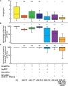
Rate of growth and progeny production decrease with transgenic load, GCaMP6s, NeuroPAL system, and actuator expression. a) Rate of progeny production and b) percentage of animals that reach adulthood in 70 h or c) in 94 h is reported for strains carrying various components of the TWISP system. Box shows median and 25th and 75th percentile values, whiskers show min and max values. Mean ± SD values and number of plates are reported in Supplementary Tables 3, 4, and 5. Experiments were performed in triplicates and repeated at least 3 times for all strain except AML32 and AML177 in (c). Statistical significance is with respect to WT, using Kruskal–Wallis test followed by Dunn's multiple comparisons, *<0.0332, **<0.0021, ***<0.0002, and ****<0.0001.
Fig. 6.
Fig. 6.
Locomotion decreases with transgenic load, GCaMP6s, NeuroPAL system, and actuator expression. a) Speed, b) reversal rate, c) body length, and d) fraction of time paused are reported for animals from strains containing various components of the TWISP system. The TWISP strain is measured with and without dexamethasone treatment. The combination of NeuroPAL and GCaMP6s expression decreases locomotion. Box and whisker plots report the distribution of behavior across animal tracks (not plates). The number of tracks recorded per condition, from left to right, are N = [1706, 304, 1283, 1232, 1940, 393, 1374, 723, 647]. Box indicates median and interquartile range. Whiskers indicate range excluding outliers. Mean ± SD values and number of plates are reported in Supplementary Table 6. Statistical significance is with respect to WT, using Kruskal–Wallis test followed by Dunn's multiple comparisons, **<0.0021, ****<0.0001.
Fig. 7.
Fig. 7.
TWISP worms respond to mechanosensory stimuli in a gentle-touch behavior assay. Small circles indicate responses to individual touch stimuli. Six animals were tested for each strain and three stimuli were delivered per animal.
Fig. 8.
Fig. 8.
Head and tail population calcium activity in response to neural activation measured with TWISP. Calcium activity of simultaneously recorded neurons from the head and tail is shown during targeted optogenetic stimulation of individual neurons. Calcium imaging is performed via single-photon spinning disk with 505 nm illumination while individual neurons are stimulated via 2-photon spatially restricted illumination at 850 nm. Neuron identities are listed on the left. Gray vertical line indicates times in which a stimulus was delivered (at every 30 s). Red thunderbolt indicates the stimulated neuron. The name of the neuron stimulated is listed above. Recordings of unidentified neurons or of neurons that were not well-segmented are excluded from the plot.

Update of

References

    1. Abdelfattah AS, Valenti R, Zheng J, Wong A; GENIE Project Team; Podgorski K, Koyama M, Kim DS, Schreiter ER. 2020. A general approach to engineer positive-going eFRET voltage indicators. Nat Commun. 11(1):3444. doi:10.1038/s41467-020-17322-1. - DOI - PMC - PubMed
    1. Abdelfattah AS, Zheng J, Singh A, Huang YC, Reep D, Tsegaye G, Tsang A, Arthur BJ, Rehorova M, Olson CVL, et al. 2023. Sensitivity optimization of a rhodopsin-based fluorescent voltage indicator. Neuron. 111(10):1547–1563.e9. doi:10.1016/j.neuron.2023.03.009. - DOI - PMC - PubMed
    1. Atanas AA, Kim J, Wang Z, Bueno E, Becker M, Kang D, Park J, Kramer TS, Wan FK, Baskoylu S, et al. 2023. Brain-wide representations of behavior spanning multiple timescales and states in C. elegans. Cell. 186(19):4134–4151.e31. doi:10.1016/j.cell.2023.07.035. - DOI - PMC - PubMed
    1. Azimi Hashemi N, Bergs ACF, Schuler C, Scheiwe AR, Steuer Costa W, Bach M, Liewald JF, Gottschalk A. 2019. Rhodopsin-based voltage imaging tools for use in muscles and neurons of Caenorhabditis elegans. Proc Natl Acad Sci U S A. 116(34):17051–17060. doi:10.1073/pnas.1902443116. - DOI - PMC - PubMed
    1. Beets I, Zels S, Vandewyer E, Demeulemeester J, Caers J, Baytemur E, Courtney A, Golinelli L, Hasakioğulları İ, Schafer WR, et al. 2023. System-wide mapping of peptide-GPCR interactions in C. elegans. Cell Rep. 42(9):113058. doi:10.1016/j.celrep.2023.113058. - DOI - PMC - PubMed

MeSH terms