. 2024 Jul:45:101988.
doi: 10.1016/j.tranon.2024.101988. Epub 2024 May 10.

Fractionated radiation therapy alters energy metabolism and induces cellular quiescence exit in patient-derived orthotopic xenograft models of high-grade glioma

Affiliations

Fractionated radiation therapy alters energy metabolism and induces cellular quiescence exit in patient-derived orthotopic xenograft models of high-grade glioma

Zi-Lu Huang et al. Transl Oncol. 2024 Jul.

Abstract

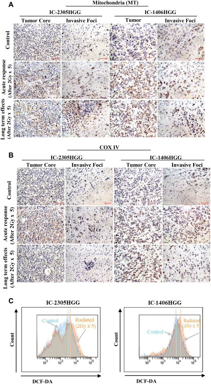
Radiation is one of the standard therapies for pediatric high-grade glioma (pHGG), of which the prognosis remains poor. To gain an in-depth understanding of biological consequences beyond the classic DNA damage, we treated 9 patient-derived orthotopic xenograft (PDOX) models, including one with DNA mismatch repair (MMR) deficiency, with fractionated radiations (2 Gy/day x 5 days). Extension of survival time was noted in 5 PDOX models (P < 0.05) accompanied by γH2AX positivity in >95 % tumor cells in tumor core and >85 % in the invasive foci as well as ∼30 % apoptotic and mitotic catastrophic cell death. The model with DNA MMR (IC-1406HGG) was the most responsive to radiation with a reduction of Ki-67(+) cells. Altered metabolism, including mitochondria number elevation, COX IV activation and reactive oxygen species accumulation, were detected together with the enrichment of CD133+ tumor cells. The latter was caused by the entry of quiescent G0 cells into cell cycle and the activation of self-renewal (SOX2 and BMI1) and epithelial mesenchymal transition (fibronectin) genes. These novel insights about the cellular and molecular mechanisms of fractionated radiation in vivo should support the development of new radio-sensitizing therapies.

Keywords: Cancer stem cells; Glioma; Mitochondrial biogenesis; Orthotopic xenograft; Radiotherapy.


Conflict of interest statement

Declaration of competing interest The authors declare that they have no known competing financial interests or personal relationships that could have appeared to influence the work reported in this paper.

Figures

Fig 1
Fig. 1
In vivo treatment of PDOX models of pHGG with fractionated radiotherapy. (A) Experiment design of the study. Tumor cells (1×10⁵ cells/2 µL) were implanted into right cerebral cortex of SCID mice. Fractionated radiation (2 Gy/day x 5 days) was administered 14 days and ∼5 weeks post tumor implantation for survival analysis and for biological examination, respectively. (B) Log rank analysis of animal survival times in 6 pHGG models. There were at least 10 mice for each control and radiation therapy (XRT) group. The model marked with * were selected in this research for further biological verify and analysis.
Fig 2
Fig. 2
In vivo responses of pHGG tumor cells toward fractionated radiation. (A) Representative images showing the detection of tumor cells with double strand breaks and apoptosis with immunohistochemical staining of γH2AX (green arrows) (left panel) and cleaved caspase-3 (green arrows) (right panel), respectively (Bar = 50 µm). Positive cells were counted from at least 5 microscopic fields (10×40) in the tumor core (Core) or in at least 8 invasive foci (INV) (5–30 cells), and represent as Mean ± SE in the graphs (lower panel), ** P < 0.01; * P < 0.05, compared with the untreated control group. (B) Representative images of H&E staining showing increased volume of cancer cells, cavity formation, nuclear heterogeneity, cell degeneration, cytoplasm dyeing change could be seen (a–d) and sings of mitotic catastrophe (green arrows and circles) (c–f) in vivo in xenograft tumors treated with or without radiation (2 Gy daily for 5 days). Quantitative data were graphed. (C) Changes of cell proliferation as detected by immunostaining of Ki-67 (red arrows) (bar = 50 µm). Mitotic catastrophe and Ki-67 positive cells were counted in at least 5 microscopic fields (10×40) (Mean ± SE), ** P < 0.01; *P < 0.05, compared with control group. Magnification, 10×40: a–d;10×100: e and f.
Fig 3
Fig. 3
Representative images of immunohistochemical detection and functional examination of mitochondria in vivo in xenograft tumors (quantification data summarized in Table 2). (A) Analysis of mitochondrial abundance in tumor core and invasive foci with immunohistochemical staining. Xenograft tumors harvested immediately at the end of fractionated radiation therapy (Acute response) and at the late phase after fractionated radiation therapy (Long term effects) were compared with that in the untreated control group. bar = 50 µm. (B) Immunohistochemical staining of mitochondria-specific protein COX IV. Xenograft tumors harvested immediately at the end of fractionated radiation therapy (Acute response) and at the late phase after fractionated radiation therapy (Long term effects) were compared with the untreated control, bar = 50 µm. (C) FCM quantitative analysis of ROS production. Xenograft cells were harvested at the end of 5-day fractionated radiation, stained with DCF-DA and analyzed.
Fig 4
Fig. 4
Flow cytometric analysis of CD133+ and CD133 tumor cells following fractionated radiation in vivo. (A) Flow chart showing the increase of CD133+ glioma cells from the acute response phase and lasted for long-term in the remanent tumors. (B) Quantification of CD133+ glioma cell after fractionated radiation in both mouse model. Data from at least three independent experiments are shown. (C) FCM gate setting for G0, G1 and S/G2M analysis. Tumor cells were sequentially incubated with Hoechst 33342 to stain DNA and Pyronin Y to stain RNA. (D) Representative flow chart showing the reduction of G0 phase cells accompanied by the increase of G1 phase cells. Tumor cells were harvested at the end of 5-day radiation (2 Gy per day). (E) Quantification of G0 cell after fractionated irradiation. Data from at least three independent experiments are shown (Mean ± SD) (P < 0.01).
Fig 5
Fig. 5
Representative images of immunohistochemical staining of PDOX tumor cells at the end of acute and long-term radiation therapy in tumor core and invasive front in two pHGG models (IC-3752HGG, IC-2305HGG) (quantification data summarized in Table 3). (A) Nuclear protein expression of BMI1(blue arrow). (B) Nuclear protein expression of COX IV (green arrow) (green arrow). Representative images were from three independent experiments. (Original magnification, 40×10). Bar = 50 µm.
Fig 6
Fig. 6
Representative images of immunohistochemical staining of PDOX tumor cells at the end of acute and long term radiation therapy in both tumor core and invasive front in two pHGG models (IC-3752HGG, IC-2305HGG). (quantification data summarized in Table 4) (A) Cytoplasmic protein expression of Fibronectin (green arrow). (B) Cytoplasmic protein expression of Vimentin (blue arrow). Representative images were from three independent experiments. (Original magnification, 40×10). Bar = 50 µm.

References

    1. Erker C., Tamrazi B., Poussaint T.Y., Mueller S., Mata-Mbemba D., Franceschi E., et al. Response assessment in paediatric high-grade glioma: recommendations from the Response Assessment in Pediatric Neuro-Oncology (RAPNO) working group. Lancet Oncol. 2020;21(6):e317–ee29. - PubMed
    1. Jones C., Karajannis M.A., Jones D.T.W., Kieran M.W., Monje M., Baker S.J., et al. Pediatric high-grade glioma: biologically and clinically in need of new thinking. Neuro Oncol. 2017;19(2):153–161. - PMC - PubMed
    1. Sturm D., Pfister S.M., Jones D.T.W. Pediatric gliomas: current concepts on diagnosis, biology, and clinical management. J. Clin. Oncol. 2017;35(21):2370–2377. - PubMed
    1. Gaspar N., Marshall L., Perryman L., Bax D.A., Little S.E., Viana-Pereira M., et al. MGMT-independent temozolomide resistance in pediatric glioblastoma cells associated with a PI3-kinase-mediated HOX/stem cell gene signature. Cancer Res. 2010;70(22):9243–9252. - PMC - PubMed
    1. Jakacki R.I., Cohen K.J., Buxton A., Krailo M.D., Burger P.C., Rosenblum M.K., et al. Phase 2 study of concurrent radiotherapy and temozolomide followed by temozolomide and lomustine in the treatment of children with high-grade glioma: a report of the Children's Oncology Group ACNS0423 study. Neuro Oncol. 2016;18(10):1442–1450. - PMC - PubMed