. 2024 Jul:73:103179.
doi: 10.1016/j.redox.2024.103179. Epub 2024 May 8.

Mitochondrial GPX4 acetylation is involved in cadmium-induced renal cell ferroptosis

Affiliations

Mitochondrial GPX4 acetylation is involved in cadmium-induced renal cell ferroptosis

Yue-Yue Guo et al. Redox Biol. 2024 Jul.

Abstract

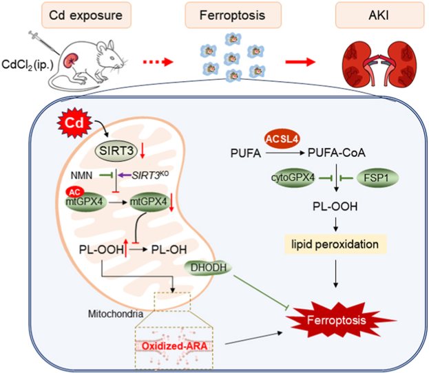
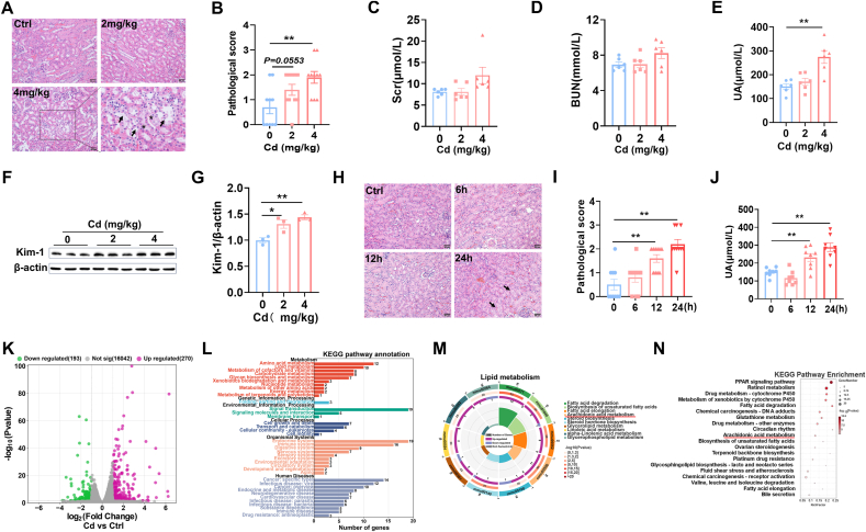
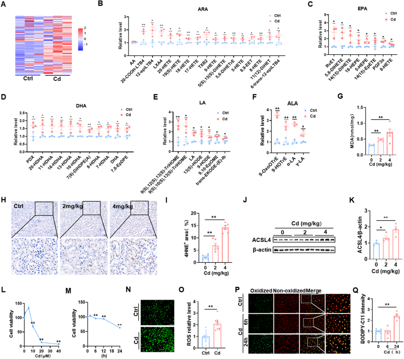
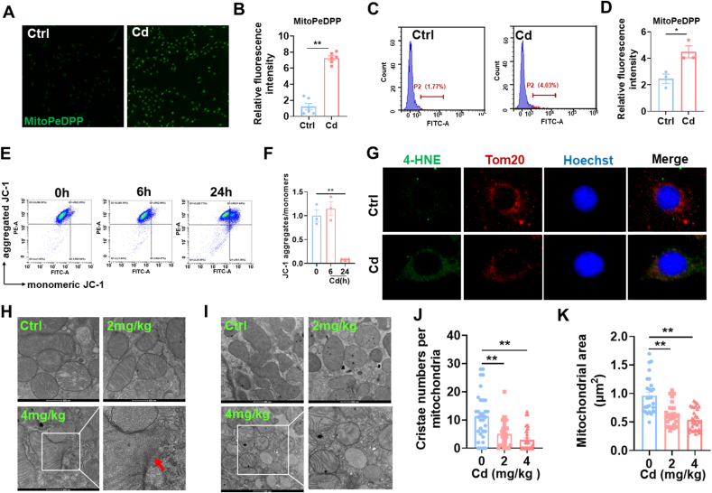
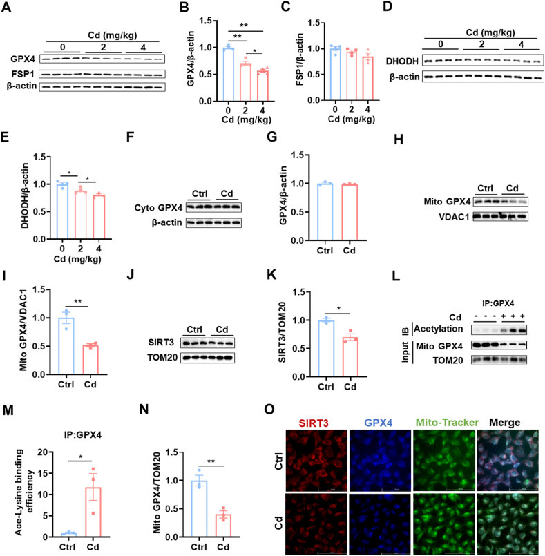
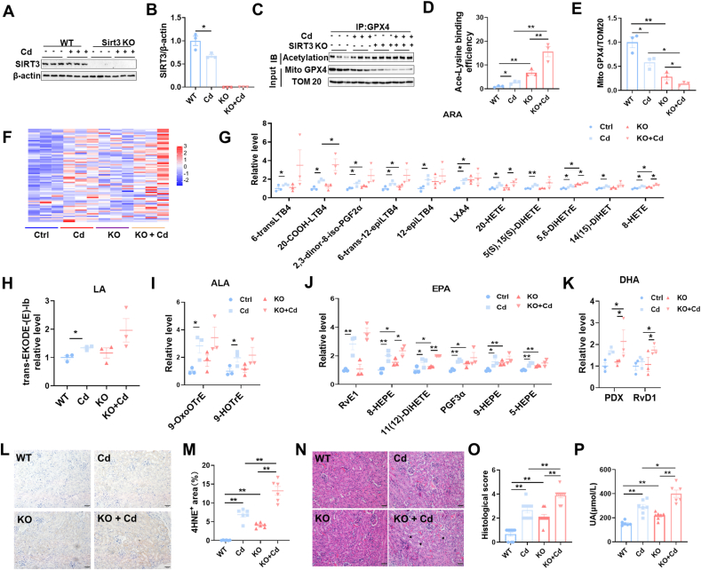
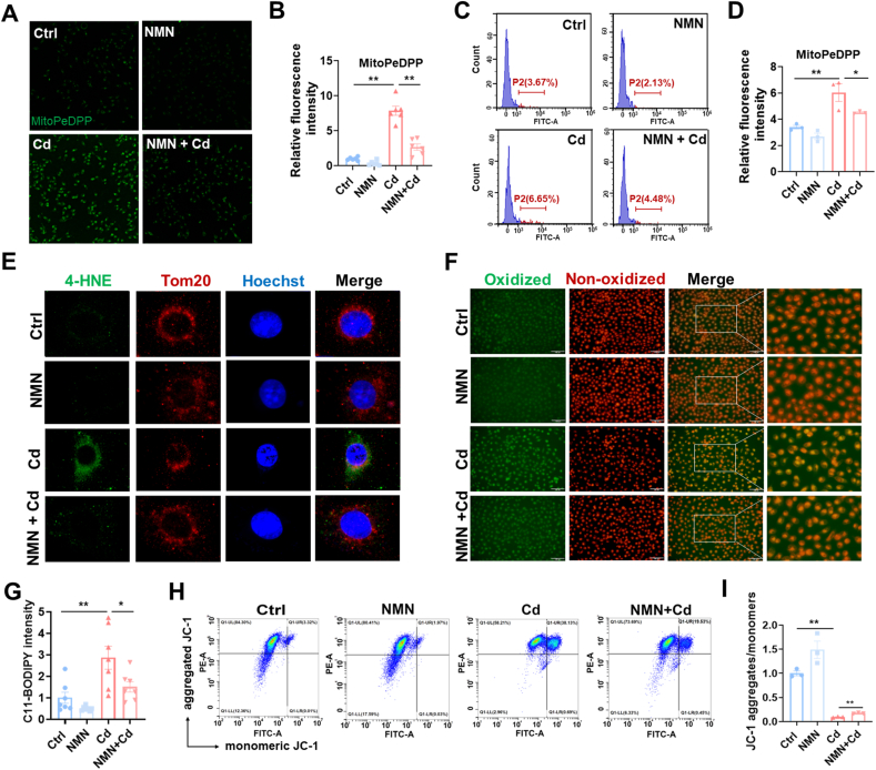
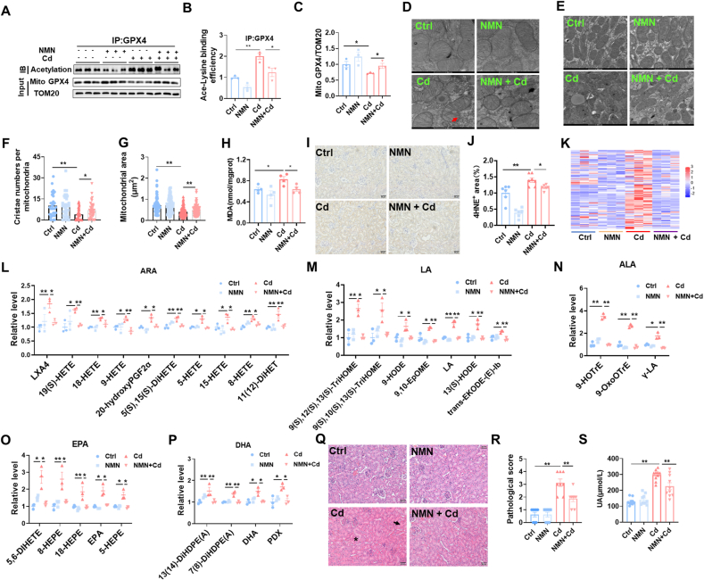
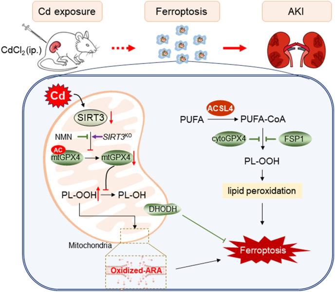
Increasing evidences demonstrate that environmental stressors are important inducers of acute kidney injury (AKI). This study aimed to investigate the impact of exposure to Cd, an environmental stressor, on renal cell ferroptosis. Transcriptomics analyses showed that arachidonic acid (ARA) metabolic pathway was disrupted in Cd-exposed mouse kidneys. Targeted metabolomics showed that renal oxidized ARA metabolites were increased in Cd-exposed mice. Renal 4-HNE, MDA, and ACSL4, were upregulated in Cd-exposed mouse kidneys. Consistent with animal experiments, the in vitro experiments showed that mitochondrial oxidized lipids were elevated in Cd-exposed HK-2 cells. Ultrastructure showed mitochondrial membrane rupture in Cd-exposed mouse kidneys. Mitochondrial cristae were accordingly reduced in Cd-exposed mouse kidneys. Mitochondrial SIRT3, an NAD+-dependent deacetylase that regulates mitochondrial protein stability, was reduced in Cd-exposed mouse kidneys. Subsequently, mitochondrial GPX4 acetylation was elevated and mitochondrial GPX4 protein was reduced in Cd-exposed mouse kidneys. Interestingly, Cd-induced mitochondrial GPX4 acetylation and renal cell ferroptosis were exacerbated in Sirt3-/- mice. Conversely, Cd-induced mitochondrial oxidized lipids were attenuated in nicotinamide mononucleotide (NMN)-pretreated HK-2 cells. Moreover, Cd-evoked mitochondrial GPX4 acetylation and renal cell ferroptosis were alleviated in NMN-pretreated mouse kidneys. These results suggest that mitochondrial GPX4 acetylation, probably caused by SIRT3 downregulation, is involved in Cd-evoked renal cell ferroptosis.

Keywords: Acute kidney injury; Ferroptosis; Mitochondrial GPX4 acetylation; Mitochondrial lipid peroxidation; Nicotinamide mononucleotide; SIRT3.


Conflict of interest statement

Declaration of competing interest The authors declare that they have no known competing financial interests or personal relationships that could have appeared to influence the work reported in this paper.

Figures

Image 1
Graphical abstract
Fig. 1
Fig. 1
Acute Cd exposure induces acute kidney injury and changes in renal lipid metabolic pathway. CdCl2 (2 mg/kg or 4 mg/kg) was injected intraperitoneally into adult BALB/c male mice. Kidney tissues and blood sera were taken 24 h after CdCl2. (A and B) Renal pathology was assessed. (A) Representative H&E images. (B) Pathological score. (C) Scr. (D) BUN. (E) Serum UA. (F and G) Kim-1 was measured by immunoblot. (F) Representative images. (G) Kim-1/β-actin. CdCl2 was injected intraperitoneally into adult BALB/c male mice for 0, 6, 12 and 24 h (4 mg/kg), respectively. Kidney tissues and blood were taken 24 h after CdCl2. (H and I) Renal pathology was assessed. (H) Representative H&E images. (I) Pathological score. (J) Serum UA. (K–N) Transcriptome analyses were performed in Cd-exposed mice kidney. (K) Scatter plot. (L) The KEGG pathway enrichment was analyzed. (M) Lipid metabolic pathways were analyzed. (N) Top of KEGG pathway was enriched. All data are presented as mean ± S.E.M. (N = 6–8). *P < 0.05, **P < 0.01.
Fig. 2
Fig. 2
Cd exposure induces lipid peroxidation in mice kidneys and HK-2 cells. (A–K) CdCl2 (2 mg/kg or 4 mg/kg) was injected intraperitoneally into adult BALB/c male mice. Kidney tissues were taken 24 h after CdCl2. (A–F) Targeted metabolomics of oxidized lipids were examined. (A) A heatmap. (B–G) Renal oxidized metabolites of (B) ARA, (C) EPA, (D) DHA, (E) LA and (F) ALA. (G) Renal MDA content. (H and I) Renal 4-HNE+ area was evaluated by IHC. (H) Representative images. (I) Renal 4-HNE+ area. (J–K) ACSL4 was detected by immunoblot. (J) Representative images. (K) ACSL4/β-actin. HK-2 cells were co-cultured different doses with CdCl2 for 24 h. (L) Cell viability. HK-2 cells were co-cultured for different times with CdCl2 (10 μM). (M) Cell viability. (N and O) HK-2 cells were co-cultivated with CdCl2 (10 μM) for 24 h. ROS was detected by immunofluorescence. (N) Representative images. (O) Quantitative analysis. (P and Q) HK-2 cells were co-cultivated with CdCl2 (10 μM) for 6 or 24 h. C11-BODIPY was used for detection of oxidized lipids. (P) Representative images. (Q) Quantitative analysis. All data are presented as mean ± S.E.M. (N = 6). *P < 0.05, **P < 0.01.
Fig. 3
Fig. 3
Cd exposure evokes mitochondrial lipid peroxidation and mitochondrial dysfunction. HK-2 cells were co-cultivated with CdCl2 (10 μM) for 24 h. (A and B) Mitochondrial oxidized lipids were determined by MitoPeDPP. (A) Representative pictures. (B) Statistical analysis. (C and D) Mitochondrial oxidized lipids were evaluated by flow cytometry. (C) Representative pictures. (D) Quantitative analysis. (E and F) MMPs were detected by JC-1 staining. (E) Representative images. (F) Statistical analysis. (G) Co-localization of 4-HNE with TOM20. (H–K) CdCl2 (2 mg/kg or 4 mg/kg) was injected intraperitoneally into adult BALB/c male mice. Mitochondrial ultrastructure was evaluated by electron microscopy. (H) The integrity of mitochondrial membrane. Arrow indicates mitochondrial membrane rupture. (I–K) Mitochondrial area and cristae were analyzed. (I) Representative pictures. (J) Cristae number per mitochondrion. (K) Mitochondrial area. All data are presented as mean ± S.E.M. (N = 3). *P < 0.05, **P < 0.01.
Fig. 4
Fig. 4
Cd induces mitochondrial SIRT3 downregulation and GPX4 acetylation. CdCl2 (2 mg/kg or 4 mg/kg) was injected intraperitoneally into adult BALB/c male mice. Kidney tissues were reserved 24 h after CdCl2. (A–C) GPX4 and FSP1 were detected by immunoblot. (A) Representative images. (B) GPX4/β-actin; (C) FSP1/β-actin. (D–E) DHODH was detected by immunoblot. (D) Representative images. (E) DHODH/β-actin. (F–I) CdCl2 (4 mg/kg) was injected intraperitoneally into adult BALB/c male mice. Kidney tissues were reserved 24 h after CdCl2. Renal mitochondria and cytoplasm were extracted. (F and G) Cytoplasmic GPX4 was measured by immunoblot. (F) Representative images. (G) GPX4/β-actin. (H and I) Mitochondrial GPX4 was measured by immunoblot. (H) Representative images. (I) GPX4/VDAC1. (J and K) Mitochondrial SIRT3 was measured by immunoblot. (J) Representative images. (K) SIRT3/TOM20. (L–N) Mitochondrial GPX4 acetylation was analyzed by Co-IP. (L) Representative images. (M) Ace-Lysine binding efficiency. (N) GPX4/TOM20. (O) HK-2 cells were co-cultivated with CdCl2 (10 μM) for 24 h. Co-localization of SIRT3, GPX4 and mitochondrial Tracker was observed by immunofluorescence. Red indicates SIRT3; Blue represents GPX4; Green represents Mito-Tracker. All data are presented as mean ± S.E.M. (N = 3–4). *P < 0.05, **P < 0.01. (For interpretation of the references to colour in this figure legend, the reader is referred to the Web version of this article.)
Fig. 5
Fig. 5
Sirt3 knockout exacerbates Cd-induced mitochondrial GPX4 acetylation, renal cell ferroptosis and AKI. Wild type and Sirt3−/− male mice intraperitoneally injected with CdCl2 (4 mg/kg). Kidney tissues were collected 24 h after CdCl2. (A) SIRT3 was detected using immunoblot; (B) SIRT3/β-actin. (C–E) Mitochondrial GPX4 acetylation was analyzed by Co-IP. (C) Representative images. (D) Ace-Lysine binding efficiency. (E) GPX4/TOM20. (F–K) Targeted metabolomics of oxidized lipids were examined by LC-MS/MS. Renal oxidized metabolites of (F) A heatmap. (G)ARA, (H) LA, (I) ALA, (J) EPA and (K) DHA are shown. (L and M) Renal 4-HNE+ area was measured by IHC. (L) Representative images. (M) Quantitative analysis. (N and O) Renal pathology was evaluated. (N) Representative H&E images. (O) Pathological score. (P) Serum UA. All data are presented as mean ± S.E.M. (N = 6–8). *P < 0.05, **P < 0.01.
Fig. 6
Fig. 6
Pretreatment with NMN attenuates Cd-induced mitochondrial lipid peroxidation in HK-2 cells. HK-2 cells were co-cultivated with CdCl2 (10 μM) for 24 h. Some HK-2 cells were pretreated with NMN (1 mM) for 2 h before Cd exposure. (A and B) Mitochondrial oxidized lipids were measured using confocal microscopy. (A) Representative pictures. (B) Statistical analysis. (C and D) Mitochondrial oxidized lipids were evaluated using flow cytometry. (C) Representative pictures. (D) Statistical analysis. (E) Co-localization of 4-HNE with TOM20 was evaluated using immunofluorescence. (F and G) Oxidized lipids were measured by C11-BODIPY staining. (F) Representative pictures. (G) Statistical analysis. (H and I) MMPs were detected using JC-1 staining. (H) Representative pictures. (I) Statistical analysis. All data are presented as mean ± S.E.M. (N = 3). *P < 0.05, **P < 0.01.
Fig. 7
Fig. 7
Pretreatment with NMN attenuates Cd-induced mitochondrial GPX4 acetylation, renal cell ferroptosis and AKI. All mice except controls were intraperitoneally injected with CdCl2 (4 mg/kg). Some mice were pretreated with NMN (500 mg/kg) for 5 consecutive days. Kidney tissues were reserved 24 h after CdCl2. (A–C) Renal mitochondria was extracted. Mitochondrial GPX4 acetylation was analyzed by Co-IP. (A) Representative images. (B) Ace-Lysine binding efficiency. (C) GPX4/TOM20. (D–G) Mitochondrial ultrastructure was evaluated by electron microscopy. (D) The integrity of mitochondrial membrane. Arrow indicates mitochondrial membrane rupture. (E–G) Mitochondrial area and cristae were analyzed. (E) Representative images. (F) Cristae number per mitochondrion. (G) Mitochondrial area. (H)Renal MDA content. (I and J) Renal 4-HNE+ area was measured by IHC. (I) Representative images. (J) Quantitative analysis. (K–P) Targeted metabolomics of oxidized lipids were examined by LC-MS/MS. Renal oxidized metabolites of (K) A heatmap. (L) ARA, (M) LA, (N) ALA, (O) EPA and (P) DHA were shown. (Q and R) Renal pathology was evaluated. (Q) Representative H&E images. (R) Pathological scores. (S) Serum UA. All data are presented as mean ± S.E.M. (N = 6–8). *P < 0.05, **P < 0.01.
Fig. 8
Fig. 8
Role of mitochondrial GPX4 acetylation in Cd-induced renal cell ferroptosis and acute kidney injury. Briefly, acute Cd exposure reduces mitochondrial SIRT3, resulting in mitochondrial GPX4 acetylation and mitochondrial GPX4 reduction in mouse kidney. Mitochondrial GPX4 reduction induces elevation of mitochondrial oxidized lipids, mainly oxidized ARA, subsequently renal cell ferroptosis and acute kidney injury. Mitochondrial GPX4 acetylation, probably caused by SIRT3 reduction, is partially involved in Cd-induced renal cell ferroptosis. ACSL4: Acyl-CoA synthetase long chain family member 4; AKI: Acute kidney injury; ARA: arachidonic acid; CdCl2: Cadmium dichloride; DHODH: Dihydroorotate dehydrogenase; FSP1: Ferroptosis suppressor protein 1; GPX4: Glutathione peroxidase 4; NMN: Beta-Nicotinamide Mononucleotide; PUFA: Polyunsaturated fatty acids; SIRT3: Sirtuin 3.

References

    1. Pickkers P., Darmon M., Hoste E., Joannidis M., Legrand M., Ostermann M., Prowle J.R., Schneider A., Schetz M. Acute kidney injury in the critically ill: an updated review on pathophysiology and management. Intensive Care Med. 2021;47(8):835–850. - PMC - PubMed
    1. Scholz H., Boivin F.J., Schmidt-Ott K.M., Bachmann S., Eckardt K.U., Scholl U.I., Persson P.B. Kidney physiology and susceptibility to acute kidney injury: implications for renoprotection. Nat. Rev. Nephrol. 2021;17(5):335–349. - PubMed
    1. Peerapornratana S., Manrique-Caballero C.L., Gómez H., Kellum J.A. Acute kidney injury from sepsis: current concepts, epidemiology, pathophysiology, prevention and treatment. Kidney Int. 2019;96(5):1083–1099. - PMC - PubMed
    1. Perazella M.A., Rosner M.H. Drug-induced acute kidney injury. Clin. J. Am. Soc. Nephrol.: CJASN. 2022;17(8):1220–1233. - PMC - PubMed
    1. Min J., Kang D.H., Kang C., Bell M.L., Kim H., Yang J., Gasparrini A., Lavigne E., Hashizume M., Kim Y., Fook Sheng Ng C., Honda Y., das Neves Pereira da Silva S., Madureira J., Leon Guo Y., Pan S.C., Armstrong B., Sera F., Masselot P., Schwartz J., Lee W. Fluctuating risk of acute kidney injury-related mortality for four weeks after exposure to air pollution: a multi-country time-series study in 6 countries. Environ. Int. 2024;183 - PubMed

MeSH terms

LinkOut - more resources