. 2024 May 11;25(1):375.
doi: 10.1186/s12891-024-07491-1.

Correlation of meniscus tear type with synovial inflammation and the therapeutic potential of docosapentaenoic acid

Affiliations

Correlation of meniscus tear type with synovial inflammation and the therapeutic potential of docosapentaenoic acid

Lichuang Wu et al. BMC Musculoskelet Disord. .

Abstract

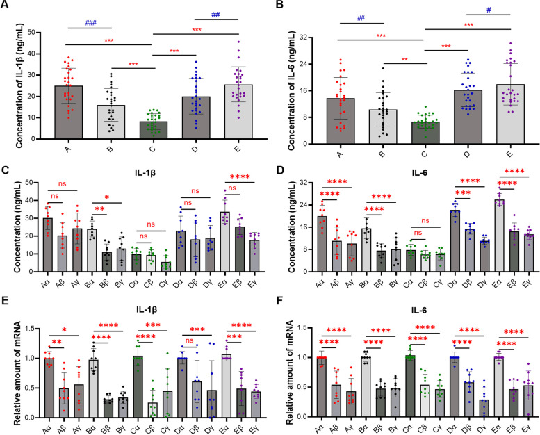
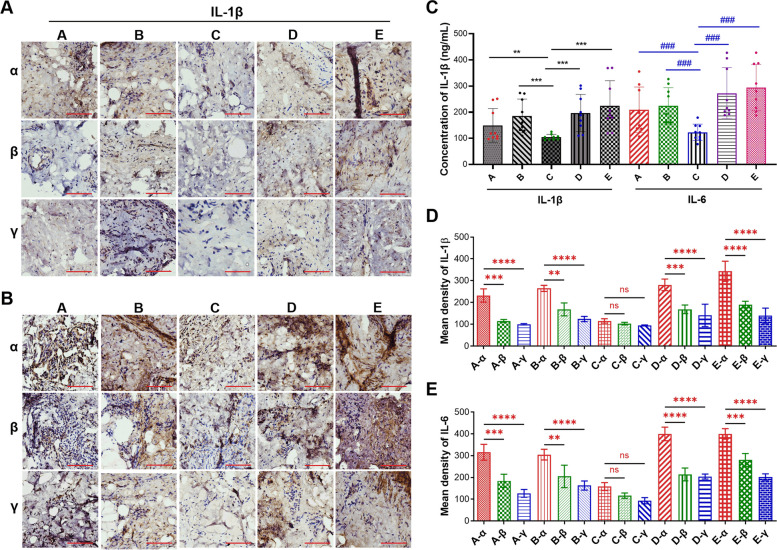
Background: Synovitis, characterized by inflammation of the synovial membrane, is commonly induced by meniscus tears. However, significant differences in inflammatory responses and the key inflammatory mediators of synovium induced by different types of meniscal tears remain unclear.

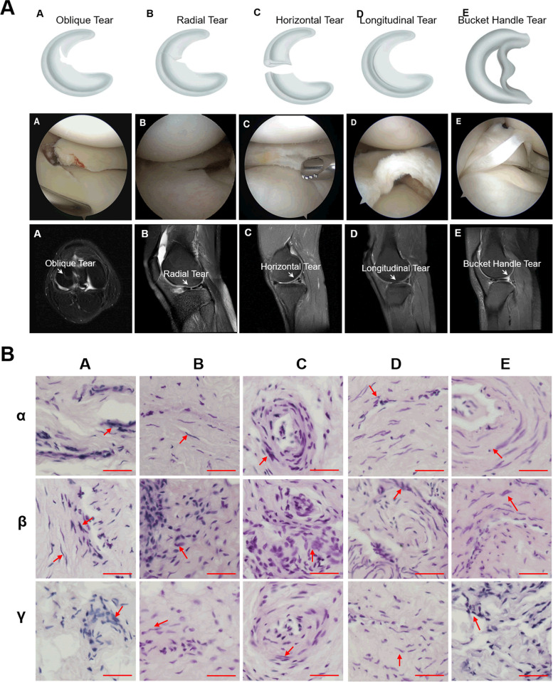
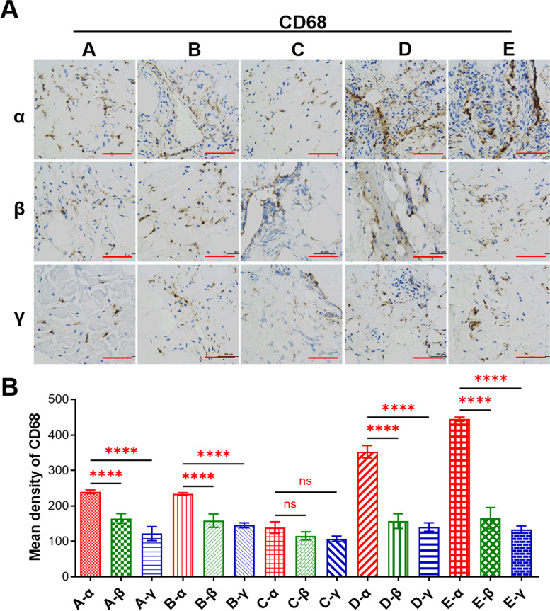
Methods: Magnetic resonance imaging (MRI) was employed to identify the type of meniscus tear, and the quantification of synovial inflammation was assessed through H&E staining assay. Transcription and expression levels of IL-1β and IL-6 were evaluated using bioinformatics, ELISA, RT-qPCR, and IHC of CD68 staining assays. The therapeutic potential of Docosapentaenoic Acid (DPA) was determined through network pharmacology, ELISA, and RT-qPCR assays. The safety of DPA was assessed using colony formation and EdU staining assays.

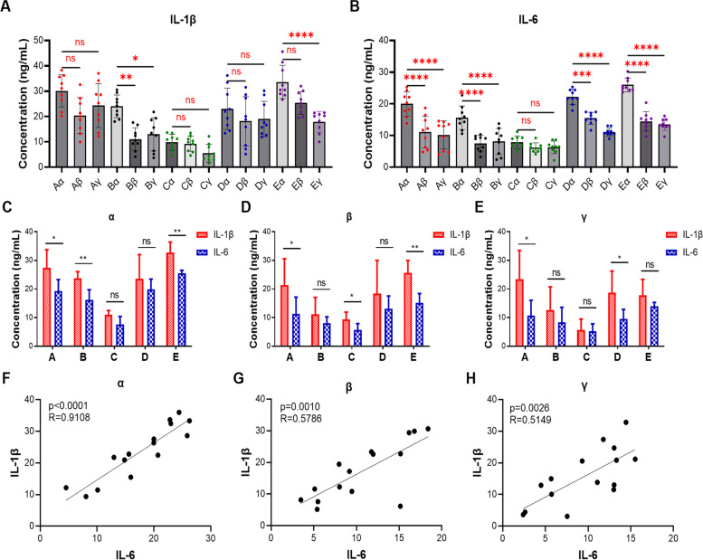
Results: The results indicate that both IL-1β and IL-6 play pivotal roles in synovitis pathogenesis, with distinct expression levels across various subtypes. Among tested meniscus tears, oblique tear and bucket handle tear induced the most severe inflammation, followed by radial tear and longitudinal tear, while horizontal tear resulted in the least inflammation. Furthermore, in synovial inflammation induced by specific meniscus tears, the anterior medial tissues exhibited significantly higher local inflammation than the anterior lateral and suprapatellar regions, highlighting the clinical relevance and practical guidance of anterior medial tissues' inflammatory levels. Additionally, we identified the essential omega-3 fatty acid DPA as a potential therapeutic agent for synovitis, demonstrating efficacy in blocking the transcription and expression of IL-1β and IL-6 with minimal side effects.

Conclusion: These findings provide valuable insights into the nuanced nature of synovial inflammation induced by various meniscal tear classifications and contribute to the development of new adjunctive therapeutic agents in the management of synovitis.

Keywords: Docosapentaenoic acid; Inflammation; Meniscus tears; Omega-3; Synovitis.


Conflict of interest statement

The authors declare no competing interests.

Figures

Fig. 1
Fig. 1
Classification of meniscal tears. A Meniscus tear can be divided into five types according to the different types of tear sites, namely oblique tear (A), radial tear (B), horizontal tear (C); longitudinal tear (D), and bucket handle tear (E). B Representative images of H&E staining assay of synovial inflammation induced by different meniscal injuries caused by oblique tear (A), radial tear (B), horizontal tear (C); longitudinal tear (D), and bucket handle tear (E), respectively. Additionally, in the same synovitis clinical sample, α represents the anterior medial region, β represents the anterior lateral region, and γ represents the suprapatellar region. Red arrow indicated cell nuclei. Scale bar, 100 µm
Fig. 2
Fig. 2
Degree of macrophage infiltration in clinical synovitis tissue. A Representative images of IHC of CD68 in clinical synovitis tissue. A refers to synovitis are caused by oblique tear in the meniscus, B refers to radial tear, C refers to horizontal tear, D refers to longitudinal tear, and E refers to bucket handle tear. Additionally, in the same synovitis clinical sample, α represents the anterior medial region, β represents the anterior lateral region, and γ represents the suprapatellar region. B Quantification of data in A. Values are the average ± SD of three independent experiments. p values were calculated using the two-way analysis of variance (ANOVA) (ns, not significant, ****p < 0.001). Scale bar, 100 µm
Fig. 3
Fig. 3
GO and KEGG analyses of GSE46750 and GSE198520 dataset. A Top 10 enriched GO terms for biological process of GSE46750 dataset. The color of each bubble represents the p-value, while the bubble size represents the gene number. B Top 10 enriched KEGG terms of GSE46750 dataset. The color of each bubble represents the p-value, while the bubble size represents the gene number. C Significant variations in the expression levels of IL-1β and IL-6 were observed before and after the clinical treatment among 42 genes exhibiting the most robust correlation with synovitis treatment targets according to the GSE46750 dataset. D Top 10 enriched GO terms of GSE198520 dataset. The color of each bubble represents the p-value, while the bubble size represents the gene number. E The expression of IL-1β and IL-6 in synovial tissue before and after treatment were statistically significant in GSE198520 dataset. Values are the average ± SD of three independent experiments. p values were calculated using the unpaired student’s t-test (**p < 0.01)
Fig. 4
Fig. 4
Transcription and expression levels of IL-1β and IL-6 in clinical synovitis tissue. A and B ELISA determined the abundance of IL-1β protein (A) and IL-6 protein (B) in clinical synovitis tissues caused by different types of meniscal tears. C and D ELISA determined the protein abundance of IL-1β (C) and IL-6 (D) in different region. E and F RT-qPCR determined the transcription levels of IL-1β and IL-6 in clinical samples. A refers to synovitis is caused by oblique tear in the meniscus, B refers to radial tear, C refers to horizontal tear; D refers to longitudinal tear, and E refers to bucket handle tear. Additionally, in the same synovitis clinical sample, α represents the anterior medial region, β represents the anterior lateral region, and γ represents the suprapatellar region. Values are the average ± SD of three independent experiments. p values were calculated using the two-way analysis of variance (ANOVA) (ns, not significant, *p < 0.05, **p < 0.01, ***p < 0.001, ****p < 0.0001 or #p < 0.05, ##p < 0.01, ###p < 0.001)
Fig. 5
Fig. 5
The abundance of IL-1β and IL-6 in clinical synovitis tissue. A and B Representative images of IHC of IL-1β (A) and IL-6 (B) in clinical synovitis tissue. C-E Quantification of data in A and B respectively. Values are the average ± SD of three independent experiments. p values were calculated using the two-way analysis of variance (ANOVA) (ns, not significant, **p < 0.01, ***p < 0.001, ****p < 0.0001 or ###p < 0.001). Scale bar, 100 µm
Fig. 6
Fig. 6
The correlation between IL-1β and IL-6 in different region of clinical synovitis tissue. A and B ELISA determined the expression levels of IL-1β (A) and IL-6 (B) in different region of clinical synovitis tissue causing by different meniscus tears. C The abundance of IL-1β and IL-6 in the anterior medial region of synovitis induced by different meniscal tears were determined by ELISA assay. D The abundance of IL-1β and IL-6 in the anterior lateral region of synovitis induced by different meniscal tears were determined by ELISA assay. E The abundance of IL-1β and IL-6 in the suprapatellar region of synovitis induced by different meniscus tears were determined by ELISA assay. F The correlation of IL-1β and IL-6 in the anterior medial region of synovitis. G The correlation of IL-1β and IL-6 in the anterior lateral region of synovitis. H The correlation of IL-1β and IL-6 in the suprapatellar region of synovitis. Values are the average ± SD of three independent experiments. p values were calculated using the unpaired student’s t-test and one-way analysis of variance (ANOVA) (ns, not significant, *p < 0.05, **p < 0.01, ***p < 0.001, ****p < 0.0001). The R values in F, G and H were calculated using the Pearson correlation analysis
Fig. 7
Fig. 7
Docosapentaenoic acid (DPA) inhibited the transcription and expression of IL-1β and IL-6 without any side-effect. A The molecular structure of DPA. B Venn diagram depicting overlaps of common targets between synovitis and DPA. C The drug–common targets interconnection network of DPA against synovitis. D Protein–protein interaction network of IL-6. E and F DPA decreased the abundance of IL-1β and IL-6 protein in a dose-dependent manner. G and H DPA inhibited the transcription of IL-1β and IL-6 in a dose-dependent manner. I DPA had no inhibitory effect on the colony formation of human normal liver MIHA cells. J Quantitative analysis of the results shown in I. K DPA had no inhibitory effect on the DNA synthesis of human normal liver MIHA cells. L Quantitative analysis of the results shown in K. Values are the average ± SD of three independent experiments. p values were calculated using the one-way analysis of variance (ANOVA) (ns means no significant with p > 0.05, *p < 0.05, **p < 0.01). Scale bar, 100 µm

References

    1. Vaquero-Picado A, Rodriguez-Merchan EC. Arthroscopic repair of the meniscus: surgical management and clinical outcomes. EFORT Open Rev. 2018;3(11):584–594. doi: 10.1302/2058-5241.3.170059. - DOI - PMC - PubMed
    1. Markes AR, Hodax JD, Ma CB. Meniscus form and function. Clin Sports Med. 2020;39(1):1–12. doi: 10.1016/j.csm.2019.08.007. - DOI - PubMed
    1. Adams BG, Houston MN, Cameron KL. The epidemiology of meniscus injury. Sports Med Arthrosc Rev. 2021;29(3):e24–e33. doi: 10.1097/JSA.0000000000000329. - DOI - PubMed
    1. Manzi JE, Trauger N, Quan T, Ciccotti MC, Ruzbarsky JJ, Dines JS. Increased knee and meniscal injury incidence in professional baseball pitchers with wider, positive stride width. Sports Health. 2023;15(1):36–44. doi: 10.1177/19417381221092024. - DOI - PMC - PubMed
    1. Katz JN, Brophy RH, Chaisson CE, de Chaves L, Cole BJ, Dahm DL, Donnell-Fink LA, Guermazi A, Haas AK, Jones MH, et al. Surgery versus physical therapy for a meniscal tear and osteoarthritis. N Engl J Med. 2013;368(18):1675–1684. doi: 10.1056/NEJMoa1301408. - DOI - PMC - PubMed

MeSH terms