. 2024 Sep;245(3):405-419.
doi: 10.1111/joa.14055. Epub 2024 May 12.

An ovine model for investigation of the microenvironment of the male mammary gland

Affiliations

An ovine model for investigation of the microenvironment of the male mammary gland

Benjamin P Davies et al. J Anat. 2024 Sep.

Abstract

The specific biology of the male breast remains relatively unexplored in spite of the increasing global prevalence of male breast cancer. Delineation of the microenvironment of the male breast is restricted by the low availability of human samples and a lack of characterisation of appropriate animal models. Unlike the mouse, the male ovine gland persists postnatally. We suggest that the male ovine mammary gland constitutes a promising adjunctive model for the male breast. In this study, we evaluate the male ovine mammary gland microenvironment, comparing intact and neutered males. Assessment of the glandular histo-anatomy highlights the resemblance of the male gland to that of neonatal female sheep and confirms the presence of rudimentary terminal duct lobular units. Irrespective of neutered status, cell proliferation in epithelial and stromal compartments is similarly low in males, and cell proliferation in epithelial cells and in the intralobular stroma is significantly lower than in pubertal female sheep. Between 42% and 72% of the luminal mammary epithelial cells in the male gland express the androgen receptor and expression is significantly reduced by neutering. Luminal epithelial cells within the intact and neutered male gland also express oestrogen receptor alpha, but minimal progesterone receptor expression is observed. The distribution of leukocytes within the ducts and stroma is similar to the mammary gland of female sheep and females of other species. Both macrophages and T lymphocytes are intercalated in the epithelial bilayer and are more abundant in the intralobular stroma than the interlobular stroma, suggesting that they may have a protective immunological function within the vestigial glandular tissue of the male sheep. Mast cells are also observed within the stroma. These cells cluster near the glandular tissue and are frequently located adjacent to blood vessels. The abundance of mast cells is significantly higher in intact males compared to neutered males, suggesting that hormone signalling may impact mast cell recruitment. In this study, we demonstrate the utility of the male ovine mammary gland as a model for furthering our knowledge of postnatal male mammary biology.

Keywords: male; mammary gland; microenvironment; model; sheep; udder.


Figures

FIGURE 1
FIGURE 1
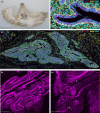
Macro and histo‐anatomy of the male ovine mammary gland. (a) A sub‐gross image of fixed male ovine mammary tissue. Arrow indicates mammary gland. (b, c) Immunofluorescence staining for luminal epithelial cells, using E‐cadherin (magenta), myoepithelial cells, using alpha‐smooth muscle actin (yellow) and DNA, using DAPI (cyan). Red asterisks indicate areas of intralobular mammary stroma. White asterisks indicate areas of interlobular mammary stroma. (d, e) 3D maximum intensity projections of optically cleared male ovine mammary tissue, using confocal microscopy. Immunofluorescence staining for alpha‐smooth muscle actin (magenta). Arrows indicate terminal duct lobular units. Images are representative of three biological repeats Scale bar = 1 cm (a); 50 μm (b); 1 mm (c); 200 μm (d); 100 μm (e).
FIGURE 2
FIGURE 2
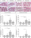
Proliferation dynamics within the intact male, neutered male, pubertal female and mature female mammary gland. (a–h) Dual immunohistochemical staining for Ki67 (brown) and alpha‐SMA (magenta) in the mammary gland of intact males (a, e), neutered males (b, f), pubertal females (c, g) and mature females (d, h). (i–l) Bar graphs illustrating differences in mean luminal cell proliferation (i), myoepithelial cell proliferation (j), intralobular stroma proliferation (k) and interlobular stroma proliferation (l) per mm2 of glandular tissue, intra‐ or interlobular mammary stroma + standard deviation (*p < 0.05, **p < 0.01, ***p < 0.001, N = 6 for intact males, N = 9 for neutered males, N = 7 for pubertal females, N = 7 for mature females, using Kruskal–Wallis test). Dots represent individual sheep. Images representative of 6 (a, e), 9 (b, f) and 7 (c, g, d, h) biological repeats. All IHC is shown with a haematoxylin counterstain. Scale bar = 100 μm (a–d); 50 μm (e–h).
FIGURE 3
FIGURE 3
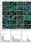
Differences in mammary epithelial hormone receptor expression between intact males, neutered males, pubertal females and mature females. (a–x) Immunofluorescence staining for androgen receptor (AR) (a–h), oestrogen receptor alpha (ERα) (i–p), progesterone receptor A/B (PR) (q–x) (magenta), alpha‐SMA (yellow) and DAPI (cyan) in the mammary gland of intact males (a, e, i, m, q, u), neutered males (b, f, j, n, r, v), pubertal females (c, g, k, o, s, w) and mature females (d, h, l, p, t, x). (y–aa) Bar graphs illustrating differences in the mean percentage of AR (y), ERα (z) or PR (aa) positive luminal cell nuclei + standard deviation (**p < 0.01, ***p < 0.001, N = 5 for intact males, N = 9 for neutered males, N = 7 for pubertal females, N = 7 for mature females, using Kruskal–Wallis test). Dots represent individual sheep. Images representative of a minimum of 5 biological repeats. Scale bar = 100 μm (a–d, i–l, q–t); 50 μm (e–h, m–p, u–x).
FIGURE 4
FIGURE 4
3D Macrophage distribution within the male ovine mammary gland. A 3D maximum intensity projection of optically cleared male ovine mammary tissue, using confocal microscopy. Immunofluorescence staining for IBA1 (magenta), alpha‐SMA (yellow) and DAPI (cyan). Image representative of 3 biological repeats. Scale bar = 100 μm.
FIGURE 5
FIGURE 5
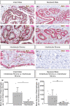
Macrophage abundance and localisation within the intact and neutered male ovine mammary gland. (a–f) Dual immunohistochemical staining for IBA1 (brown) and alpha‐SMA (magenta) in the mammary gland of intact males (a, c) and neutered males (b, d). (e–f) The abundance of macrophages in the intralobular (e) and interlobular mammary stroma (f). (g, h) Bar graphs illustrating differences in the mean number of macrophages per mm2 of mammary stroma in the intralobular and interlobular stroma of intact (g) and neutered males (h) + standard deviation (*p < 0.05, **p < 0.01, N = 6 for intact males, N = 10 for neutered males, using Wilcoxon signed‐rank test). Dots represent individual sheep. Images representative of 6 (a, c), 10 (b, d–f) biological repeats. Scale bar = 100 μm (a, b); 50 μm (c–f).
FIGURE 6
FIGURE 6
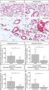
T‐lymphocyte abundance within the intact and neutered male ovine mammary gland. (a–f) Dual immunohistochemical staining for CD3 (brown) and CD20 (magenta) in the mammary gland of intact males (a, c) and neutered males (b, d) and within the intralobular (e) and interlobular mammary stroma (f). (g, h) Bar graphs illustrating differences in the mean number of T‐lymphocytes per mm2 of mammary stroma in the intralobular and interlobular stroma of intact (g) and neutered males (h) + standard deviation (**p < 0.01, N = 5 for intact males, N = 9 for neutered males, using Wilcoxon signed‐rank test). Dots represent individual sheep. Images representative of 5 (a, c, e, f) and 9 (b, d) biological repeats. All IHC is shown with a haematoxylin counterstain. Scale bar = 100 μm (a, b); 50 μm (c, f).
FIGURE 7
FIGURE 7
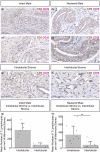
Mast cell abundance is significantly higher in intact males. (a–c) Dual immunohistochemical staining for c‐Kit (brown) and alpha‐SMA (magenta) in the mammary gland of intact males (a) and neutered males (b). Arrows indicate positive staining for mast cells. (c) Mast cells are located in close proximity to blood vessels in the male ovine mammary gland. Arrows indicate positive mast cell staining. (d, e) Bar graphs illustrating differences in the mean number of mast cells per mm2 of intralobular (d) and interlobular (e) stroma in intact and neutered males + standard deviation (*p < 0.05, N = 6 for intact males, N = 7 for neutered males, using Mann–Whitney U test). (f, g) Bar graphs illustrating differences in the mean number of mast cells per mm2 of mammary stroma in the intralobular and interlobular stroma of intact (f) and neutered males (g) + standard deviation (*p < 0.05, N = 6 for intact males, N = 7 for neutered males, using Wilcoxon signed‐rank test). Dots represent individual sheep. Images representative of 6 (a, c) and 7 (b) biological repeats. All IHC is shown with a haematoxylin counterstain. Scale bar = 50 μm (a, b); 25 μm (c).

References

    1. Atwood, C.S. , Hovey, R.C. , Glover, J.P. , Chepko, G. , Ginsburg, E. , Robison, W.G. et al. (2000) Progesterone induces side‐branching of the ductal epithelium in the mammary glands of peripubertal mice. Journal of Endocrinology, 167, 39–52. Available from: 10.1677/joe.0.1670039 - DOI - PubMed
    1. Beaudry, K.L. , Parsons, C.L.M. , Ellis, S.E. & Akers, R.M. (2016) Localization and quantitation of macrophages, mast cells, and eosinophils in the developing bovine mammary gland1. Journal of Dairy Science, 99, 796–804. Available from: 10.3168/jds.2015-9972 - DOI - PubMed
    1. Brisken, C. , Park, S. , Vass, T. , Lydon, J.P. , O'Malley, B.W. & Weinberg, R.A. (1998) A paracrine role for the epithelial progesterone receptor in mammary gland development. Proceedings of the National Academy of Sciences of the United States of America, 95, 5076–5081. - PMC - PubMed
    1. Cardiff, R.D. , Jindal, S. , Treuting, P.M. , Going, J.J. , Gusterson, B. & Thompson, H.J. (2018) 23. Mammary gland. In: Treuting, P.M., Dintzis, S.M. & Montine, K.S. (Eds.) Comparative anatomy and histology, 2nd edition. San Diego: Academic Press, pp. 487–509. Available from: 10.1016/B978-0-12-802900-8.00023-3 - DOI
    1. Cardoso, F. , Bartlett, J.M.S. , Slaets, L. , van Deurzen, C.H.M. , van Leeuwen‐Stok, E. , Porter, P. et al. (2018) Characterization of male breast cancer: results of the EORTC 10085/TBCRC/BIG/NABCG International Male Breast Cancer Program. Annals of Oncology, Incorporating Blood‐Based Liquid Biopsy Information into Cancer Staging, 29, 405–417. Available from: 10.1093/annonc/mdx651 - DOI - PMC - PubMed

Publication types