. 2024 Jun;76(3):313-327.
doi: 10.1007/s10616-024-00621-6. Epub 2024 Mar 26.

LncRNA MIR181A2HG negatively regulates human keratinocytes proliferation by binding SRSF1

Affiliations

LncRNA MIR181A2HG negatively regulates human keratinocytes proliferation by binding SRSF1

Xiaomei Fan et al. Cytotechnology. 2024 Jun.

Abstract

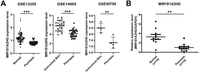
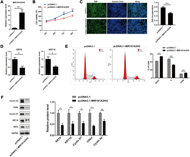
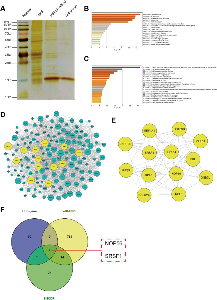
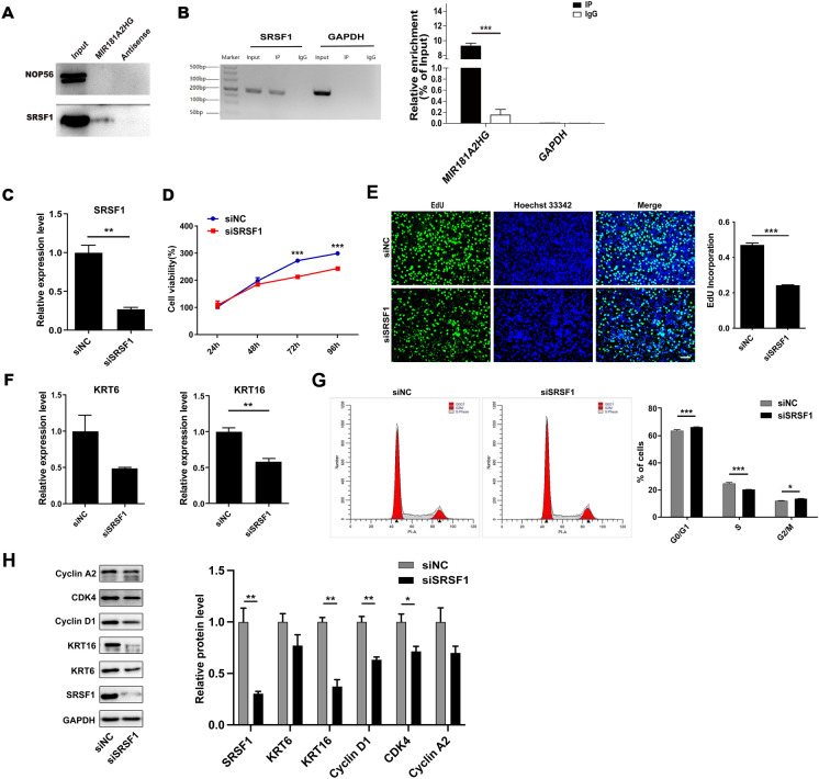
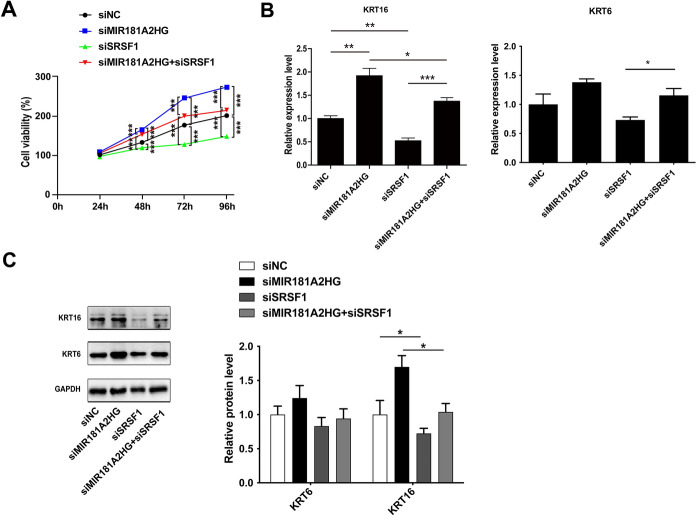
Psoriasis is a common chronic inflammatory skin disease. Abnormal proliferation of keratinocytes plays an important role in the pathogenesis of psoriasis. Long non-coding RNAs (lncRNAs) are involved in the regulation of a variety of cell biological processes. The purpose of this study was to investigate the potential role of lncRNA MIR181A2HG in the proliferation of human keratinocytes. qRT-PCR and Western blotting were performed to measure the expression levels of MIR181A2HG, SRSF1, KRT6, and KRT16 in tissue specimens and HaCaT keratinocytes. The effects of MIR181A2HG on HaCaT keratinocytes proliferation were evaluated using Cell Counting Kit-8 (CCK-8) assays, 5-Ethynyl-2'-deoxyuridine (EdU) incorporation, and cell-cycle assays. RNA pulldown-mass spectrometry (MS) was applied to identify the proteins interacting with MIR181A2HG. RNA pull-down-Western blotting and RNA immunoprecipitation coupled with real-time quantitative reverse transcription-PCR (RIP-qRT-PCR) assays were used to determine the interactions between MIR181A2HG and its RNA-binding proteins (RBPs). MIR181A2HG was down-regulated in psoriasis tissues. MIR181A2HG overexpression induced G0/G1 and G2/M phase cell cycle arrest and decreased the protein levels of KRT6, KRT16, Cyclin D1, CDK4, and Cyclin A2 in HaCaT keratinocytes. MIR181A2HG knockdown showed the opposite effect. By using RNA pulldown-MS, 356 proteins were identified to interact with MIR181A2HG potentially. Bioinformatics analysis showed that NOP56 and SRSF1 may be RNA binding proteins (RBPs) that may be interact with MIR181A2HG. Furthermore, by using RNA pull-down-Western blotting and RIP-qRT-PCR, SRSF1 was determined to interact with MIR181A2HG. Moreover, silencing of SRSF1 inhibited keratinocytes proliferation, which could be reversed with the knockdown of MIR181A2HG. Our findings indicated that MIR181A2HG can negatively regulate HaCaT keratinocytes proliferation by binding SRSF1, suggesting that MIR181A2HG and SRSF1 may serve as potential targets for the treatment of psoriasis.

Supplementary information: The online version contains supplementary material available at 10.1007/s10616-024-00621-6.

Keywords: Keratinocytes; LncRNAs; MIR181A2HG; Proliferation; Psoriasis; SRSF1.


Conflict of interest statement

Competing interestsThe authors declare no competing interests.

Figures

Fig. 1
Fig. 1
The expression of MIR181A2HG in psoriatic lesions and normal tissues. A The expression of MIR181A2HG in normal skin and psoriatic lesions was analyzed using publicly available GEO datasets (GSE13355, GSE14905 and GSE50790). ***P < 0.001, **P < 0.01. B qRT-PCR was applied to measure the expression of MIR181A2HG in the psoriatic lesions and normal skin. GAPDH served as an internal reference. **P < 0.01
Fig. 2
Fig. 2
MIR181A2HG was predicted to be involved in regulating the cell cycle. A LncATLAS was used to explore the localization of MIR181A2HG. B Nuclear/cytoplasmic separation qRT-PCR was performed to detect the nuclear and cytoplasmic distribution of MIR181A2HG. GAPDH served as extranuclear RNA control. MALAT1 and U3 served as intranuclear RNA controls. C KEGG pathway enrichment analysis of MIR181A2HG-related genes. D The intersection of KEGG analysis results in three GEO datasets
Fig. 3
Fig. 3
Effect of MIR181A2HG overexpression on proliferation of HaCaT keratinocytes. A HaCaT keratinocytes were seeded in 12-well plates and transfected with pcDNA3.1 or pcDNA3.1-MIR181A2HG for 48 h. qRT-PCR was applied to measure the expression of MIR181A2HG. **P < 0.01. B CCK-8 assays were carried out to record the growth curves of HaCaT keratinocytes transfected with pcDNA3.1 or pcDNA3.1-MIR181A2HG for 24–96 h. **P < 0.01. C EdU assays were used to detect the proliferation rate of HaCaT keratinocytes transfected with pcDNA3.1 or pcDNA3.1-MIR181A2HG for 48 h. Scale bars, 100 μm. ***P < 0.001. D KRT6 and KRT16 expression were measured by qRT-PCR in HaCaT keratinocytes transfected with pcDNA3.1 or pcDNA3.1-MIR181A2HG for 72 h. *P < 0.05. E The cell cycle distribution of HaCaT keratinocytes at 48 h transfected with pcDNA3.1 or pcDNA3.1-MIR181A2HG was analyzed by flow cytometry. ***P < 0.001. F Cells were collected and total proteins were extracted for Western blotting to detect KRT6, KRT16, Cyclin D1, CDK4 and Cyclin A2 protein levels. GAPDH served as an internal reference. **P < 0.01, *P < 0.05
Fig. 4
Fig. 4
Effect of MIR181A2HG knockdown on the proliferation of HaCaT keratinocytes. A HaCaT keratinocytes were seeded in 12-well plates and transfected with 50 nM scramble siRNA (siNC) or MI181A2HG siRNA (siMIR181A2HG) for 48 h. qRT-PCR was performed to measure the expression of MIR181A2HG. *P < 0.05. B CCK-8 assays were carried out to record the growth curves of HaCaT keratinocyte transfected with 50 nM siNC or siMIR181A2HG for 24–96 h. *P < 0.05. C EdU assays were used to detect the proliferation rate of HaCaT keratinocytes transfected with siNC or siMIR181A2HG for 48 h. Scale bars, 100 μm. **P < 0.01. D KRT6 and KRT16 expression were measured by qRT-PCR in HaCaT keratinocytes transfected with siNC or si-MIR181A2HG for 72 h. *P < 0.05. E The cell cycle distribution of HaCaT keratinocytes at 48 h transfected with 50nM siNC or siMIR181A2HG was analyzed by flow cytometry. ***P < 0.001, **P < 0.01. F Cells were collected and total proteins were extracted for Western blotting to detect KRT6, KRT16, Cyclin D1, CDK4 and Cyclin A2 protein levels. GAPDH served as an internal reference. *P < 0.05
Fig. 5
Fig. 5
Screening of MIR181A2HG interaction protein. A The silver staining result of RNA pulldown proteins. B Pathway analysis of MIR181A2HG interacting proteins. C GO biological process analysis of MIR181A2HG interacting protein. D The construction of protein interaction network of MIR181A2HG interaction protein. E The interaction network of 13 key genes. F The intersection of 13 key proteins with the proteins predicted by catRAPID and ENCORI
Fig. 6
Fig. 6
MIR181A2HG could bind to SRSF1. A RNA pulldown-Western blotting assay was used to verify the interaction of MIR181A2HG and NOP56/SRSF1. B RIP-qRT-PCR assay was performed to confirm the interaction between MIR181A2HG and SRSF1. C HaCaT keratinocytes were seeded in 12-well plates and transfected with 50 nM scramble siRNA (siNC) or SRSF1 siRNA (siSRSF1) for 48 h. qRT-PCR was applied to measure the expression of SRSF1. **P < 0.01. D CCK-8 assays were carried out to record the growth curves of HaCaT keratinocytes transfected with 50nM siNC or siSRSF1 for 24–96 h. ***P < 0.001. E EdU assays were used to detect the proliferation rate of HaCaT keratinocytes transfected with 50nM siNC or siSRSF1 for 48 h. Scale bars, 100 μm. ***P < 0.001. F KRT6 and KRT16 expression were measured by qRT-PCR in HaCaT keratinocytes transfected with 50nM siNC or siSRSF1 for 72 h. **P < 0.01. G The cell cycle distribution of keratinocytes at 48 h transfected with 50nM siNC or siSRSF1 was analyzed by flow cytometry. ***P < 0.001, *P < 0.05. H Cells were collected and total proteins were extracted for Western blotting to detect KRT6, KRT16, Cyclin D1, CDK4 and Cyclin A2 protein levels. GAPDH served as an internal reference. **P < 0.01, *P < 0.05
Fig. 7
Fig. 7
The knockdown of MIR181A2HG could partially restore the inhibitory effect of cell proliferation caused by SRSF1 knockdown. A CCK-8 assay for cell viability in HaCaT keratinocytes transfected with 50 nM siRNA (siNC), MIR181A2HG siRNA (siMIR181A2HG) or SRSF1 siRNA (siSRSF1) for 24–96 h. ***P < 0.001. B KRT6 and KRT16 expression were measured by qRT-PCR in HaCaT keratinocytes transfected with 100nM siNC, siMIR181A2HG or siSRSF1. ***P < 0.001, **P < 0.01, *P < 0.05. C Cells were collected and total proteins were extracted for Western blotting to detect KRT6 and KRT16 protein levels. GAPDH served as an internal reference. *P < 0.05

Similar articles

Cited by

References

    1. Afonina IS, Van Nuffel E, Beyaert R. Immune responses and therapeutic options in psoriasis. Cell Mol Life Sci. 2021;78:2709–2727. doi: 10.1007/s00018-020-03726-1. - DOI - PMC - PubMed
    1. Albanesi C, Madonna S, Gisondi P, Girolomoni G. The interplay between keratinocytes and Immune cells in the pathogenesis of psoriasis. Front Immunol. 2018;9:1549. doi: 10.3389/fimmu.2018.01549. - DOI - PMC - PubMed
    1. Anczuków O, Akerman M, Cléry A, Wu J, Shen C, Shirole NH, Krainer AR. SRSF1-regulated alternative splicing in breast cancer. Mol Cell. 2015;60:105–117. doi: 10.1016/j.molcel.2015.09.005. - DOI - PMC - PubMed
    1. Beermann J, Piccoli MT, Viereck J, Thum T. Non-coding RNAs in development and disease: background, mechanisms, and therapeutic approaches. Physiol Rev. 2016;96:1297–1325. doi: 10.1152/physrev.00041.2015. - DOI - PubMed
    1. Billi AC, Gudjonsson JE, Voorhees JJ. Psoriasis: past, present, and future. J Invest Dermatol. 2019;139:e133–e142. doi: 10.1016/j.jid.2019.08.437. - DOI - PMC - PubMed

LinkOut - more resources