. 2024 Sep;20(37):e2400815.
doi: 10.1002/smll.202400815. Epub 2024 May 13.

Preformed Vesicle Approach to LNP Manufacturing Enhances Retinal mRNA Delivery

Affiliations

Preformed Vesicle Approach to LNP Manufacturing Enhances Retinal mRNA Delivery

Yulia Eygeris et al. Small. 2024 Sep.

Abstract

Complete encapsulation of nucleic acids by lipid-based nanoparticles (LNPs) is often thought to be one of the main prerequisites for successful nucleic acid delivery, as the lipid environment protects mRNA from degradation by external nucleases and assists in initiating delivery processes. However, delivery of mRNA via a preformed vesicle approach (PFV-LNPs) defies this precondition. Unlike traditional LNPs, PFV-LNPs are formed via a solvent-free mixing process, leading to a superficial mRNA localization. While demonstrating low encapsulation efficiency in the RiboGreen assay, PFV-LNPs improved delivery of mRNA to the retina by up to 50% compared to the LNP analogs across several benchmark formulations, suggesting the utility of this approach regardless of the lipid composition. Successful mRNA and gene editors' delivery is observed in the retinal pigment epithelium and photoreceptors and validated in mice, non-human primates, and human retinal organoids. Deploying PFV-LNPs in gene editing experiments result in a similar extent of gene editing compared to analogous LNP (up to 3% on genomic level) in the Ai9 reporter mouse model; but, remarkably, retinal tolerability is significantly improved for PFV-LNP treatment. The study findings indicate that the LNP formulation process can greatly influence mRNA transfection and gene editing outcomes, improving LNP treatment safety without sacrificing efficacy.

Keywords: IRD; gene delivery; lipid nanoparticle; mRNA; retinal degeneration.


Conflict of interest statement

Conflicts of Interest

G.S. is a co-founder of EnterX Bio and RNAvax Bio, and has an advisory role to Rare Air Inc. and Serina Tx. Y.E. has stock options and an advisory role to EnterX Bio. Other authors have no conflicts to declare.

Figures
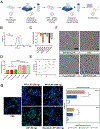
Figure 1.
Figure 1.. Characterization of lipid nanoparticles (LNP).
(A) Formulation workflow of pre-formed vesicle (PFV) intermediates and mRNA-loaded PFV-LNPs. (B) Representative shift in size distribution for PFV-LNPs compared to unloaded PFV. (C) Zeta potential for PFV, LNP, and PFV-LNP formulations (n=3). (D) Encapsulation efficiency in mRNA-loaded LNPs and PFV-LNPs (n=3). PFV-LNPs consistently have <50% encapsulation efficiency; therefore, the dose is calculated by total mRNA found in sample. (E) Laurdan hydration assay of LNP, PFV, and PFV-LNP formulations, where generalized polarization (GP) is indicative of changes in lipid packing (higher = tighter lipid packing; n=3). (F) Representative cryo-TEM micrographs of standard LNPs, PFV, or mRNA loaded LNPs with two distinct cargos (FLuc or EGFP mRNA respectively). Gold boxes highlight typical PFV or PFV-LNP morphologies. Scale bars are 50 nm. (G) Gal9 recruitment in modified HEK293T/17 cells treated with LNPs at 24 hours after 50 ng or 200 ng treatment with LNPs. Images presented at maximum intensity projection, DAPI – blue and Gal9-GFP - green. Scale bars represent 50 μm. Quantification includes normalizing Gal9 puncta counts by the number of cells present (from DAPI; n=6). Data are presented as means ± standard deviation. Statistical analyses were performed using ANOVA with Tukey multiple comparison test. n.s.: not significant; *: p < 0.05; **: p < 0.01; ****: p < 0.001. PFV – preformed vesicles approach, no cargo; PFV-LNP – preformed vesicle approach, loaded with mRNA; LNP – standard microfluidic mixing, loaded with mRNA
Figure 2.
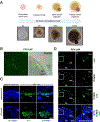
Figure 2.. Localization of tdTomato expression after Cre mRNA delivery in Ai9 murine retina.
Nanoparticles were administered via subretinal injection at 400 ng mRNA. Eyes were harvested at the 7-day post-injection and stained with DAPI. (A) Mechanism of Cre-mediated recombination. (B) Representative brightfield (top) and tdTomato (bottom) fundus images for each treatment groups (n=4 eyes each). (C) Representative immunofluorescence images showing tdTomato expression in the RPE and ONL. Gold box indicates the area of photoreceptor transfection. Scale bars represent 75 μm. RPE: retinal pigment epithelium; ONL: outer nuclear layer; INL: inner nuclear layer; GCL: ganglion cell layer.
Figure 3.
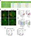
Figure 3.. EGFP expression after EGFP mRNA LNP or PFV-LNP treatment in 129X1/SvJ murine retina.
Nanoparticles were administered via subretinal injection at 200 ng mRNA. Eyes were imaged via fundoscopy and harvested at the 48 hours post-injection and processed for IF and imaging. (A) Representative bright field (top) and GFP (bottom) fundus images for each treatment group 48 hours post-injection. (B) Representative immunofluorescence images demonstrate GFP expression in the RPE for LNP groups. (C) Quantification of GFP expression in fundus images (n=4) and in immunofluorescence images (n=4 eyes, with 3–4 individual images per eye). (D) Representative confocal images of IF with showing GFP expression in the RPE and ONL for PFV-LNP group. Data are presented as means ± standard deviation with statistical significance reported using an ordinary one-way ANOVA test. n.s.: not significant; *: p < 0.05; **: p < 0.01; ****: p < 0.001. GFP: green fluorescent protein; RPE: retinal pigment epithelium; ONL: outer nuclear layer; INL: inner nuclear layer; GCL: ganglion cell layer.
Figure 4.
Figure 4.. EGFP expression after EGFP mRNA PFV-LNP treatment in non-human primate retina.
Nanoparticles were administered via subretinal injection at 25μg total mRNA dose. NHP eye (N=1) was harvested at the 48 hours post-injection and processed for IF imaging. (A) Representative fundus autofluorescence image 48 hours post-injection indicates prominent EGFP signal (area of the injection bleb highlighted by the dashed circle). (B-D) Representative IF images highlighting transfection in ONL. Transfection of the cone photoreceptors can be confirmed by colocalization of EGFP and cone arrestin signal. (E-G) Representative IF images highlighting transfection in RPE; transfection of RPE can be confirmed by colocalization of EGFP and RPE65 signals. Gross retinal health is visualized by OCT (H; the scanning plane is shown in panel A by yellow line and the bracket demonstrates the area of the injection bleb). (I) DAPI and EGFP composite IF image demonstrates the transfection through the entire injection bleb, with stronger signal observed in RPE. (J-L) Mild immune infiltration through RPE and ONL was observed, as suggested by the CD3 and IBA staining of the retina sections. GFP: green fluorescent protein; RPE: retinal pigment epithelium; ONL: outer nuclear layer.
Figure 5.
Figure 5.. EGFP delivery to human iPSC-derived retinal organoids and RPE cells.
(A) The schematic and brightfield images provide an overview of the differentiation process going from iPSCs to mature retinal organoids. (B) Native EGFP expression (green) is shown in a live control retinal organoid, with and without brightfield overlay, at 48 hours post-PFV-LNP transfection with 2μg EGFP mRNA. At 48 hours post-transfection with LNPs or PFV-LNPs carrying EGFP mRNA, retinal organoids were fixed, cryosectioned, and immunolabeled. (C) Representative images of organoids or RPE cell clusters after immunolabeling of GFP (green) shows abundant signal in RPE for standard LNPs and PFV-LNPs. Immunolabeling with antibodies targeting G/RCO (white) shows sparse labeling in cone photoreceptors in retinal organoids (D) transfected with 10μg EGFP mRNA in PFV-LNPs. Nuclei are counterstained with DAPI (blue). Panel ii. is the magnified area highlighted in Panel i. GFP: green fluorescent protein; OS: outer segment; PR: photoreceptor; RPE: retinal pigment epithelium; ONL: outer nuclear layer; INL: inner nuclear layer; G/RCO: green/red cone opsin. Scale bars = 50μm.
Figure 6.
Figure 6.. Comparison of gene editing outcomes after PFV-LNP and LNP treatments in Ai9 mice.
(A) Comparison of LP01 PFV-LNP and LNP nanoparticle characteristics. LNPs were administered subretinally at 200 ng total nucleic acid (1:1 wt/wt Cas9 mRNA:sgRNA) and the eyes were harvested 7 days after injection for flatmount and NGS analysis; n = 7–9 in each group. (B) Representative RPE flatmount images (green: ZO-1 staining, red: tdTomato; background removed for visual clarity). Dashed red circles indicate the transfected area. Image-based quantification of gene editing efficiency (C) and retinal health (D) was performed within whole RPE flatmounts and the injection site and presented as % of area. (E) Next-generation sequencing results; some outliers were removed via ROUT method (Q=1%). No outliers were noted in image-based analysis. Data are presented as means ± standard deviation. Statistical significance reported using ANOVA for image analysis of the whole flatmounts and NGS data, and Welch’s t-test for image analysis within the treated areas. n.s.: not significant; *: p < 0.05; **: p < 0.01; ****: p < 0.001. RPE: retinal pigment epithelium, EE: encapsulation efficiency, PDI: polydispersity index, ZP: zeta potential, RPE: retinal pigment epithelium, sgGFP: non-targeting sgRNA, sgAi9: targeting sgRNA.

References

    1. Boye SE; Boye SL; Lewin AS; Hauswirth WW A Comprehensive Review of Retinal Gene Therapy. Mol. Ther. 2013, 21 (3), 509–519. 10.1038/mt.2012.280. - DOI - PMC - PubMed
    1. Russell S; Bennett J; Wellman JA; Chung DC; Yu Z-F; Tillman A; Wittes J; Pappas J; Elci O; McCague S; Cross D; Marshall KA; Walshire J; Kehoe TL; Reichert H; Davis M; Raffini L; George LA; Hudson FP; Dingfield L; Zhu X; Haller JA; Sohn EH; Mahajan VB; Pfeifer W; Weckmann M; Johnson C; Gewaily D; Drack A; Stone E; Wachtel K; Simonelli F; Leroy BP; Wright JF; High KA; Maguire AM Efficacy and Safety of Voretigene Neparvovec (AAV2-hRPE65v2) in Patients with RPE65-Mediated Inherited Retinal Dystrophy: A Randomised, Controlled, Open-Label, Phase 3 Trial. The Lancet 2017, 390 (10097), 849–860. 10.1016/S0140-6736(17)31868-8. - DOI - PMC - PubMed
    1. Kim J; Eygeris Y; Ryals RC; Jozić A; Sahay G Strategies for Non-Viral Vectors Targeting Organs beyond the Liver. Nat. Nanotechnol. 2023, 1–20. 10.1038/s41565-023-01563-4. - DOI - PubMed
    1. Hajj KA; Whitehead KA Tools for Translation: Non-Viral Materials for Therapeutic mRNA Delivery. Nat. Rev. Mater. 2017, 2 (10), 17056. 10.1038/natrevmats.2017.56. - DOI
    1. Zulliger R; Conley SM; Naash MI Non-Viral Therapeutic Approaches to Ocular Diseases: An Overview and Future Directions. J. Control. Release Off. J. Control. Release Soc 2015, 219, 471–487. 10.1016/j.jconrel.2015.10.007. - DOI - PMC - PubMed

Publication types

LinkOut - more resources