. 2024 Aug;27(3):475-499.
doi: 10.1007/s10456-024-09922-y. Epub 2024 May 13.

CCL4 contributes to aging related angiogenic insufficiency through activating oxidative stress and endothelial inflammation

Affiliations

CCL4 contributes to aging related angiogenic insufficiency through activating oxidative stress and endothelial inflammation

Ting-Ting Chang et al. Angiogenesis. 2024 Aug.

Erratum in

Abstract

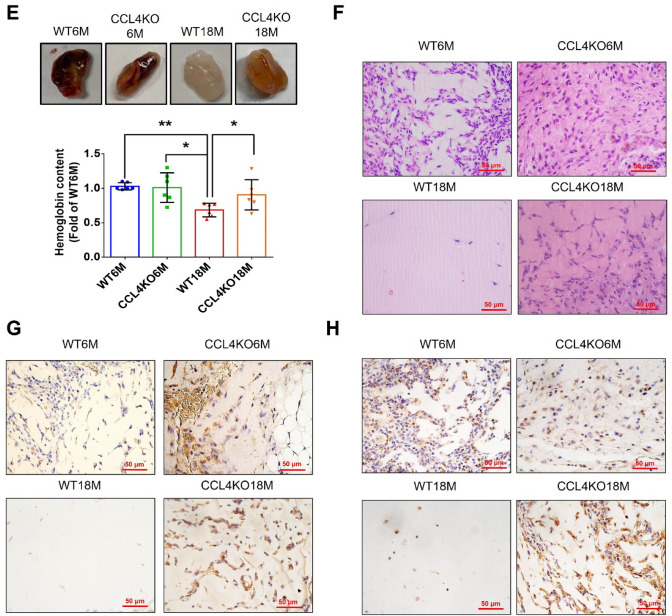
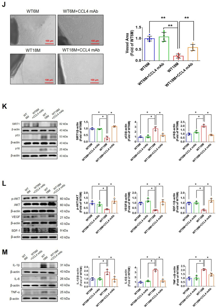
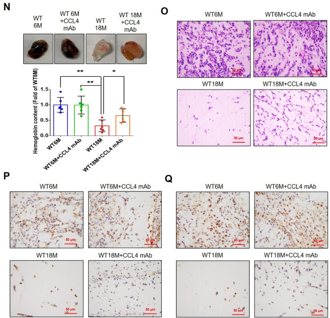
Aging is a natural process associated with chronic inflammation in the development of vascular dysfunction. We hypothesized that chemokine C-C motif ligands 4 (CCL4) might play a vital role in aging-related vascular dysfunction. Circulating CCL4 was up-regulated in elderly subjects and in aged animals. CCL4 inhibition reduced generation of reactive oxygen species (ROS), attenuated inflammation, and restored cell functions in endothelial progenitor cells from elderly subjects and in aged human aortic endothelial cells. CCL4 promoted cell aging, with impaired cell functioning, by activating ROS production and inflammation. CCL4 knockout mice and therapeutic administration of anti-CCL4 neutralizing antibodies exhibited vascular and dermal anti-aging effects, with improved wound healing, via the down-regulation of inflammatory proteins and the activation of angiogenic proteins. Altogether, our findings suggested that CCL4 may contribute to aging-related vascular dysfunction via activating oxidative stress and endothelial inflammation. CCL4 may be a potential therapeutic target for vascular protections during aging.

Keywords: Aging; CCL4; Inflammation; Oxidative stress; Vascular dysfunction.


Conflict of interest statement

The authors declare that they have no competing interests.

Figures

Fig. 1
Fig. 1
Inhibition of CCL4 reversed cell aging and inflammation in EPCs from aged subjects. A Elderly subjects (> 55 years old; n = 8) had higher plasma CCL4 concentrations compared to the young subjects (< 30 years old; n = 7). B Inhibition of CCL4 reduced senescence of EPCs from aged subjects. C Inhibition of CCL4 reduced ROS productions in EPCs from aged subjects (n = 4). D and F Western blotting and statistical analysis of CCL4, xanthine oxidase, p47, and p-p65 in primary cultured EPCs (n = 4). G and H Western blotting and statistical analysis of SIRT1, p53, p16, IL-1β, IL-6, and TNF-α expression in primary cultured EPCs (n = 4). I Inhibition of CCL4 did not affect cell proliferation in HAECs (n = 4). J and K Inhibition of CCL4 improved tube formation and migration abilities in EPCs from aged subjects (n = 4). L Western blotting and statistical analysis of eNOS, p-AKT, VEGF, and SDF-1 expression in primary cultured EPCs (n = 4). *P < 0.05, **P < 0.01
Fig. 1
Fig. 1
Inhibition of CCL4 reversed cell aging and inflammation in EPCs from aged subjects. A Elderly subjects (> 55 years old; n = 8) had higher plasma CCL4 concentrations compared to the young subjects (< 30 years old; n = 7). B Inhibition of CCL4 reduced senescence of EPCs from aged subjects. C Inhibition of CCL4 reduced ROS productions in EPCs from aged subjects (n = 4). D and F Western blotting and statistical analysis of CCL4, xanthine oxidase, p47, and p-p65 in primary cultured EPCs (n = 4). G and H Western blotting and statistical analysis of SIRT1, p53, p16, IL-1β, IL-6, and TNF-α expression in primary cultured EPCs (n = 4). I Inhibition of CCL4 did not affect cell proliferation in HAECs (n = 4). J and K Inhibition of CCL4 improved tube formation and migration abilities in EPCs from aged subjects (n = 4). L Western blotting and statistical analysis of eNOS, p-AKT, VEGF, and SDF-1 expression in primary cultured EPCs (n = 4). *P < 0.05, **P < 0.01
Fig. 2
Fig. 2
Administration of CCL4 caused cell aging and ROS generation in HAECs. A and B Cell senescence and ROS productions were increased in CCL4-treated HAECs (n = 4). C Western blotting and statistical analyses of ROS production proteins, such as xanthine oxidase and p47 (n = 4). D Western blotting and statistical analyses of aging factors, such as SIRT1, p53, and p16 (n = 4). E Western blotting and statistical analyses of angiogenic factors, such as eNOS, p-AKT, VEGF, and SDF-1 (n = 4). F Western blotting and statistical analyses of inflammatory factors, such as IL-1β, IL-6, and TNF-α (n = 4). *P < 0.05, **P < 0.01
Fig. 3
Fig. 3
Inhibition of CCL4 reversed cell aging and inflammation in HAECs. A Inhibition of CCL4 reduced senescence of aged HAECs (n = 4). B Inhibition of CCL4 reduced ROS productions in aged HAECs (n = 4). C Western blotting and statistical analysis of CCL4 in HAECs (n = 4). D Western blotting and statistical analysis of xanthine oxidase and p47 in HAECs (n = 4). E Western blotting and statistical analysis of p-p65 and p65 in HAECs (n = 4). F, G Western blotting and statistical analysis of SIRT1, p53, p16, TNF-α, IL-1β and IL-6 expression in HAECs (n = 4). H, I Western blotting and statistical analysis of p-p65, CCL4, SIRT1, p53, p16, IL-1β, IL-6, and TNF-α expression after administration of MCI186 in aged HAECs (n = 4). J Western blotting and statistical analysis of p-p65 and CCL4 after administration of p65 siRNA in aged HAECs (n = 4). P4, passage 4; P9, passage 9; siCCL4, siRNA of CCL4; MCI186, a free radical scavenger. *P < 0.05, **P < 0.01
Fig. 3
Fig. 3
Inhibition of CCL4 reversed cell aging and inflammation in HAECs. A Inhibition of CCL4 reduced senescence of aged HAECs (n = 4). B Inhibition of CCL4 reduced ROS productions in aged HAECs (n = 4). C Western blotting and statistical analysis of CCL4 in HAECs (n = 4). D Western blotting and statistical analysis of xanthine oxidase and p47 in HAECs (n = 4). E Western blotting and statistical analysis of p-p65 and p65 in HAECs (n = 4). F, G Western blotting and statistical analysis of SIRT1, p53, p16, TNF-α, IL-1β and IL-6 expression in HAECs (n = 4). H, I Western blotting and statistical analysis of p-p65, CCL4, SIRT1, p53, p16, IL-1β, IL-6, and TNF-α expression after administration of MCI186 in aged HAECs (n = 4). J Western blotting and statistical analysis of p-p65 and CCL4 after administration of p65 siRNA in aged HAECs (n = 4). P4, passage 4; P9, passage 9; siCCL4, siRNA of CCL4; MCI186, a free radical scavenger. *P < 0.05, **P < 0.01
Fig. 4
Fig. 4
Inhibition of CCL4 reversed aging-induced cell dysfunction in HAECs. A Inhibition of CCL4 did not affect cell proliferation in HAECs (n = 4). B and C Inhibition of CCL4 improved tube formation and migration abilities in aged HAECs (n = 4). D Western blotting and statistical analysis of p-eNOS, p-AKT, VEGF, and SDF-1 expression in HAECs (n = 4). P4, passage 4; P9, passage 9; siCCL4, siRNA of CCL4. *P < 0.05, **P < 0.01
Fig. 5
Fig. 5
Deletion of CCL4 improved aging-induced delayed wound healing. A Aged mice have higher serum CCL4 concentrations compared to young mice (n = 6). B Representative photographs of wound healing in each group at various time points after wounding. The wound closure results were quantified on days 7 after wounding (n = 6). C Representative images with H&E staining. D and E Representative images with immunostaining of CD31 and Ki67. Deletion of CCL4 enhanced both CD31 and Ki67 positive areas in the 18 months old mice compared to the aged wild-type mice. F Representative images with Masson’s trichrome staining. Deletion of CCL4 enhanced collagen deposition in the 18 months old mice compared to the aged wild-type mice. G Western blotting and statistical analyses of aging factors, such as SIRT1, p53, and p16 (n = 4). H Western blotting and statistical analyses of angiogenic factors, such as p-AKT, VEGF, and SDF-1 (n = 4). I Western blotting and statistical analyses of inflammatory factors, such as IL-1β, IL-6, and TNF-α (n = 4). WT6M, wild-type mice at 6 months old; CCL4KO6M, CCL4 knockout mice at 6 months old; WT18M, wild-type mice at 18 months old; CCL4KO18M, CCL4 knockout mice at 18 months old. *P < 0.05, **P < 0.01
Fig. 5
Fig. 5
Deletion of CCL4 improved aging-induced delayed wound healing. A Aged mice have higher serum CCL4 concentrations compared to young mice (n = 6). B Representative photographs of wound healing in each group at various time points after wounding. The wound closure results were quantified on days 7 after wounding (n = 6). C Representative images with H&E staining. D and E Representative images with immunostaining of CD31 and Ki67. Deletion of CCL4 enhanced both CD31 and Ki67 positive areas in the 18 months old mice compared to the aged wild-type mice. F Representative images with Masson’s trichrome staining. Deletion of CCL4 enhanced collagen deposition in the 18 months old mice compared to the aged wild-type mice. G Western blotting and statistical analyses of aging factors, such as SIRT1, p53, and p16 (n = 4). H Western blotting and statistical analyses of angiogenic factors, such as p-AKT, VEGF, and SDF-1 (n = 4). I Western blotting and statistical analyses of inflammatory factors, such as IL-1β, IL-6, and TNF-α (n = 4). WT6M, wild-type mice at 6 months old; CCL4KO6M, CCL4 knockout mice at 6 months old; WT18M, wild-type mice at 18 months old; CCL4KO18M, CCL4 knockout mice at 18 months old. *P < 0.05, **P < 0.01
Fig. 6
Fig. 6
Deletion of CCL4 improved neovascularization in aged mice. A Representative images with endothelial sprouting in aortic rings. Aged CCL4 knockout mice had increased sprouting vessel number compared to the aged wild-type mice (n = 4). B Western blotting and statistical analyses of aging factors, such as SIRT1, p53, and p16 (n = 4). C Western blotting and statistical analyses of angiogenic factors, such as p-AKT, VEGF, and SDF-1 (n = 4). D Western blotting and statistical analyses of inflammatory factors, such as IL-1β, IL-6, and TNF-α (n = 4). E Representative Matrigel plug and the analysis of hemoglobin contents (n = 4). F Representative images with H&E staining. G and H Representative images with immunostaining of CD31 and Ki67. Deletion of CCL4 enhanced both CD31 and Ki67 positive areas in the 18 months old mice compared to the aged wild-type mice. WT6M, wild-type mice at 6 months old; CCL4KO6M, CCL4 knockout mice at 6 months old; WT18M, wild-type mice at 18 months old; CCL4KO18M, CCL4 knockout mice at 18 months old. *P < 0.05, **P < 0.01
Fig. 6
Fig. 6
Deletion of CCL4 improved neovascularization in aged mice. A Representative images with endothelial sprouting in aortic rings. Aged CCL4 knockout mice had increased sprouting vessel number compared to the aged wild-type mice (n = 4). B Western blotting and statistical analyses of aging factors, such as SIRT1, p53, and p16 (n = 4). C Western blotting and statistical analyses of angiogenic factors, such as p-AKT, VEGF, and SDF-1 (n = 4). D Western blotting and statistical analyses of inflammatory factors, such as IL-1β, IL-6, and TNF-α (n = 4). E Representative Matrigel plug and the analysis of hemoglobin contents (n = 4). F Representative images with H&E staining. G and H Representative images with immunostaining of CD31 and Ki67. Deletion of CCL4 enhanced both CD31 and Ki67 positive areas in the 18 months old mice compared to the aged wild-type mice. WT6M, wild-type mice at 6 months old; CCL4KO6M, CCL4 knockout mice at 6 months old; WT18M, wild-type mice at 18 months old; CCL4KO18M, CCL4 knockout mice at 18 months old. *P < 0.05, **P < 0.01
Fig. 7
Fig. 7
CCL4 inhibition improved wound healing and neovascularization in aged mice. A The CCL4 neutralizing antibody-injected group had lower serum CCL4 concentrations (n = 6). B Representative photographs of wound healing. The wound closure results were quantified on days 7 after wounding (n = 6). C and O Representative images with H&E staining. D and P Representative images with immunostaining of CD31. E and Q Representative images with immunostaining of Ki67. Inhibition of CCL4 enhanced both CD31 and Ki67 positive areas in the 18 months old mice. F Inhibition of CCL4 enhanced collagen deposition in the 18 months old mice. G and K Western blotting and statistical analyses of aging factors, such as SIRT1, p53, and p16 (n = 4). H and L Western blotting and statistical analyses of angiogenic factors, such as p-AKT, VEGF, and SDF-1 (n = 4). I and M Western blotting and statistical analyses of inflammatory factors, such as IL-1β, IL-6, and TNF-α (n = 4). J Aged mice treated with anti-CCL4 antibodies had increased sprouting vessel number (n = 4). N Representative Matrigel plug and the analysis of hemoglobin contents (n = 4). WT6M, wild-type mice at 6 months old; WT18M, wild-type mice at 18 months old. *P < 0.05, **P < 0.01
Fig. 7
Fig. 7
CCL4 inhibition improved wound healing and neovascularization in aged mice. A The CCL4 neutralizing antibody-injected group had lower serum CCL4 concentrations (n = 6). B Representative photographs of wound healing. The wound closure results were quantified on days 7 after wounding (n = 6). C and O Representative images with H&E staining. D and P Representative images with immunostaining of CD31. E and Q Representative images with immunostaining of Ki67. Inhibition of CCL4 enhanced both CD31 and Ki67 positive areas in the 18 months old mice. F Inhibition of CCL4 enhanced collagen deposition in the 18 months old mice. G and K Western blotting and statistical analyses of aging factors, such as SIRT1, p53, and p16 (n = 4). H and L Western blotting and statistical analyses of angiogenic factors, such as p-AKT, VEGF, and SDF-1 (n = 4). I and M Western blotting and statistical analyses of inflammatory factors, such as IL-1β, IL-6, and TNF-α (n = 4). J Aged mice treated with anti-CCL4 antibodies had increased sprouting vessel number (n = 4). N Representative Matrigel plug and the analysis of hemoglobin contents (n = 4). WT6M, wild-type mice at 6 months old; WT18M, wild-type mice at 18 months old. *P < 0.05, **P < 0.01
Fig. 8
Fig. 8
Summary of vital role of CCL4 in aging-impaired vascular diseases. A Aging related effect of ROS and CCL4 on the related the molecules and pathways and their effects on endothelial function. B The effects of CCL4 inhibition on the related the molecules and pathways and their effects on endothelial function. CCL4 may be a potential therapeutic target for vascular protections during aging

References

    1. Green DR, Galluzzi L, Kroemer G (2011) Mitochondria and the autophagy-inflammation-cell death axis in organismal aging. Science 333(6046):1109–1112. 10.1126/science.1201940 - PMC - PubMed
    1. Bachmann MC, Bellalta S, Basoalto R, Gómez-Valenzuela F, Jalil Y, Lépez M, Matamoros A, von Bernhardi R (2020) The challenge by multiple environmental and biological factors induce inflammation in aging: their role in the promotion of chronic disease. Front Immunol 11:570083. 10.3389/fimmu.2020.570083 - PMC - PubMed
    1. Finkel T (2015) The metabolic regulation of aging. Nat Med 21(12):1416–1423. 10.1038/nm.3998 - PubMed
    1. Gencer S, Evans BR, van der Vorst EPC, Döring Y, Weber C (2021) Inflammatory chemokines in atherosclerosis. Cells. 10.3390/cells10020226 - PMC - PubMed
    1. Lodi PJ, Garrett DS, Kuszewski J, Tsang ML, Weatherbee JA, Leonard WJ, Gronenborn AM, Clore GM (1994) High-resolution solution structure of the beta chemokine hMIP-1 beta by multidimensional NMR. Science 263(5154):1762–1767. 10.1126/science.8134838 - PubMed

LinkOut - more resources